
Nerve impulses - AL only (3.6.2.1)
On this page
Nerve cells are called neurones.
They have a cell body, dendrites which carry nerve impulses towards the cell body, and an axon that carries nerve impulses away from the cell body.
Nerves are bundles of axons.
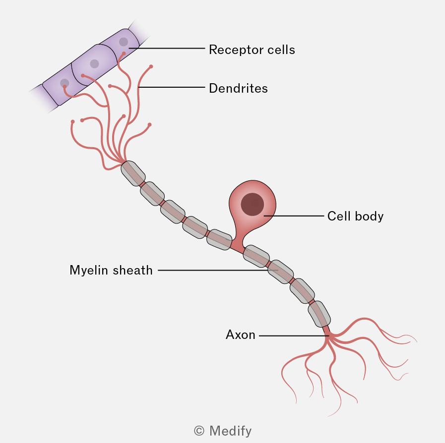
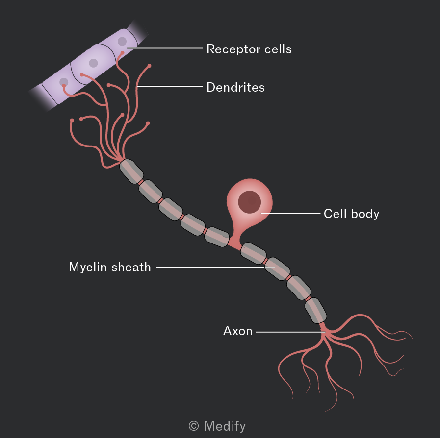
Sensory neurones carry impulses from receptors to the central nervous system, where they synapse with relay or motor neurones.
They have long dendrons that carry an impulse from the receptor to the cell body. The short axon transmits an impulse to the CNS.
Relay neurones connect sensory neurones to motor neurones within the CNS.
They have short dendrites that carry nerve impulses towards the cell body and a short axon that carries nerve impulses away from the cell body. Their short length allows rapid transmission of nerve impulses over the short distances within the CNS.
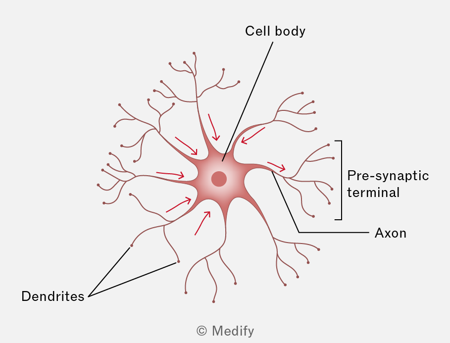
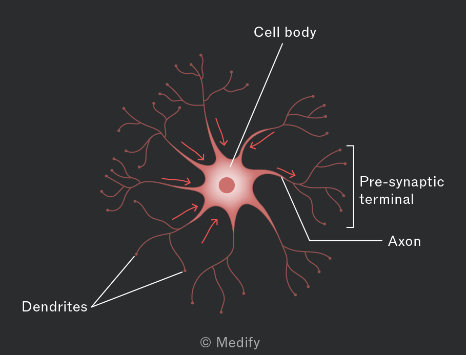
Motor neurones carry impulses from relay neurones (and sometimes directly from sensory neurones) to effectors such as muscles or glands.
They have many short dendrites that carry nerve impulses towards the cell body. They have a single long axon that carries nerve impulses away from the cell body in the CNS to an effector.
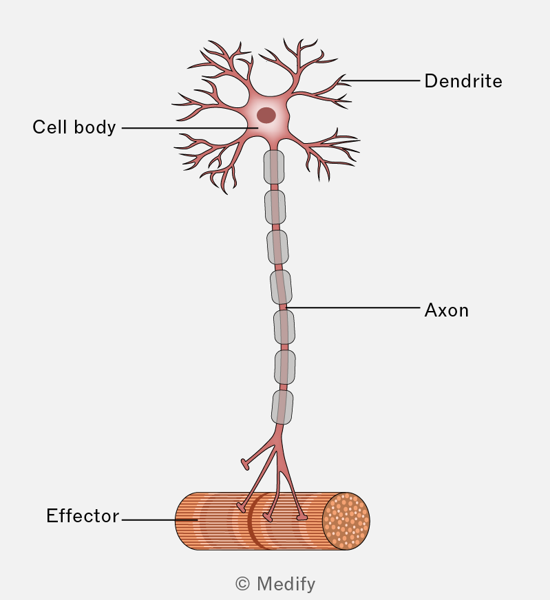
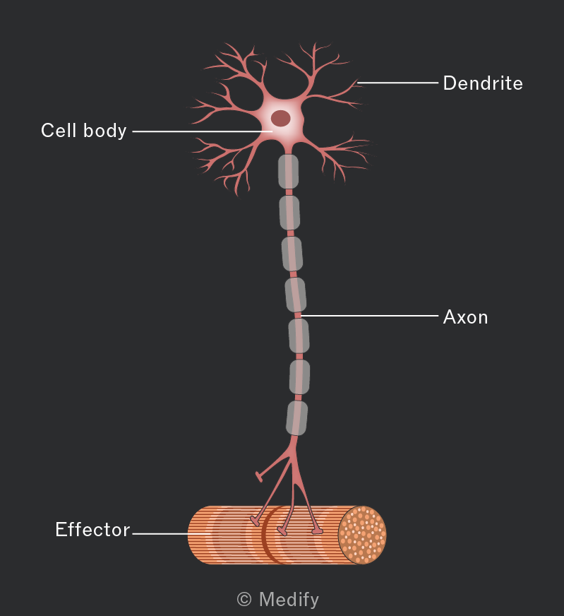
Myelin (layers of plasma membrane) is an electrical insulator.
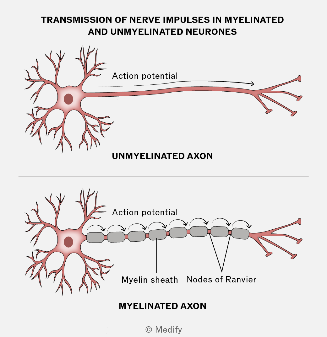
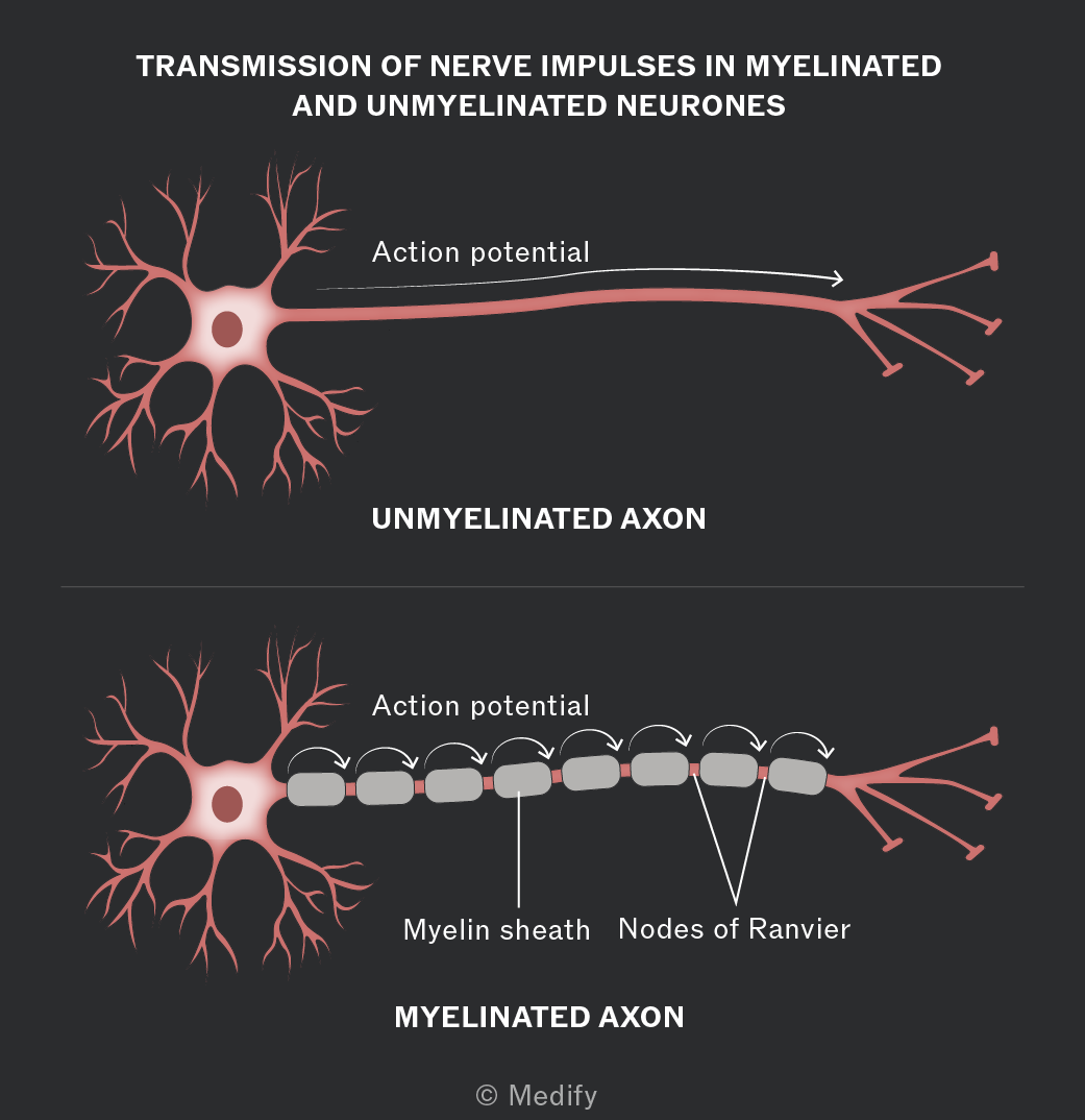
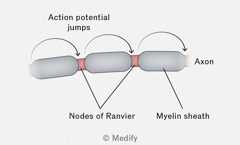
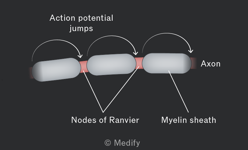
The myelin sheath is made up of Schwann cell membranes wrapped multiple times around the neurone’s axon. It insulates the axon and speeds up impulse transmission.
Schwann cells also perform phagocytosis and play a role in the regeneration of nerve cells.
Nodes of Ranvier are gaps between the Schwann cells making up the myelin sheath.
Electrical impulses jump between nodes, which speeds up signal transmission. This is known as saltatory conduction.
Electrical impulses move along a neurone by changing the voltage across its membrane.
The membrane is in one of two possible states, either resting potential or action potential.
When a neurone is not transmitting an action potential, it is at its resting potential. Neurones maintain a resting potential of around (in humans). In this state, the axon is polarised.
The inside of the axon is more negatively charged than the outside. This forms an electrochemical gradient.
Some ion channels are gated, and others are always open across the cell membrane of neurones.
The sodium–potassium pump uses ATP to move three ions out for every two ions pumped in, resulting in more sodium outside the cell and more inside the cytoplasm.
ions diffuse out of the neurone down the electrochemical gradient, but ions cannot diffuse in because the gated sodium ion channels are closed.
More positively charged ions are found outside the axon, so the inside of the neurone is negatively charged relative to the outside at resting potential.
At the resting potential, voltage–gated sodium channels are closed, and potassium ion channels are open.
Energy from a stimulus opens voltage–gated sodium channels, allowing to enter the axon down the electrochemical gradient. This triggers more sodium channels to open (positive feedback), and the membrane’s potential difference reaches
The voltage–gated sodium channels close and voltage–gated potassium channels open, so diffuses out of the axon. continues to move out of the axon causing hyperpolarisation. Potassium channels then close, and the sodium–potassium pump restores the resting potential (repolarisation) by moving out and into the axon.
In unmyelinated neurones, at the resting potential, there are ions outside and ions inside the axon.
An action potential depolarises the membrane by allowing ions to enter the axon. The influx of creates a localised electric current, causing voltage–gated sodium ion channels further along the axon to open.
More enters, causing further depolarisation and propagating the wave along the axon. Behind the depolarised region, sodium ion channels close and potassium ion channels open, allowing to leave the axon. Repolarisation occurs, restoring the neurone to a state ready for a new stimulus.
After an action potential, voltage–gated sodium channels remain closed for a short time so there can’t be another action potential. This is called the refractory period.
Action potentials travel in one direction and cannot overlap because of the refractory period.
In a myelinated neurone, depolarisation of an axon membrane occurs at the nodes of Ranvier.
The action potentials jump between nodes; this is known as saltatory conduction. This is more efficient and speeds up transmission as it is faster than a wave of depolarisation going along the whole length of the axon.
Repolarisation needs ATP, so this also reduces the amount of ATP that is required.
There are several factors that can affect the speed of an impulse, including:
- Axon diameter: the larger the diameter, the faster the transmission, as there is less resistance to the movement of ions in the axon.
- Temperature: the higher the temperature, the faster the transmission, as the rate of diffusion is increased.
- Myelin sheath: when present, transmission is faster owing to saltatory conduction.
If the threshold value is reached by a stimulus then an action potential is triggered. If this value is not met, there is no action potential.
The size of an action potential is always the same, regardless of the stimulus size.
This is known as the all-or-nothing principle, there is either an action potential or not.
A more intense stimulus results in more frequent action potentials, not bigger ones.
Threshold value in neurones can vary, so some are easier to depolarise than others.