Topic 1: Lifestyle, health and risk›Biological molecules (1.2, 1.12, 1.13, 1.14)

Biological molecules (1.2, 1.12, 1.13, 1.14)

An overview of biological molecules (1.2, 1.12, 1.13, 1.14) from Edexcel A level Biology A including: water, carbohydrates and lipids
8 min

Water, , is a simple covalent molecule. The central oxygen shares a single covalent bond with each hydrogen.

Water is polar because the oxygen atom attracts the shared electrons more strongly, meaning that it has a partial negative charge, while the hydrogen atoms are partially positive. This polarity results in water molecules interacting with each other through hydrogen bonds.
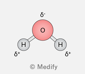
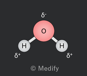
Diagram of a water molecule showing one oxygen atom (O) in the center with a red circle, connected to two hydrogen atoms (H) with gray circles. The oxygen atom has a delta negative (δ-) label above it, indicating partial negative charge. Each hydrogen atom has a delta positive (δ+) label beside it, indicating partial positive charge. The image is credited to Medify.
Add to favourites

Hydrogen bonds are strong electrostatic intermolecular interactions. They form between water molecules as the partially positively charged hydrogen atom is attracted to the lone electron pair of the electronegative oxygen atom.

Hydrogen bonds are responsible for some unique properties of water, such as cohesion, adhesion, heat capacity, and surface tension.
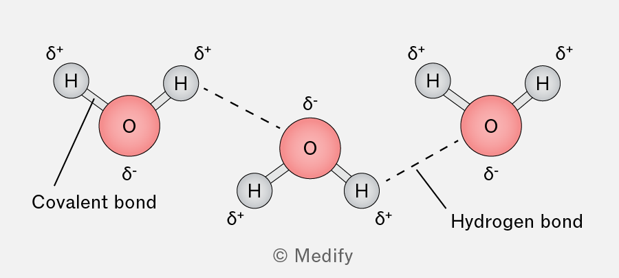
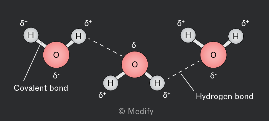
The image shows three water molecules, each with an oxygen atom labeled as 'O' and hydrogen atoms labeled as 'H'. The oxygen atoms are in red, and the hydrogen atoms are in gray. Each oxygen atom is connected to two hydrogen atoms with solid lines labeled 'Covalent bond'. The oxygen atoms have a delta negative (δ-) sign, and the hydrogen atoms have a delta positive (δ+) sign. Dashed lines labeled 'Hydrogen bond' connect the hydrogen atoms of one water molecule to the oxygen atom of another, indicating hydrogen bonding between molecules. The layout illustrates the polar nature of water molecules and the formation of hydrogen bonds between them.
Add to favourites

Water is a polar molecule, so it interacts with charged species, behaving as a solvent. This property facilitates numerous metabolic reactions that occur in aqueous environments.
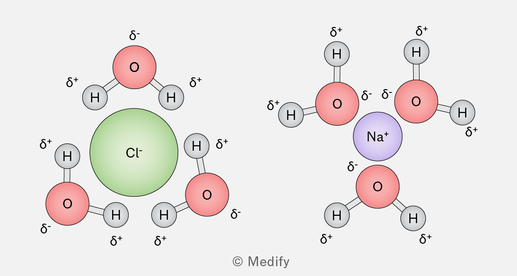
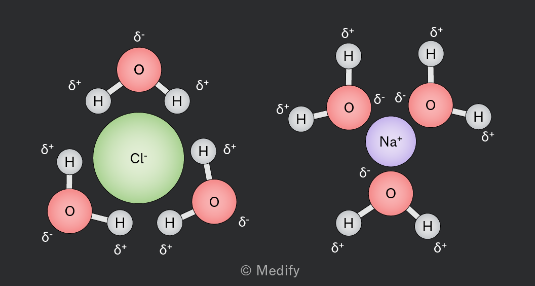
The image shows two diagrams of ions surrounded by water molecules. On the left, a green chloride ion (Cl⁻) is surrounded by three water molecules. Each water molecule has a red oxygen atom labeled with δ⁻ and two gray hydrogen atoms labeled with δ⁺. The hydrogen atoms are oriented towards the chloride ion. On the right, a purple sodium ion (Na⁺) is surrounded by three water molecules. Each water molecule has a red oxygen atom labeled with δ⁻ and two gray hydrogen atoms labeled with δ⁺. The oxygen atoms are oriented towards the sodium ion.

In ionic compounds such as sodium chloride, the positive ions are attracted to the partially negative oxygen of the water molecules, while ions are attracted to the partially positive hydrogens.

Add to favourites

As water freezes, it becomes less dense as hydrogen bonds lock the molecules into a fixed crystalline structure, where there is increased spacing between molecules. This reduced density causes ice to float.

A lot of energy is needed to break the hydrogen bonds between water molecules, which means that the structure of water also results in a high boiling point.
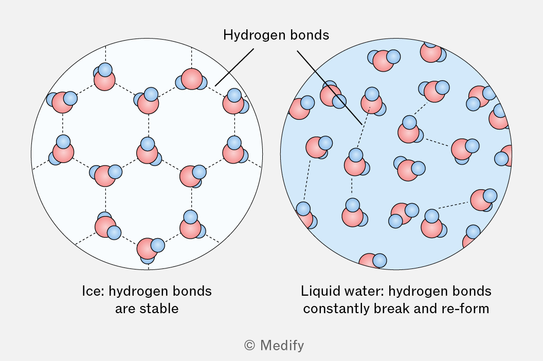
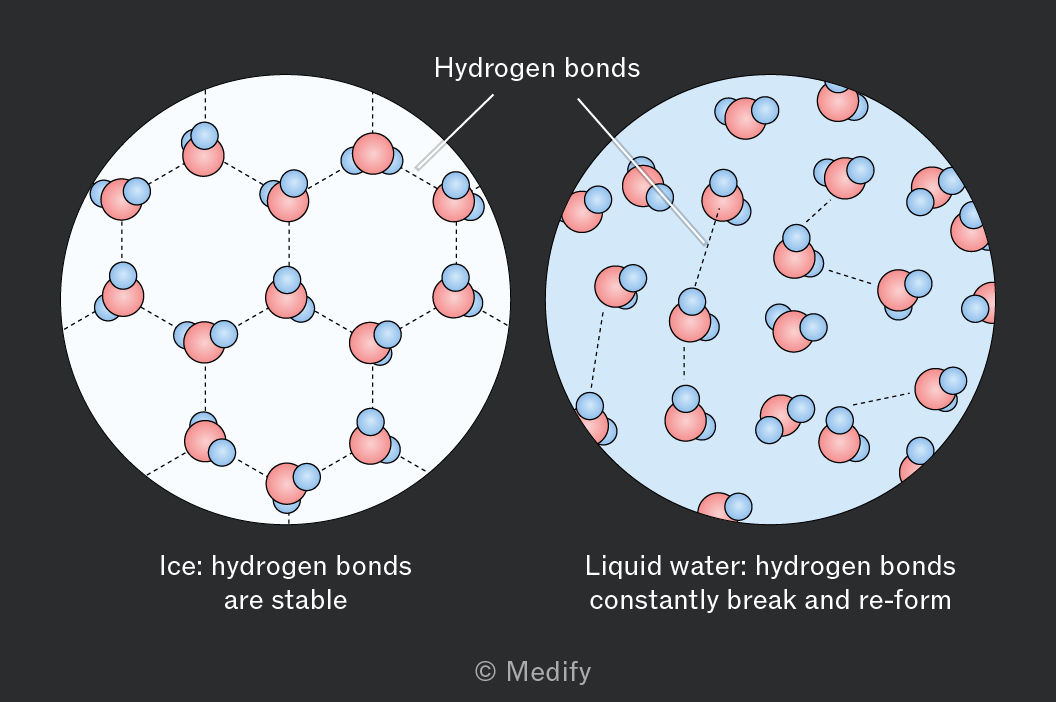
A diagram comparing the molecular structure of ice and liquid water. On the left, ice is depicted with stable hydrogen bonds between molecules, while on the right, liquid water shows hydrogen bonds that constantly break and reform.
Add to favourites

Carbohydrates are biological molecules made from sugars.

Monosaccharides are the simplest form of sugar, consisting of a single monomer. Examples include glucose, fructose, and galactose.

Two monosaccharides join via a condensation reaction to form a disaccharide. Many monosaccharides join via condensation reactions to form a polysaccharide.

Add to favourites

Monosaccharides are linked by glycosidic bonds, which form in a condensation reaction between hydroxyl () groups on adjacent monosaccharides, releasing a molecule of water.

The name of the glycosidic bond indicates the carbon atoms involved in the linkage. For example, an 1,4 glycosidic bond forms between carbon 1 of one -glucose molecule and carbon 4 of another.

Two -glucose molecules combine to form the disaccharide maltose, joined by an 1,4 glycosidic bond.
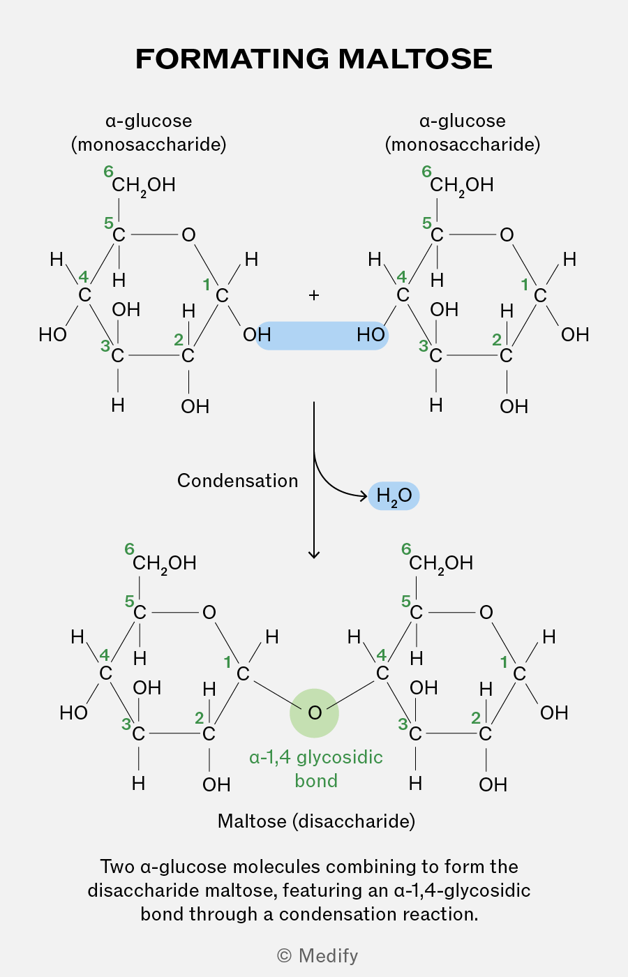
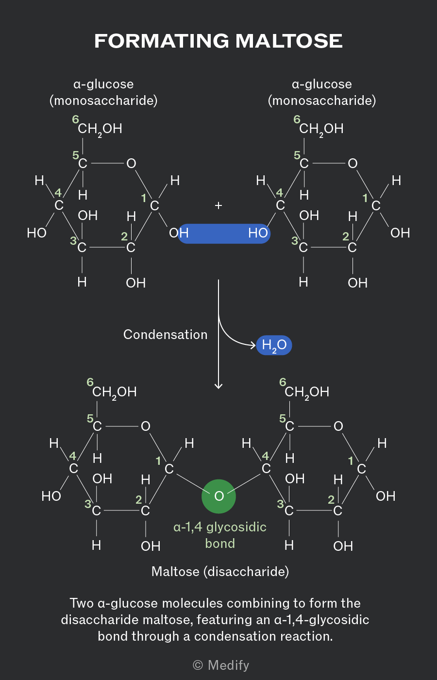
The image illustrates the formation of maltose from two alpha-glucose molecules. At the top, two alpha-glucose structures labeled as 'alpha-glucose (monosaccharide)' are shown. Each glucose molecule has carbon atoms numbered 1 through 6, with hydroxyl (OH) and hydrogen (H) groups attached. The two glucose molecules are linked by a blue highlighted area between the OH groups of one glucose's carbon 1 and the other glucose's carbon 4. An arrow labeled 'Condensation' points downward to the formation of maltose, labeled 'Maltose (disaccharide).' In the maltose structure, the glucose units are linked by an 'alpha-1,4 glycosidic bond,' highlighted in green. A blue label 'H2O' indicates the release of water during the reaction. Additional text reads, 'Two alpha-glucose molecules combining to form the disaccharide maltose, featuring an alpha-1,4-glycosidic bond through a condensation reaction.'
Add to favourites

Two monosaccharides bonded together via a glycosidic bond form disaccharides:

  • Maltose = -glucose + -glucose
  • Sucrose = -glucose + -fructose
  • Lactose = -galactose + glucose.
Add to favourites

The glycosidic bonds between monosaccharides can be broken down through hydrolysis reactions, where one molecule of water is added to break the bond.

Hydrolysis is crucial in biological processes, such as digestion and the breakdown of stored carbohydrates for cellular respiration.
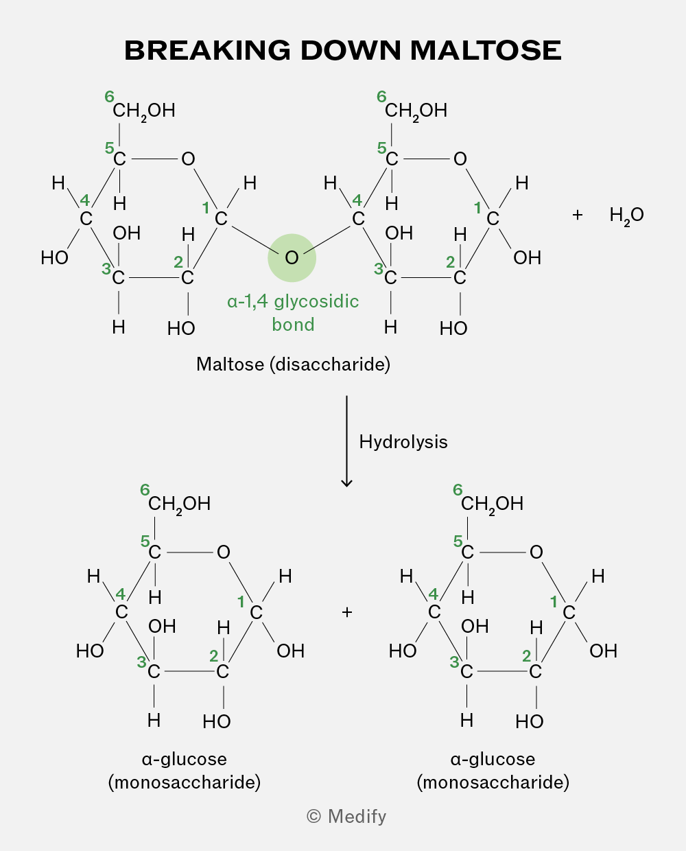
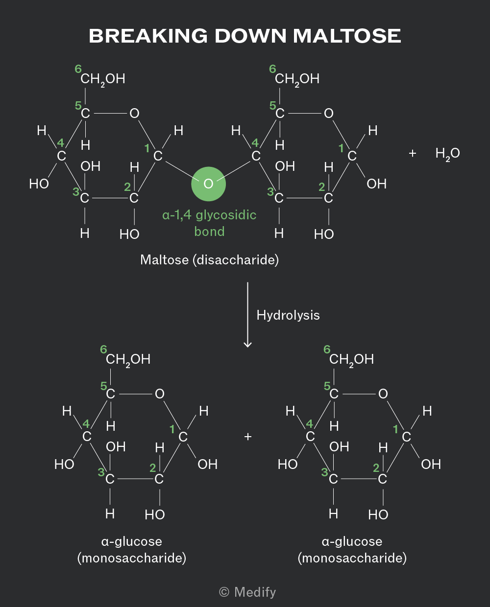
The image titled 'Breaking Down Maltose' shows the chemical structure of maltose, a disaccharide, which is composed of two α-glucose (monosaccharide) units. In the top section, two glucose rings are joined by an α-1,4 glycosidic bond, highlighted in green, between the oxygen of the first glucose and the carbon of the second glucose. Each glucose molecule has carbon atoms numbered from 1 to 6. The left glucose has a CH2OH group at carbon 6, an OH group at carbon 4, and HO at carbon 3. The right glucose is similar. The reaction shows hydrolysis, where water (H2O) is added, breaking the glycosidic bond and resulting in two separate α-glucose molecules. Each glucose molecule has the same structure, with CH2OH at carbon 6, OH at carbon 4, and HO at carbon 3. The process is indicated by a downward arrow labeled 'Hydrolysis'.
Add to favourites

Starch is the insoluble, primary storage polysaccharide in plants and is formed from a few hundred to a few thousand -glucose monosaccharides bonded together. It is made from a mixture of two polysaccharides.

Add to favourites

The two polysaccharides which form starch are:

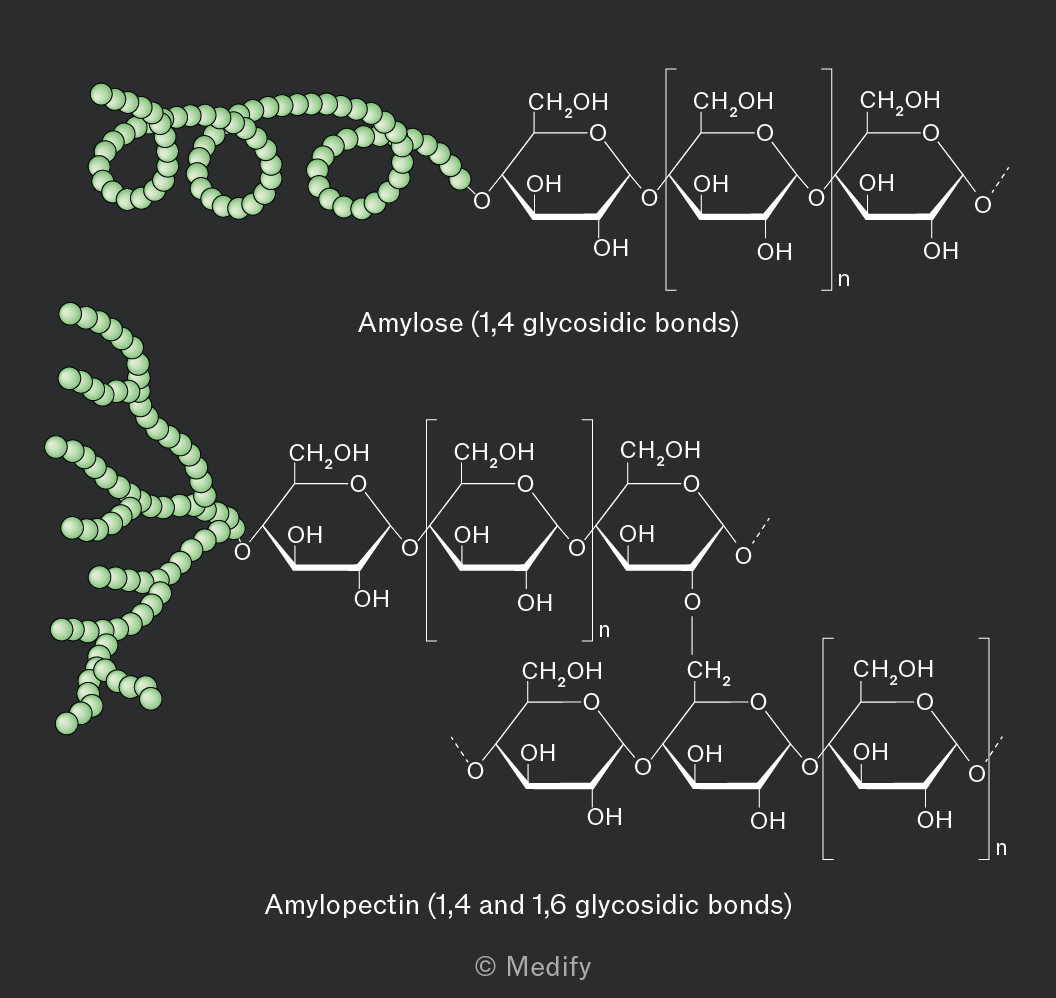
  • Amylose constitutes 10–30 of starch and has an unbranched, helix-shaped chain of -glucose molecules linked by 1,4 glycosidic bonds.
  • Amylopectin constitutes 70–90 of starch and has both 1,4 glycosidic bonds and 1,6 glycosidic bonds, creating a branched molecule. Enzymes involved in breaking down these molecules can simultaneously work at multiple branch points, leading to rapid release of glucose for energy. More accessible hydroxyl groups make amylopectin more soluble in water.
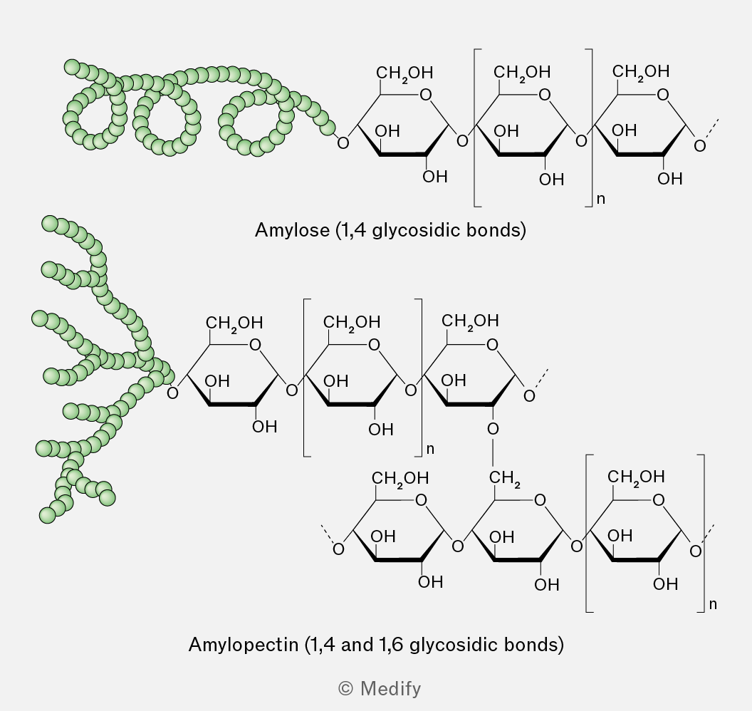
The image shows two types of polysaccharides, amylose and amylopectin. The top section illustrates amylose, labeled 'Amylose (1,4 glycosidic bonds)', with a coiled structure on the left and a linear chain of glucose units on the right. Each glucose unit is depicted with hydroxyl (OH) groups and an oxygen bridge indicating 1,4 glycosidic bonds. The bottom section shows amylopectin, labeled 'Amylopectin (1,4 and 1,6 glycosidic bonds)', with a branched structure on the left and a glucose chain on the right. The chain includes glucose units connected by 1,4 glycosidic bonds with occasional 1,6 branching points. Each glucose unit is similarly depicted with hydroxyl groups and connections indicating glycosidic bonds. The illustration uses green spheres to represent the coiled and branched structures of the polysaccharides.
Add to favourites

Glycogen is the primary storage polysaccharide in animals and fungi, characterised by a highly branched and uncoiled structure.

The highly branched structure of glycogen means there are numerous terminal glucose units leading to rapid release of glucose for energy. Branched polysaccharides with more accessible hydroxyl groups are also more soluble in water.

Glycogen’s structure is similar to amylopectin, but its branching is more extensive. This makes it more water-soluble and its terminal glucose molecules more accessible to enzymes.
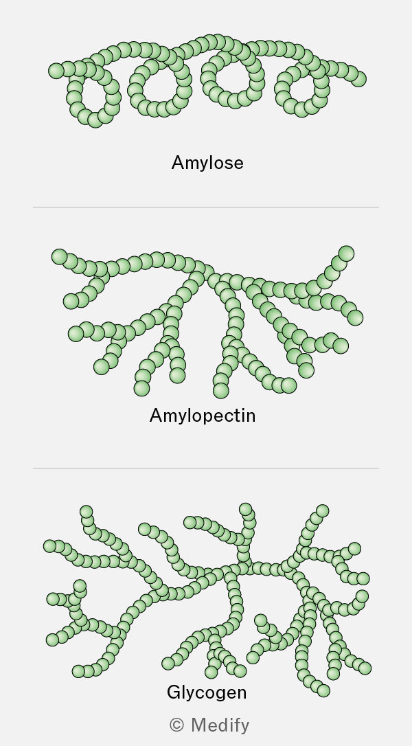
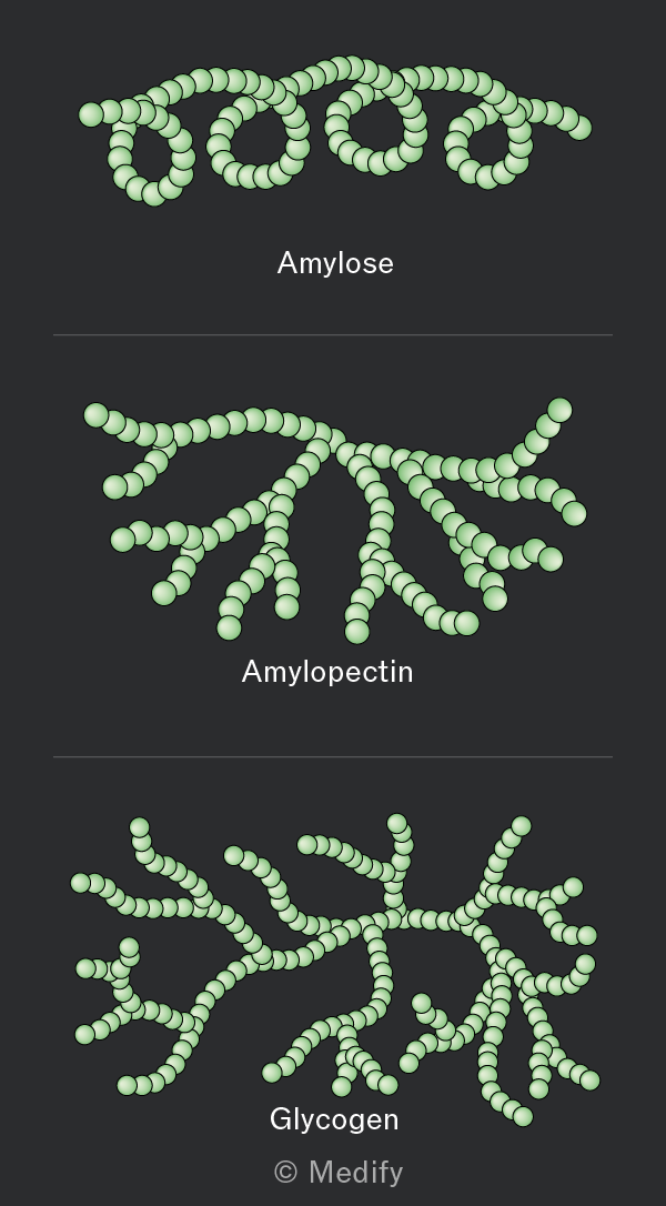
The image contains three diagrams of polysaccharide structures. The top diagram is labeled 'Amylose' and shows a linear chain of green spheres coiled into a helix. The middle diagram is labeled 'Amylopectin' and depicts a branched structure with a main chain of green spheres and several branching chains. The bottom diagram is labeled 'Glycogen' and illustrates a highly branched structure with green spheres forming multiple short, branching chains. The bottom of the image includes the copyright label '© Medify'.
Add to favourites

Amylose is coiled, making it insoluble and compact which is ideal for storage.

Amylopectin and glycogen are both highly branched molecules, making them compact and allowing more energy to be stored. Their branches provide multiple free ends where glucose molecules can be rapidly added in condensation reactions or removed in hydrolysis reactions, enabling quick adaptation to cellular energy demands.

Add to favourites

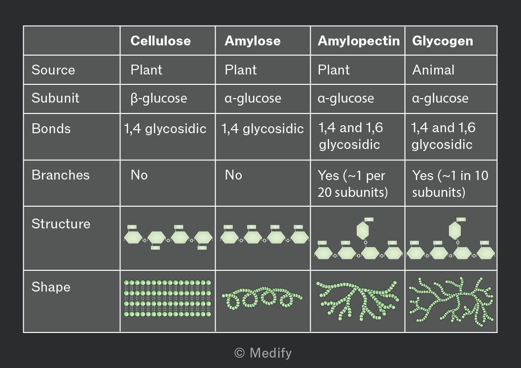
The bonding and structure of the key polysaccharides in plants and animals are summarised below.
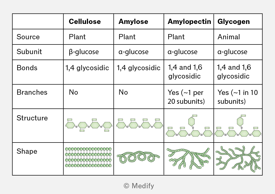
A table comparing the characteristics of cellulose, amylose, amylopectin, and glycogen. The columns are labeled: Cellulose, Amylose, Amylopectin, and Glycogen. The rows are labeled: Source, Subunit, Bonds, Branches, Structure, and Shape. 

- Source: Cellulose (Plant), Amylose (Plant), Amylopectin (Plant), Glycogen (Animal).
- Subunit: Cellulose (β-glucose), Amylose (α-glucose), Amylopectin (α-glucose), Glycogen (α-glucose).
- Bonds: Cellulose (1,4 glycosidic), Amylose (1,4 glycosidic), Amylopectin (1,4 and 1,6 glycosidic), Glycogen (1,4 and 1,6 glycosidic).
- Branches: Cellulose (No), Amylose (No), Amylopectin (Yes, approximately 1 per 20 subunits), Glycogen (Yes, approximately 1 in 10 subunits).
- Structure: Depicts linear chains for cellulose and amylose, branched chains for amylopectin and glycogen.
- Shape: Cellulose (parallel lines of circles), Amylose (spiral of circles), Amylopectin (branched cluster of circles), Glycogen (highly branched cluster of circles).
Add to favourites

Triglycerides are non-polar, hydrophobic molecules consisting of one glycerol molecule (an alcohol) and three fatty acid chains. They are the main components of fats and oils and are insoluble in water, but soluble in organic solvents, e.g., ethanol.

Fatty acids have a methyl group () at one end of a hydrocarbon chain (R group) and a carboxyl group () at the other end.
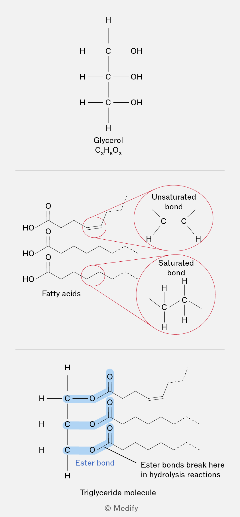
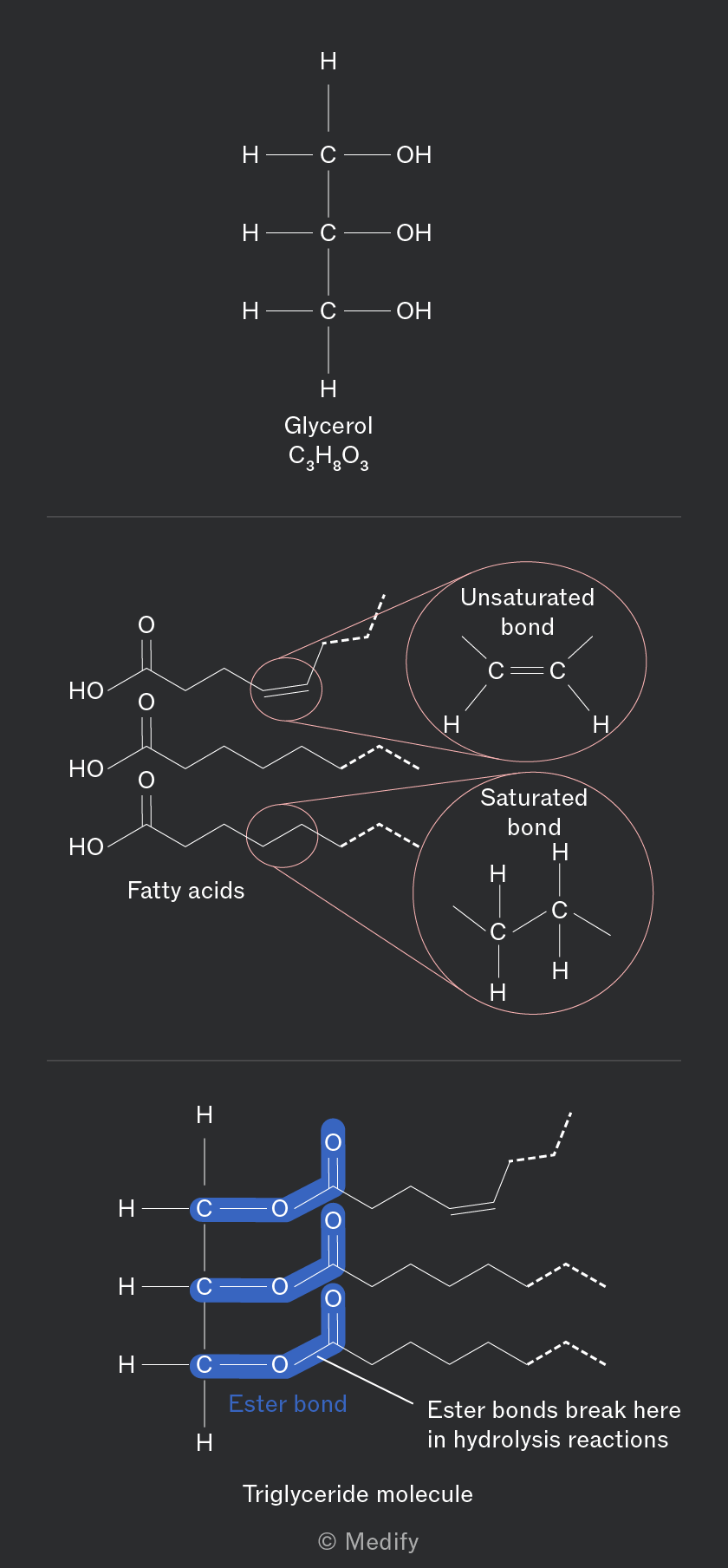
The image is divided into three sections. The top section shows the structure of glycerol with three carbon atoms each bonded to a hydroxyl group (OH) and hydrogen atoms, labeled as 'Glycerol C3H8O3'. The middle section depicts fatty acids with two zoomed-in circles showing an unsaturated bond (C=C) labeled 'Unsaturated bond', and a saturated bond (C-C) labeled 'Saturated bond'. The bottom section illustrates a triglyceride molecule where three fatty acid chains are connected to a glycerol backbone, highlighting 'Ester bond' in blue. The text 'Ester bonds break here in hydrolysis reactions' is annotated next to the ester bonds, and below is the label 'Triglyceride molecule'.
Add to favourites

Triglycerides contain a high ratio of hydrogen:oxygen atoms, which means that they release water when oxidised. This is called metabolic water and can be a source of hydration, especially for animals living in arid environments.

Triglycerides are a source of energy owing to the high number of bonds which store energy.

Add to favourites

Triglycerides are formed through a condensation reaction where each hydroxyl group () of glycerol reacts with a carboxyl group () of a fatty acid to form an ester bond.

Three fatty acids are attached to one glycerol molecule, three water molecules are released per triglyceride formed.

Ester bonds are broken during the digestion of the triglyceride molecule through hydrolysis reactions consuming three water molecules per triglyceride hydrolysed.
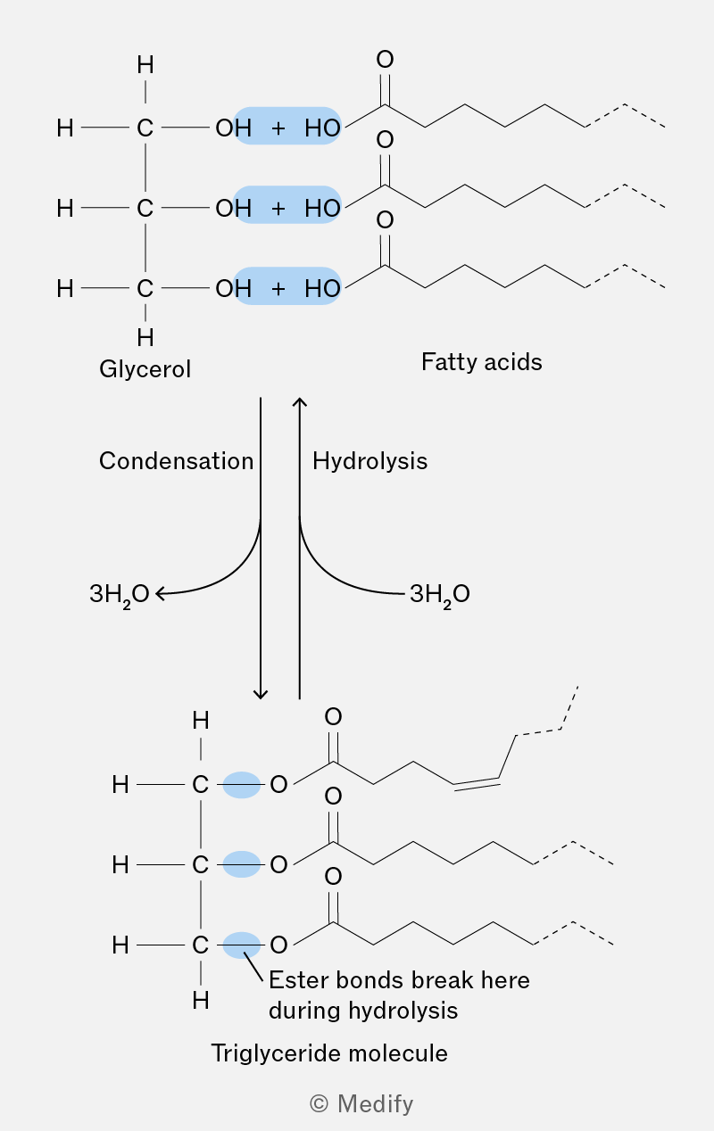
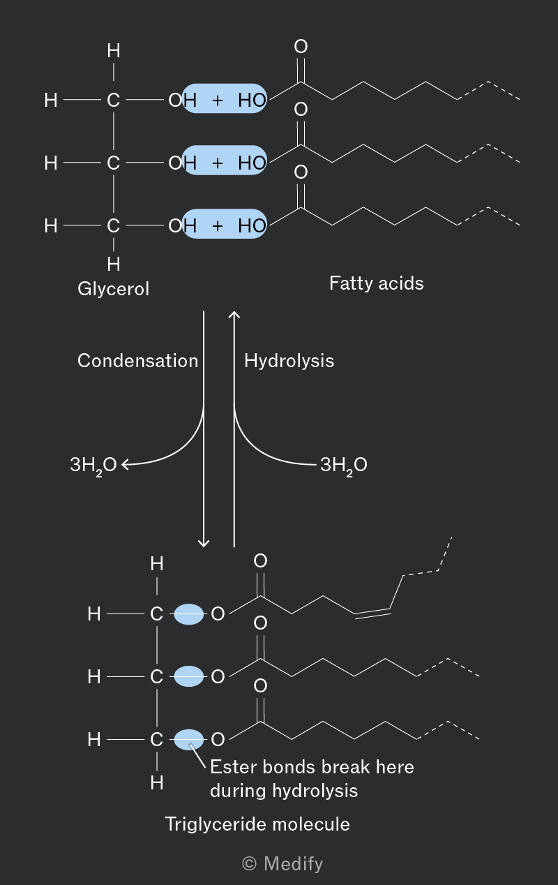
The image shows a chemical diagram illustrating the formation and breakdown of a triglyceride molecule. At the top, there are structures labeled 'Glycerol' and 'Fatty acids'. Glycerol is depicted with three hydroxyl (OH) groups, each paired with a plus sign and a hydroxyl group (HO) from the fatty acids, highlighted in blue. To the right, fatty acids are shown with long hydrocarbon chains and carboxyl groups (C=O). Below this, arrows labeled 'Condensation' and 'Hydrolysis' point downwards and upwards, respectively, with '3H2O' indicated for both processes. The bottom section shows a triglyceride molecule, where the glycerol and fatty acids are connected by ester bonds, highlighted in blue. An annotation states 'Ester bonds break here during hydrolysis'. The molecule is labeled 'Triglyceride molecule'.
Add to favourites

Fatty acids can differ in hydrocarbon chain length (R group) and in their saturation.

Saturated fatty acids have no double bonds. They form a straighter structure and pack tightly together, resulting in higher melting points and being in a solid state at room temperature. They are primarily found in animal fats.

Unsaturated fatty acids contain one (monounsaturated) or more (polyunsaturated) double bonds, leading to ‘kinks’ in the hydrocarbon chains that prevent tight packing. This lowers the melting point, so they are usually liquids at room temperature. They are typically found in vegetable oils.
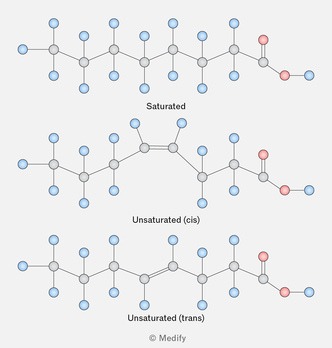
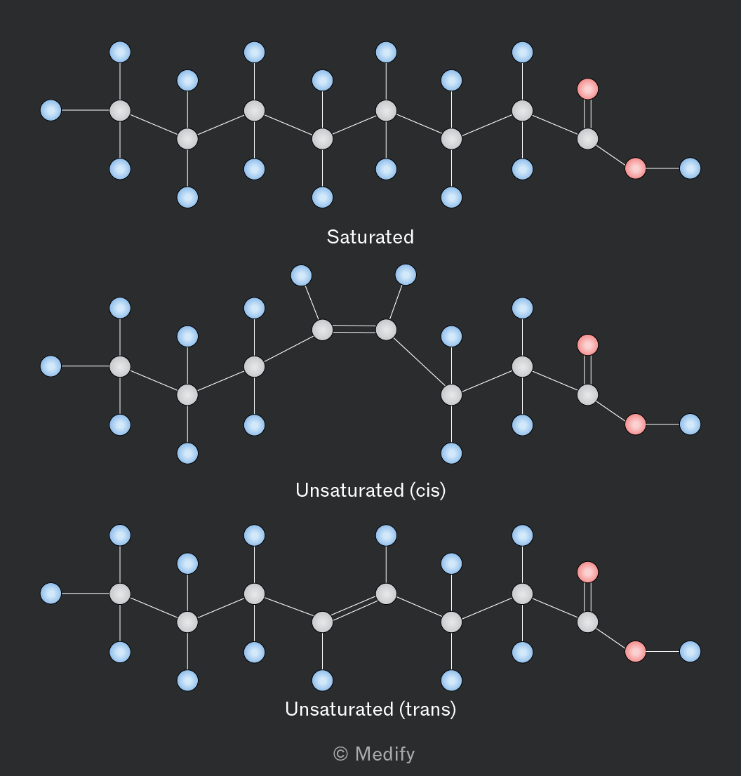
The image shows three chemical structures labeled as 'Saturated,' 'Unsaturated (cis),' and 'Unsaturated (trans).' The 'Saturated' structure is a linear chain of carbon atoms with single bonds, featuring hydrogen atoms (blue) and a carboxyl group (red) at one end. The 'Unsaturated (cis)' structure has a similar chain with one double bond creating a kink in the chain. The 'Unsaturated (trans)' structure also has a double bond but maintains a straighter chain due to the trans configuration. Each structure has labeled carbon and hydrogen atoms and a carboxyl group in red at the end.
Add to favourites