
Mass transport in animals (1.1, 1.4, 1.3, 1.17)
On this page
Both surface area and volume are crucial factors in determining how substances move into, out of, and within organisms.
Surface area is the total external area of the organism that is in contact with the environment.
Volume is the total internal space within an organism.
As organism size increases, its surface area-to-volume ratio decreases.
Single–celled organisms have a high surface area-to-volume ratio, enabling efficient diffusion of nutrients, gases, and waste owing to short diffusion distances.
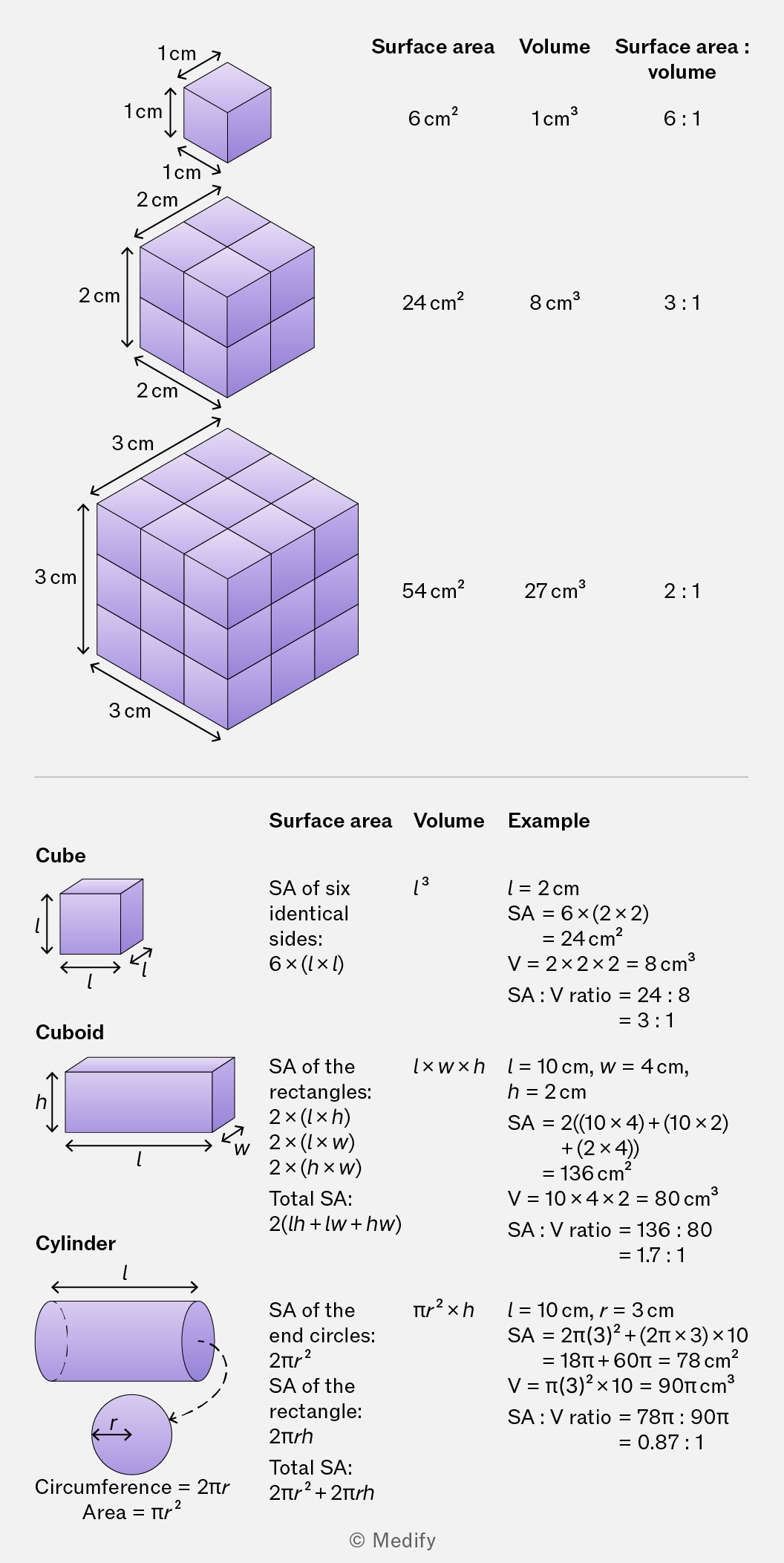
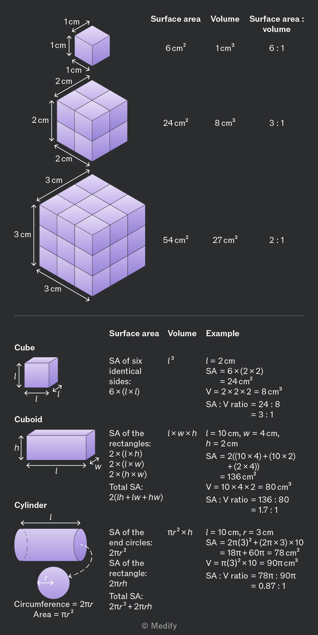
Multicellular organisms can be large and complex, with specialised tissues and organ systems. Metabolic demands include taking in nutrients, exchanging gases, and excreting waste products.
Mass transport systems are needed to overcome the limitations of diffusion in moving substances (e.g., hormones, gases, nutrients, wastes) long distances between exchange sites and cells.
Mass flow is the bulk movement of substances driven by a pressure difference. It allows rapid, directed transport over long distances in large organisms.
Diffusion occurs over short distances at exchange surfaces, where substances move down concentration gradients.
Mass transport systems link these exchange surfaces, enabling efficient distribution of gases, nutrients and wastes. They help maintain concentration gradients, ensuring effective diffusion and supporting normal cellular function.
Circulatory systems provide a means of mass transport in multicellular organisms.
They consist of a:
- transport medium
- system of vessels
- mechanism to generate pressure and drive movement.
Circulatory systems can be open or closed.
In open circulatory systems, haemolymph is pumped into body cavities (haemocoel) rather than remaining within a closed network of vessels. This occurs in arthropods and many molluscs, where flow is low-pressure and less directed than in closed systems.
In insects, a tubular heart pumps haemolymph forward. It is released into the haemocoel, where it bathes tissues before returning to the heart via valves. Flow is maintained by peristaltic contractions of the body.
Oxygen delivery is largely independent, as tracheae supply oxygen directly to tissues.
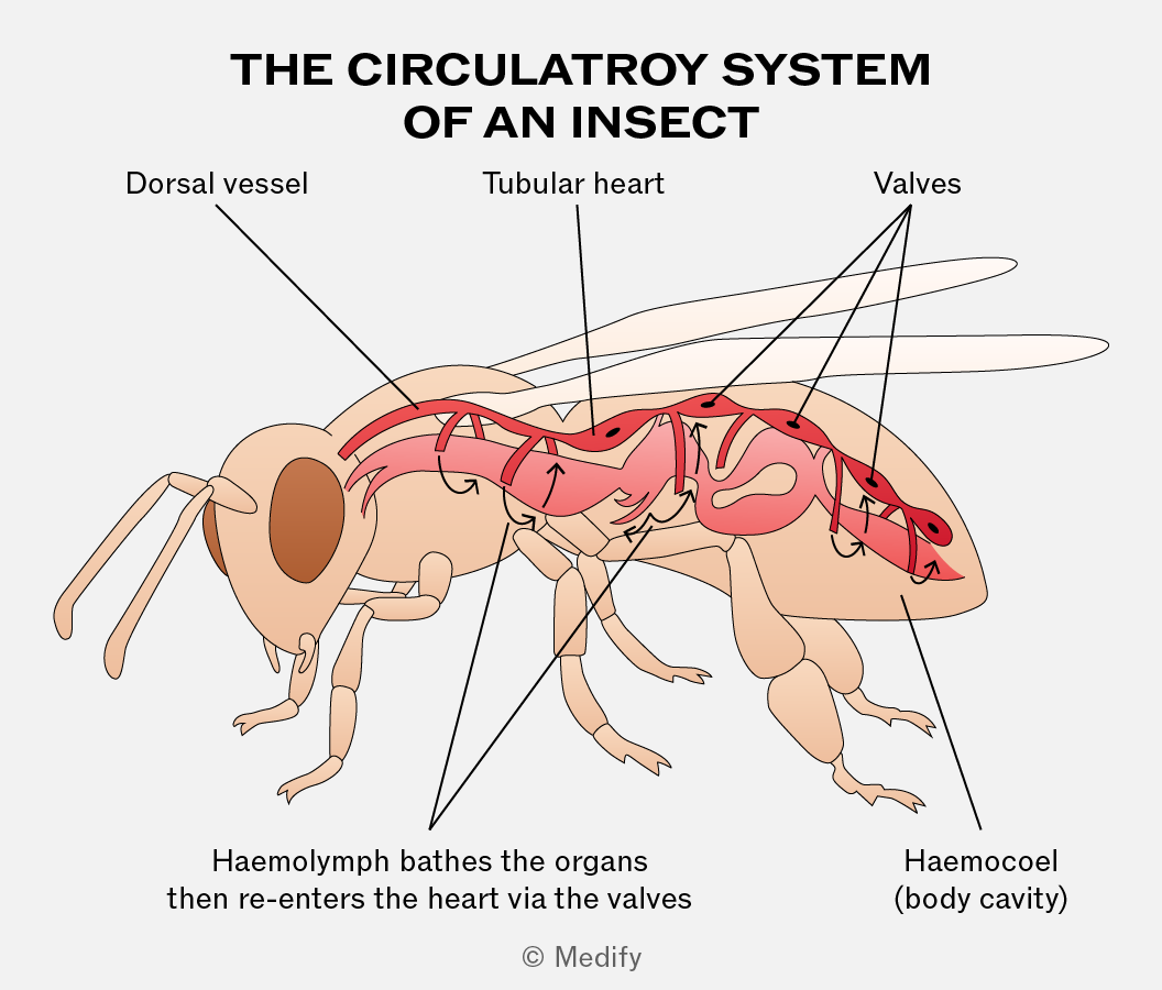
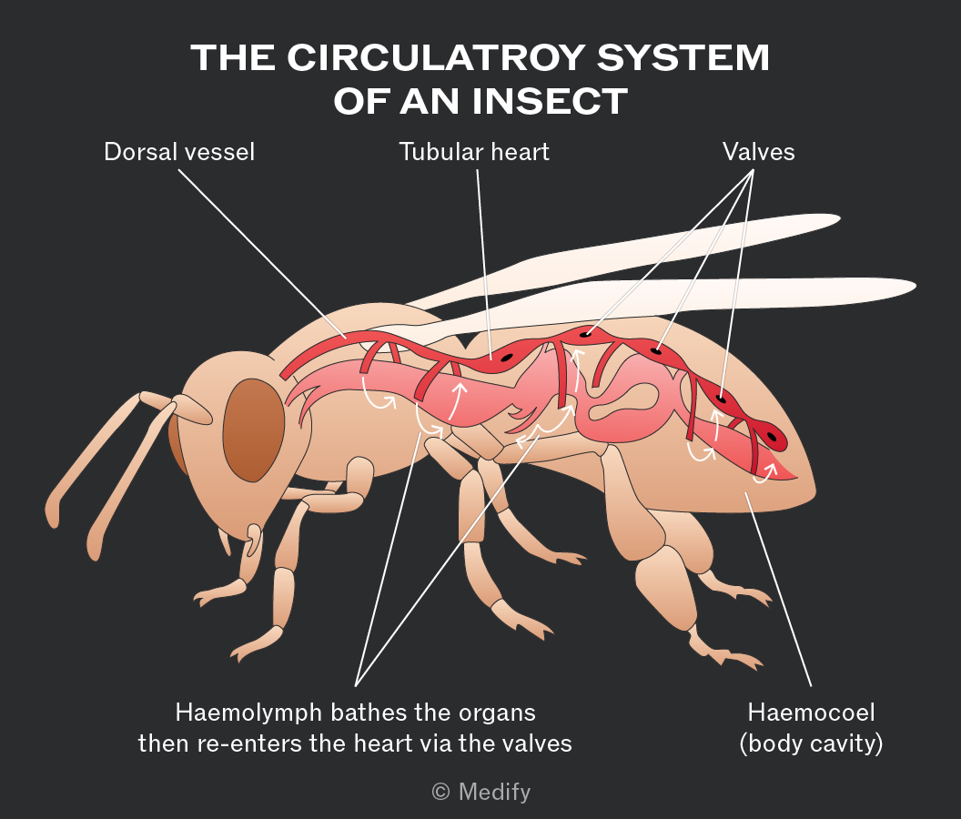
In an open circulatory system, the transport fluid (haemolymph) leaves the vessels and directly surrounds tissues.
In a closed circulatory system, the transport fluid (blood) remains within a continuous system of vessels. They work at higher pressure and are more efficient (faster and more controlled).
Closed circulatory systems can be single or double, depending on how many times blood passes through the heart in a circuit of the body.
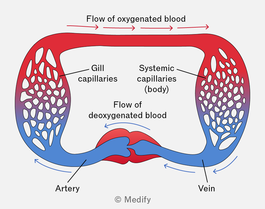
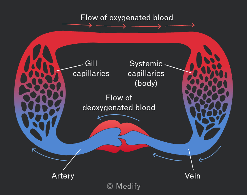
In single circulatory systems, such as in fish, blood passes once through the heart during a complete circuit of the body:
- Deoxygenated blood is pumped to the gills for gas exchange.
- Oxygenated blood then circulates through the body tissues, delivering oxygen and nutrients, and returns, deoxygenated to the heart.
It is less efficient than double circulatory systems as blood loses pressure after the gills.
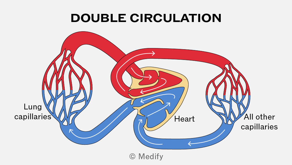
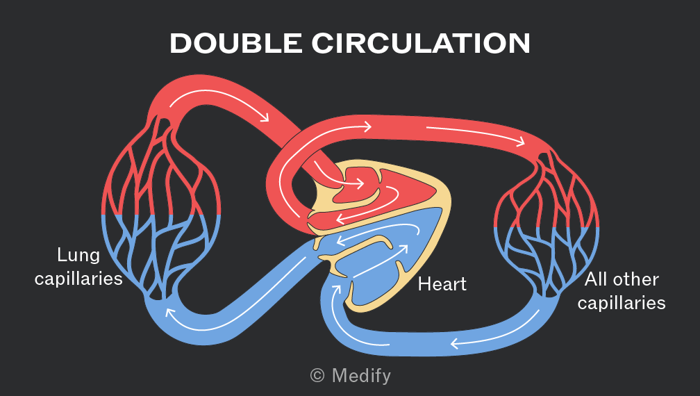
In a double circulatory system such as in mammals and birds, blood passes twice through the heart per circuit.
The right side of the heart pumps deoxygenated blood to the lungs (pulmonary circulation) while oxygenated blood returns to the left side of the heart and is pumped throughout the body (systemic circulation).
The heart pumps oxygenated blood at high pressure through a network of vessels to body tissues, ensuring large surface area for diffusion, short diffusion distances, steep diffusion gradients for fast oxygen and nutrient delivery.
The composition of blood vessels varies by function. The presence and relative thickness of each layer differs between vessels.
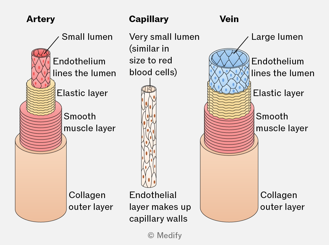
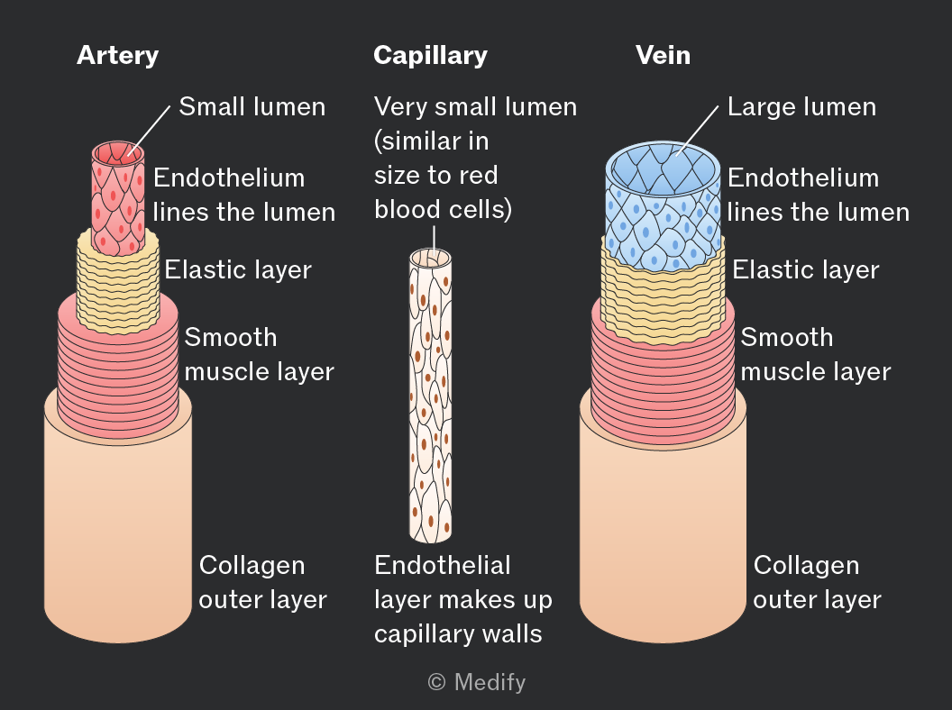
Arteries transport blood away from the heart rapidly and at high pressure.
Arteries have thick walls, muscle layers and elastic layers compared with veins:
- Collagen walls restrict the maximum stretch of the vessel as blood is pumped under high pressure.
- Smooth muscle controls blood flow through constricting and relaxing the vessel.
- Elastic fibres maintain high blood pressure throughout the arteries ensuring blood can travel long distances, smoothly and quickly.
- No valves (other than in the arteries at the point where they leave the heart).
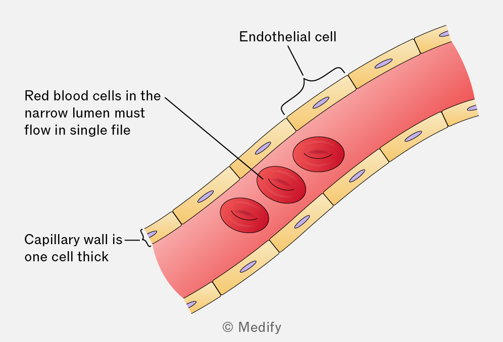
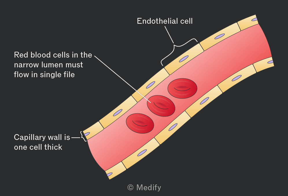
Capillaries are a network of small vessels connecting arterioles to venules and surrounding tissues.
- Their walls are a single layer of endothelial cells reducing diffusion distance between blood and tissue cells and increasing diffusion speed.
- Small diameter slows blood flow, increasing the time available for diffusion and maintaining high pressure and a steep concentration gradient.
- Gaps between endothelial cells allow cells and substances to pass out of the vessels and into the fluid surrounding tissues (e.g., white blood cells).
- Highly branched network create larger surface area for metabolite exchange.
Veins transport blood back to the heart, usually at low pressure.
- Low blood pressure means there is only a need for a thin collagen layer. This also enables compression of vessels by surrounding muscles to generate pressure for circulation.
- Thin muscle layer compared with arteries.
- Thin elastic layer owing to low pressure within the vessel meaning stretch and recoil are not needed.
- Valves are present throughout veins to ensure that blood flowing at low pressure moves only in one direction.
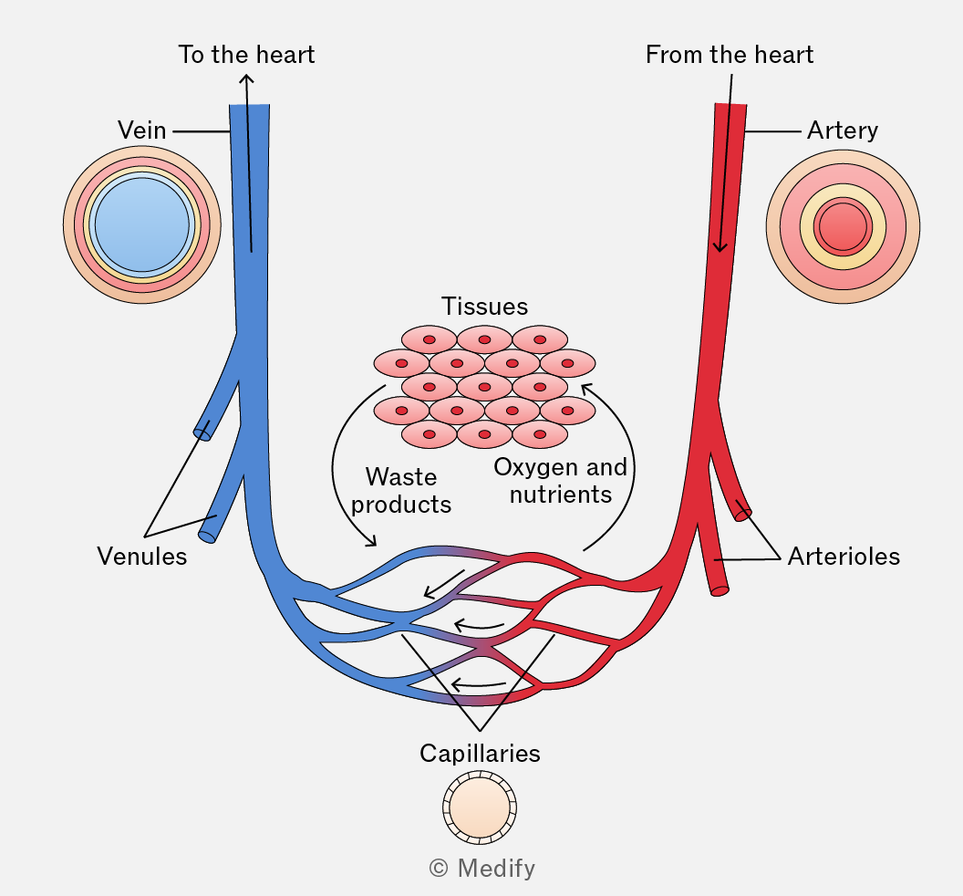
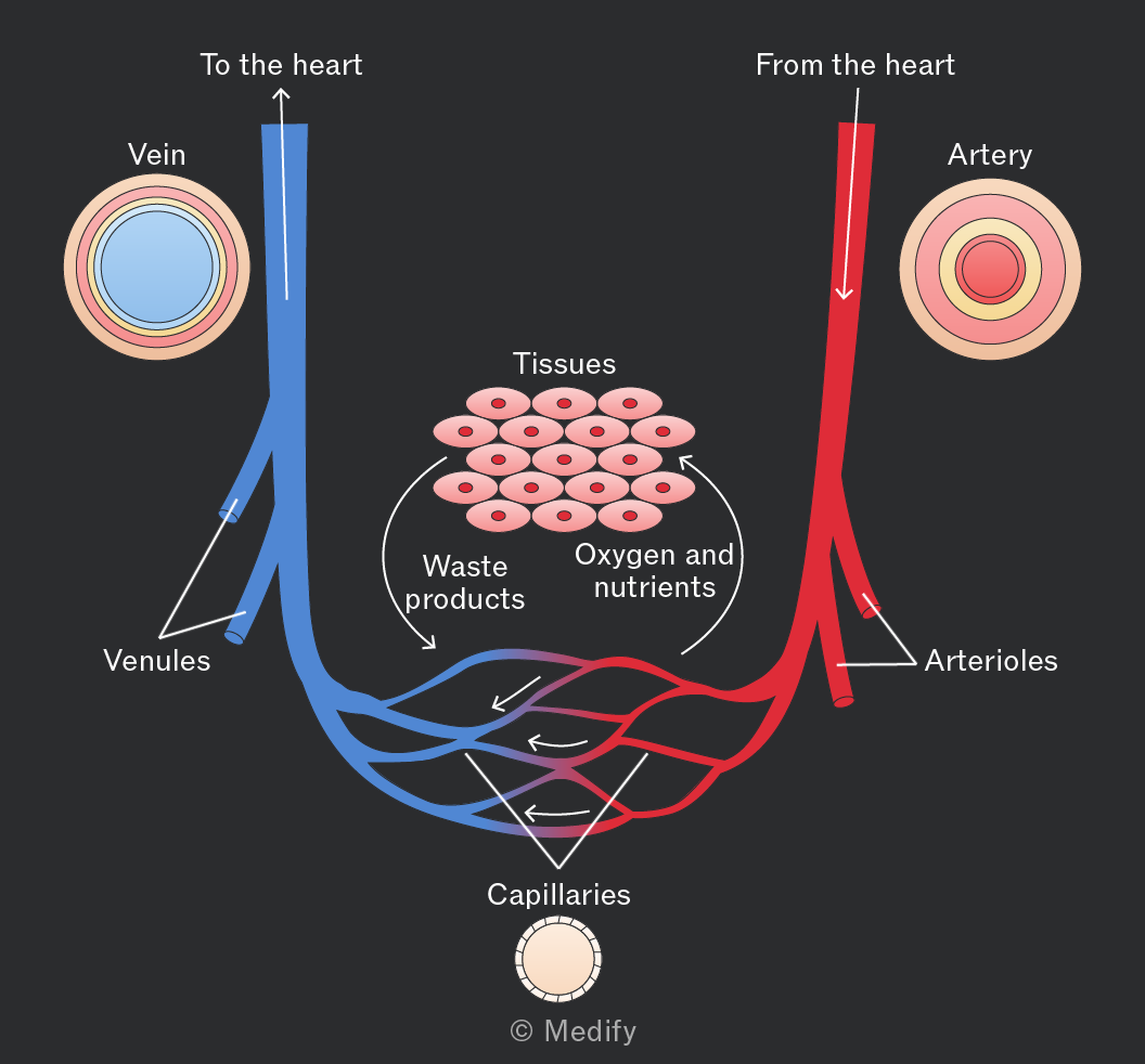
As blood flows through capillaries, some plasma leaks out through gaps in the capillary walls to surround body cells, forming tissue fluid.
Tissue fluid facilitates the exchange of substances including amino acids, ions, glucose, oxygen, carbon dioxide and other waste products between cells and the blood. Large plasma proteins are too big to pass through capillary walls and remain in the blood.
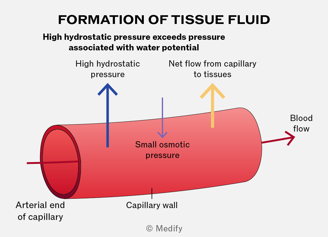
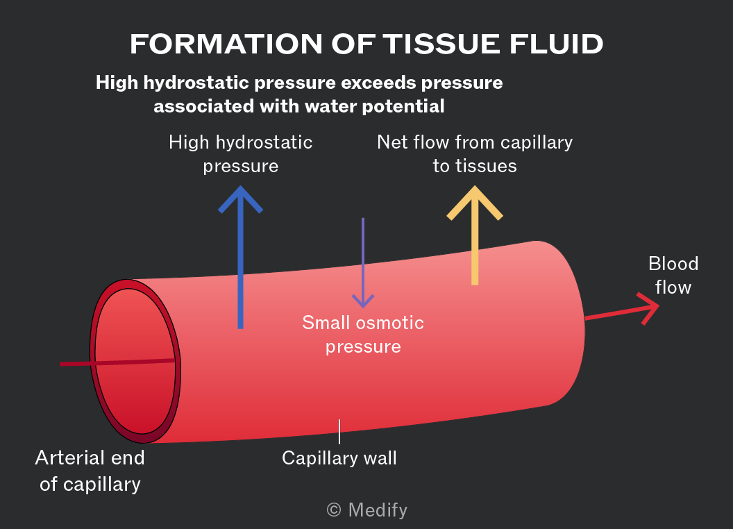
Hydrostatic pressure is exerted by blood on the walls of the capillaries as a result of heart contractions.
At the arterial end of a capillary, hydrostatic pressure is high enough to push fluid out of the capillaries to become tissue fluid. Large proteins and cells remain in the blood. This is called ultrafiltration (filtration under pressure).
After metabolites and gases have been exchanged, approximately 90 of the plasma lost as tissue fluid at the arterial end of a capillary is reabsorbed at the venous end.
The reduction in hydrostatic pressure and the low water potential of the blood (caused by plasma proteins) result in fluid moving back into the capillary.
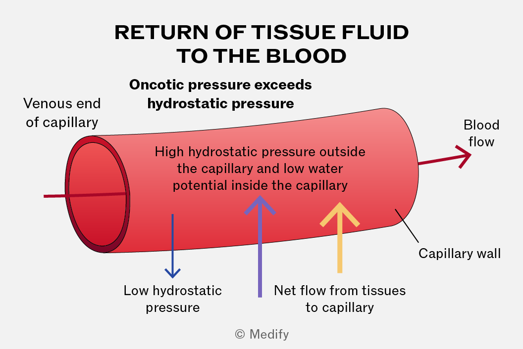
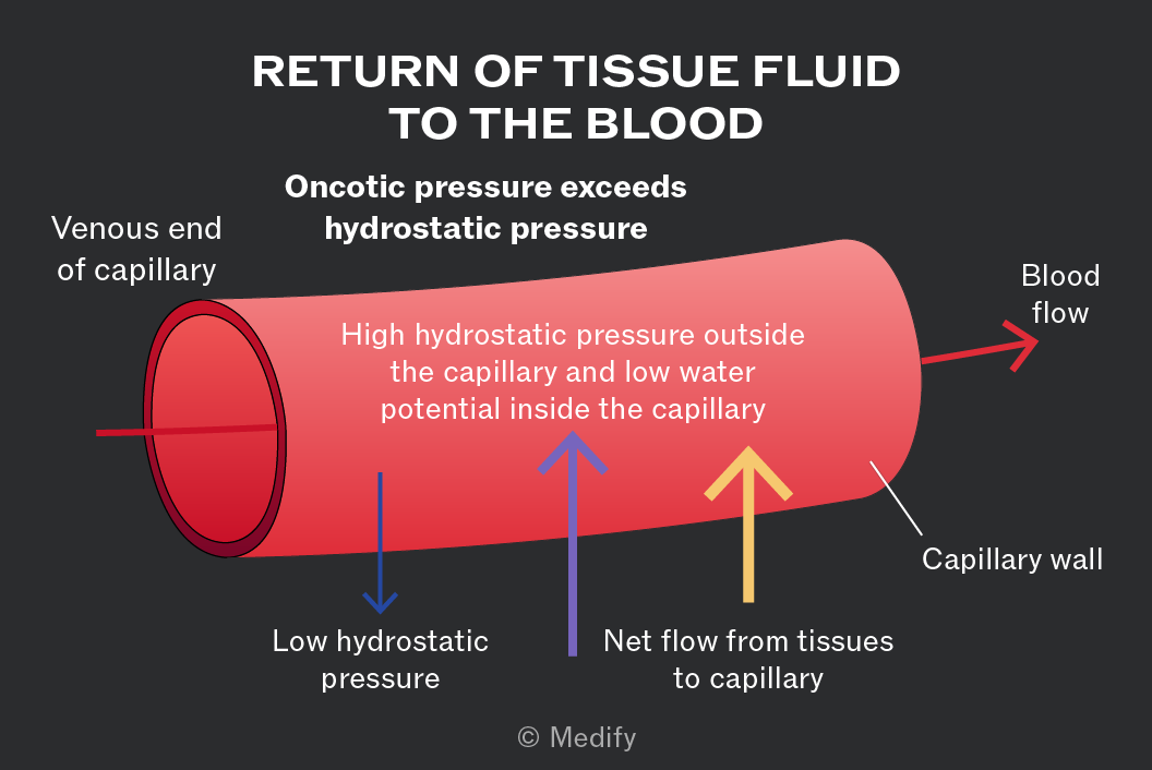
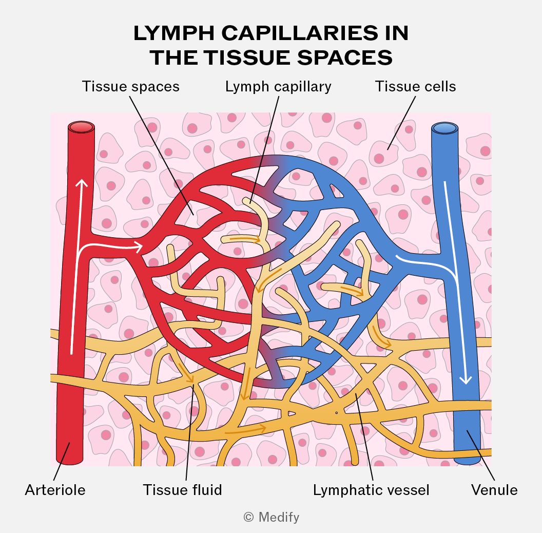
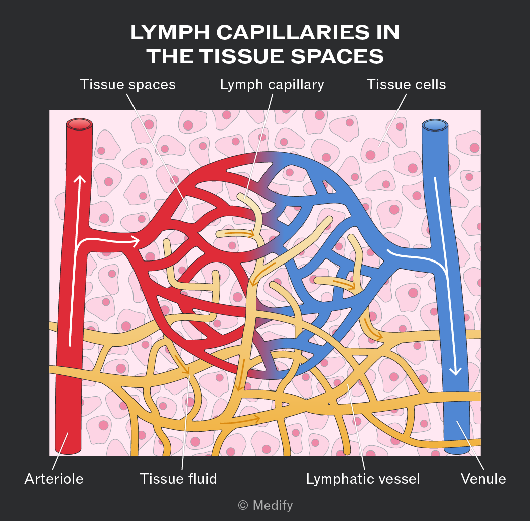
The tissue fluid that does not return to capillaries (10) drains into lymph vessels and forms lymph. The lymph vessels are like blind-ended capillaries with one-way valves and form a network over the whole body, merging with larger vessels.
Lymph is similar to plasma and tissue fluid but contains less oxygen and nutrients (which have been exchanged with tissues) and more fatty acids absorbed from the small intestine.
The lymphatic system joins the bloodstream again via ducts near the heart. Fluid is moved around the vessels by hydrostatic pressure and contraction of skeletal muscles.
The mammalian heart is a muscular organ made of two pumps and is situated in the thoracic cavity.
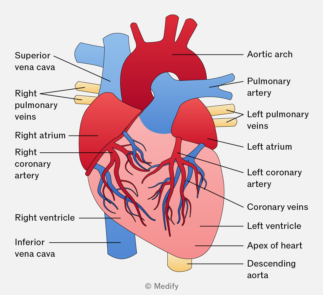
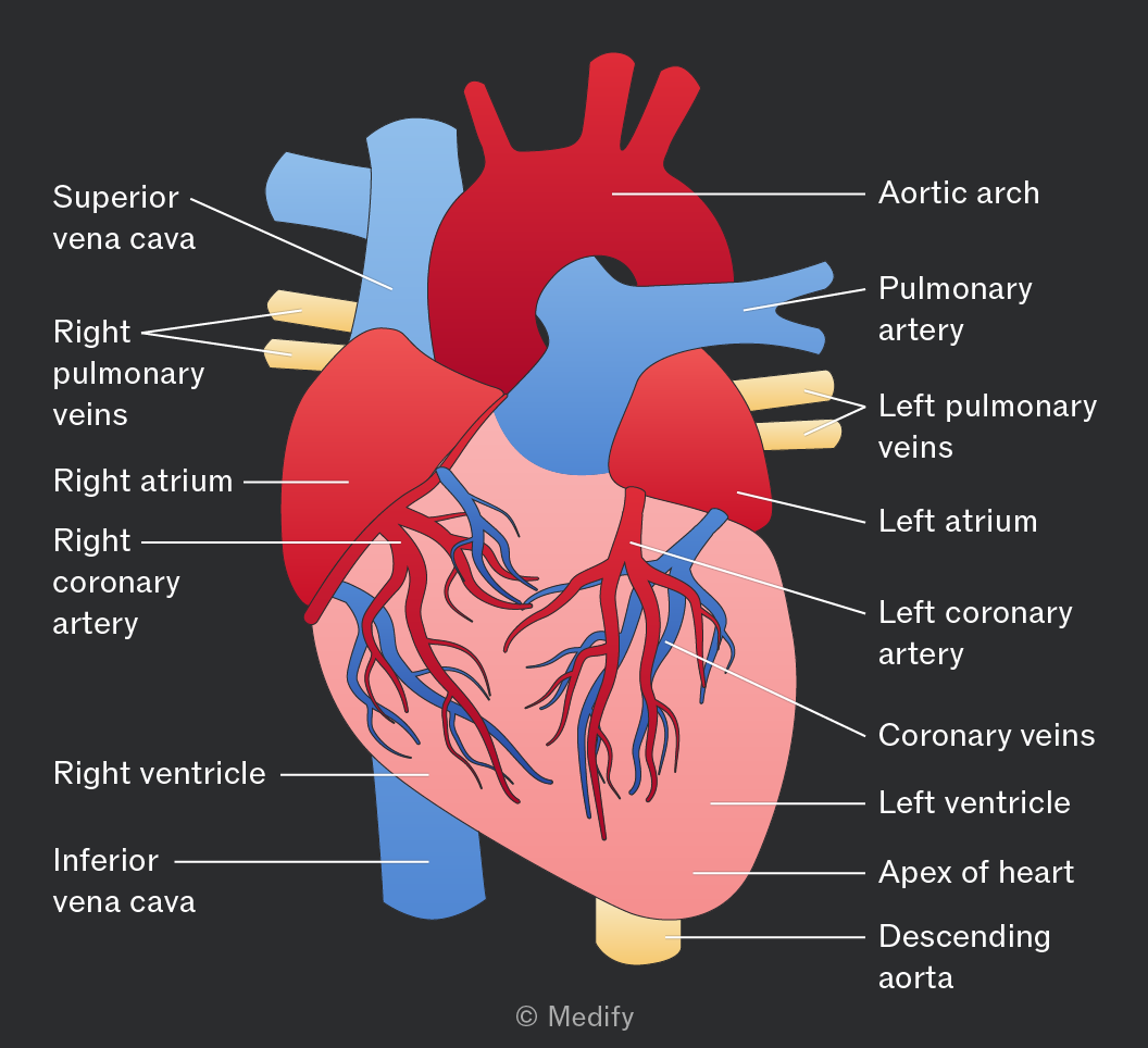
The aorta distributes oxygenated blood from the left ventricle to the rest of the body.
The vena cava are veins that carry deoxygenated blood from the head and the rest of the body back to the heart via the right atrium.
In almost all cases, veins bring deoxygenated blood back to the heart and arteries take oxygenated blood away from the heart.
One exception is the vessels taking blood between the heart and lungs:
- The pulmonary vein brings oxygenated blood from the lungs to the left atrium.
- The pulmonary artery delivers deoxygenated blood from the right ventricle to the lungs.
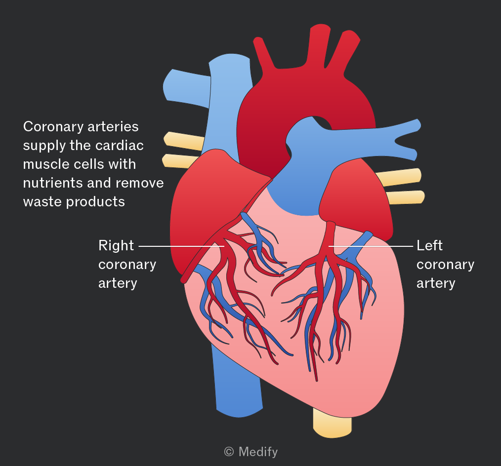
The coronary arteries are located on the surface of the heart and supply blood to the heart muscle.
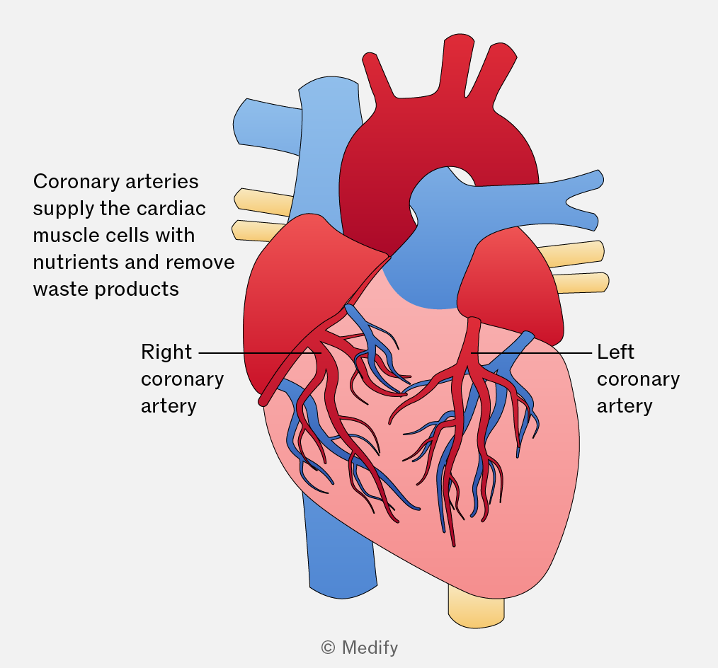
The mammalian heart consists of four chambers:
- Blood enters the heart via the atria (upper two chambers) as it returns from the body (right atrium) and the lungs (left atrium).
- The lower two chambers are the ventricles, which pump blood to the lungs (right ventricle) and to the rest of the body (left ventricle).
- The septum is a muscular wall that divides the heart into the left and right sides. This separation prevents the mixing of oxygenated (left side) and deoxygenated blood (right side).
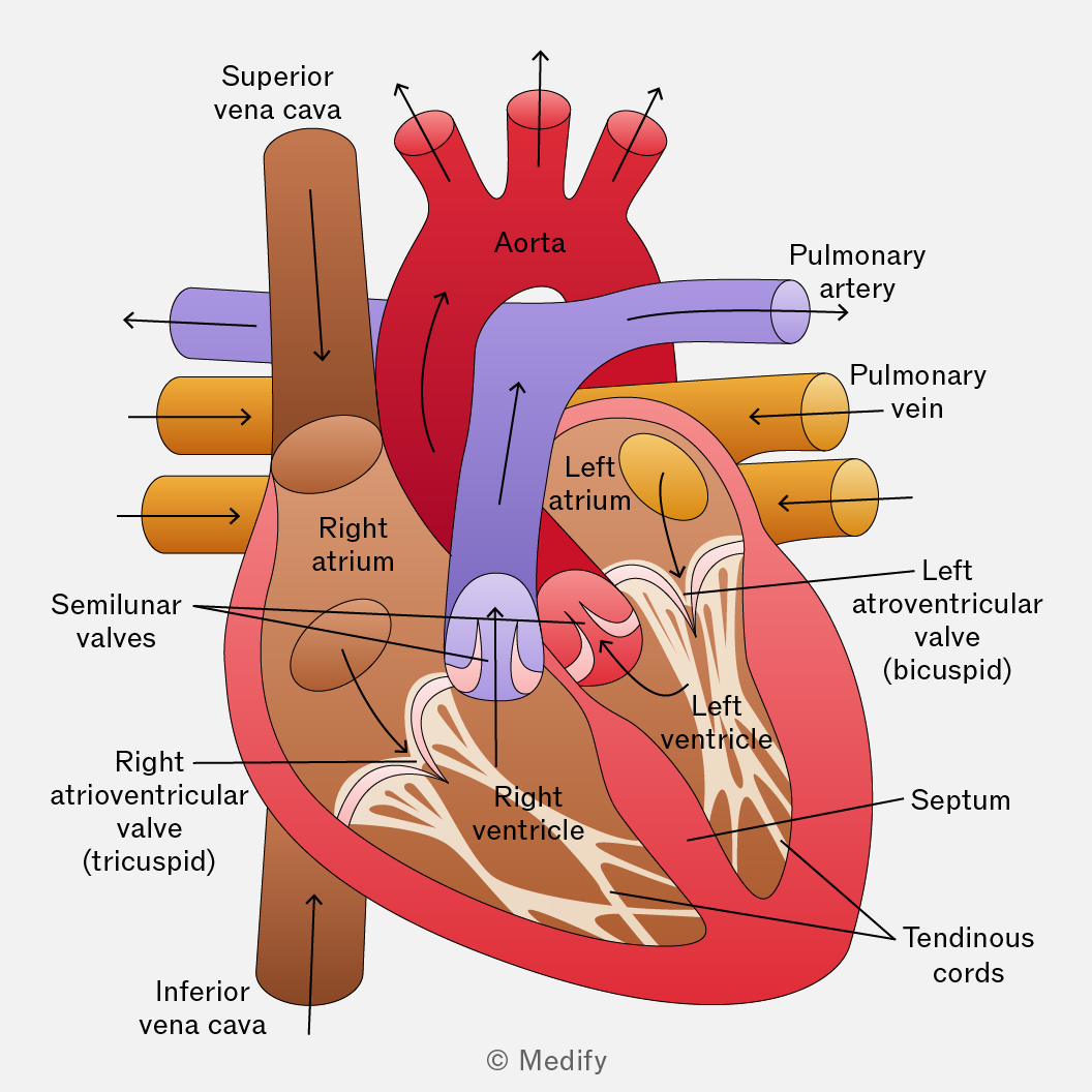
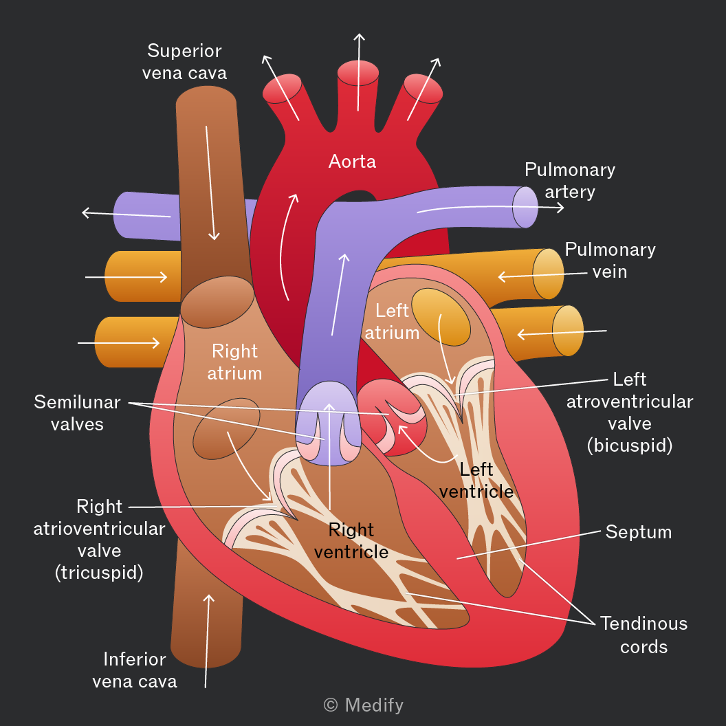
Heart valves regulate blood flow and maintain pressure within the chambers. They ensure blood travels in the correct direction, avoiding backflow.
The pressure from blood flowing into the atria opens the atrioventricular valves. When the atria and ventricles are full, the atria contract, forcing blood down into the ventricles.
The contraction of the ventricles causes the atrioventricular valves to close, preventing backflow of blood into the atria. Blood is forced out of the ventricles through the semilunar valves in the aorta and pulmonary artery. These valves prevent blood from flowing back into the heart.
Dissections are crucial for examining the internal structures of organs, allowing scientists to form theories about their functions and study anatomical relationships and comparative anatomy.
Hearts from sheep or pigs are often available from butchers and are similar in size to a human heart. They commonly have vessels and atria removed.
Ethical concerns include the treatment and sourcing of animals used for dissection, as well as adherence to cultural or religious beliefs.
The cardiac cycle is the sequence of events that occurs within one complete heartbeat:
- Atrial systole – the atria contract
- Ventricular systole – the ventricles contract
- Cardiac diastole – the heart relaxes
In diastole, the heart is relaxed as it fills with blood.
As the atria fill with blood, the pressure exceeds that in the ventricles and the atrioventricular valves open so that blood fills the ventricles.
The pressure in the relaxed ventricles is lower than in the aorta and pulmonary artery, causing the semilunar valves to close.
During atrial systole, the atria contract to force all the blood into the relaxed ventricles.
During ventricular systole, the thick ventricle walls contract and the pressure in the ventricles increases above that in the aorta and pulmonary artery. This closes the atrioventricular valves and blood is pumped to the lungs (right side) and the rest of the body (left side).
The pressure in the heart is low after ventricular systole but at a maximum in the arteries.
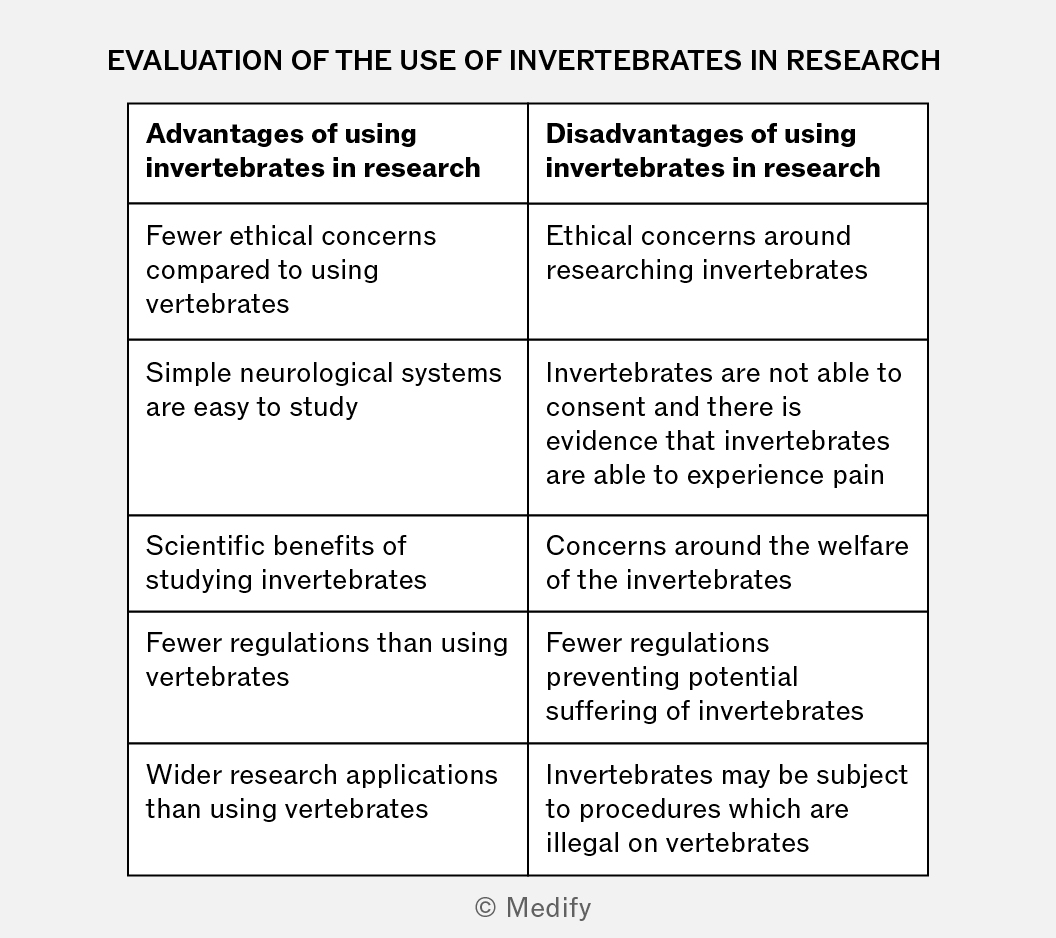
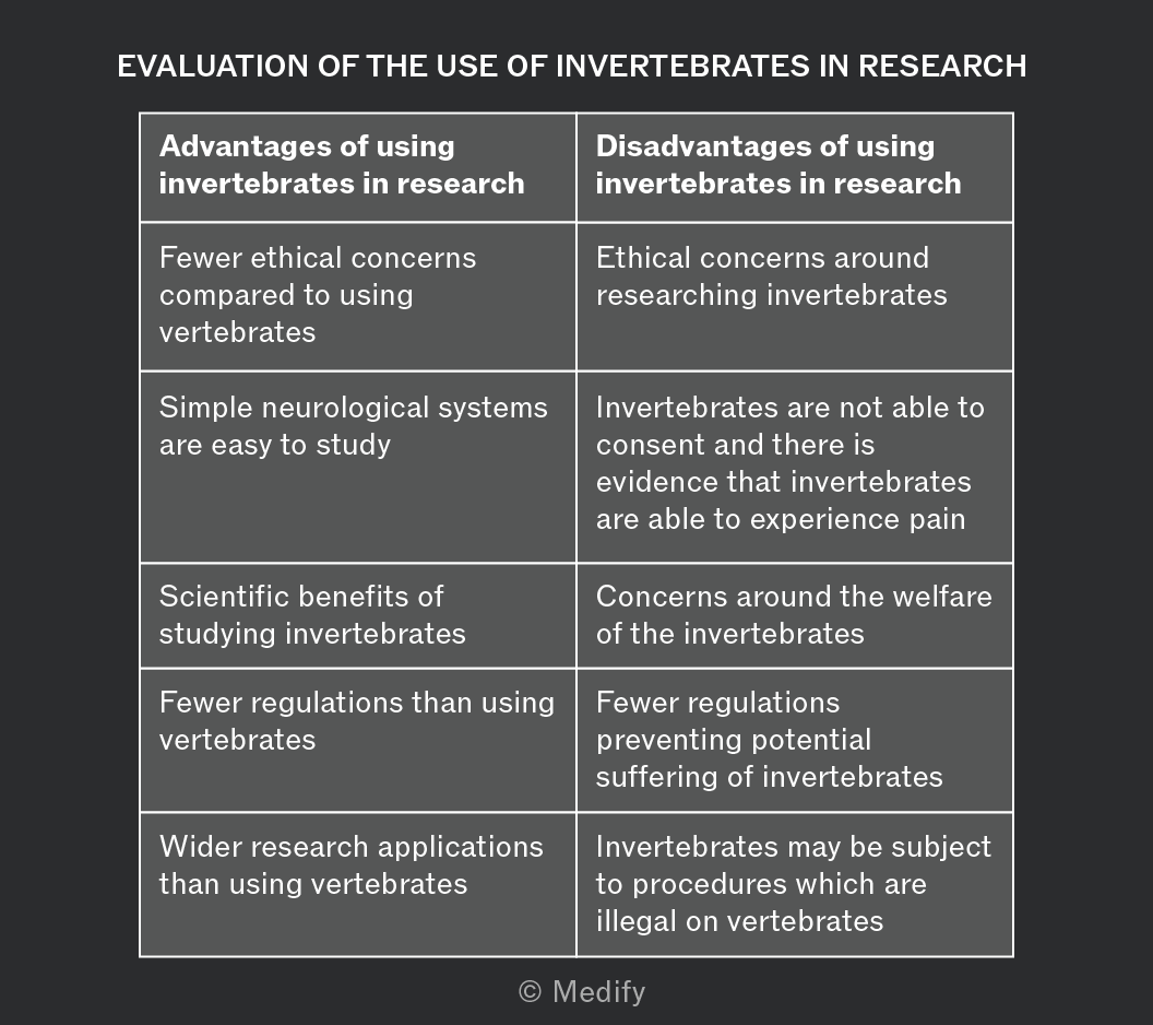
There are advantages and disadvantages to using invertebrates in research.
Some people believe that animals have rights, linking the use of animal models in research to the historic practice of using humans as slaves. Is using animals in a laboratory setting a violation of their rights?
European law has developed to protect animal welfare but does not reference rights. The legislation is designed to ensure that animals are treated ‘well’ as much as is possible.
In Europe:
“Vertebrates should not be used in medical research if there are non-animal alternatives. Where no alternative exists, strict guidelines are in place, and the research must be sufficient to warrant the use of vertebrates.”
It is debated, scientifically and philosophically, whether animals are capable of experiencing suffering and pleasure. If animals cannot “suffer”, then does their experience matter?
The arguments in favour of animal research on these grounds are most valid when discussing the use of invertebrates and fish, for which studies support the absence of these emotional responses.