Meiosis and sexual reproduction (3.9, 3.8, 3.6, 3.7)
On this page
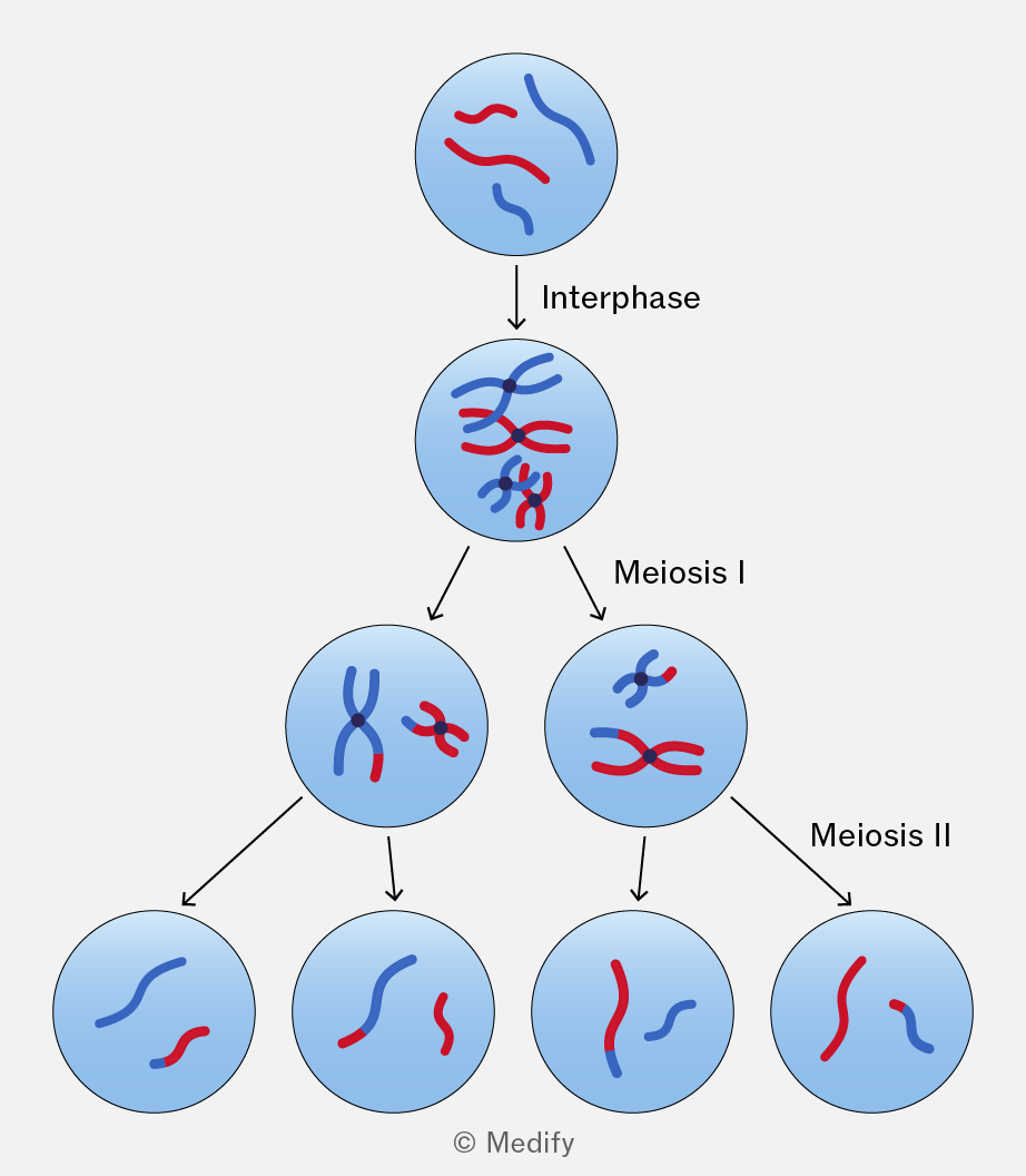
Meiosis is a type of cell division that produces four genetically distinct gametes (sperm and egg cells) and generates genetic variation through independent assortment of chromosomes and crossing over of alleles between chromatids.
It reduces the chromosome number from diploid to haploid.
Meiosis involves two sequential divisions:
- Meiosis I – separates homologous chromosomes.
- Meiosis II – separates sister chromatids.
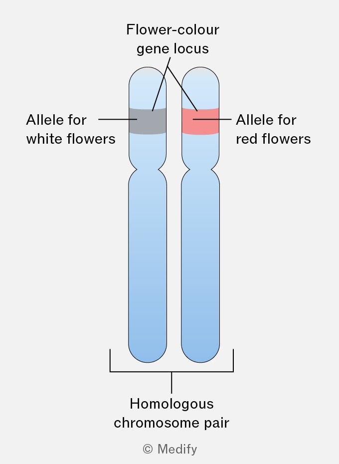
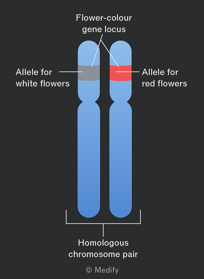
A locus (plural: loci) is the specific position of a gene on a chromosome.
Linked genes are genes located at different loci on the same chromosome. They tend to be inherited together during meiosis because they are physically linked. The closer two genes are to each other, the less likely they are to be separated by crossing over and the more likely they are to be inherited together.
Sex–linked genes are located on the sex chromosomes (X or Y). In humans, most sex-linked genes are found on the X chromosome, as the Y chromosome carries relatively few genes.
Examples of X-linked traits include red-green colour blindness and haemophilia.
Males (XY) are more likely to express X–linked recessive alleles because they have only one X chromosome and therefore no second allele to mask a recessive allele. Females (XX) may be carriers if they possess one recessive and one dominant allele.
The function of sperm cells is to deliver male genetic material to the egg for fertilisation.
Sperm cell adaptations include:
- Acrosome: a vesicle at the tip of the sperm containing enzymes that digest the zona pellucida (glycoproteins surrounding the egg) allowing sperm entry.
- Flagellum (tail): enables motility, allowing the sperm to swim towards the egg.
- Mitochondria in the midpiece: provides ATP for movement.
- Haploid nucleus: contains one set of chromosomes (23 in humans) and fuses with the haploid nucleus of the egg during fertilisation.
The function of an ovum (egg cell) is to provide female genetic material and support early embryo development.
Ovum adaptations include:
- Zona pellucida: a glycoprotein layer surrounding the egg that helps prevent polyspermy (fertilisation by more than one sperm).
- Cytoplasm: contains nutrients and organelles to support early embryonic development.
- Haploid nucleus: contains one set of chromosomes (23 in humans) that fuses with the haploid nucleus of the sperm during fertilisation.
- Cell surface membrane: can fuse with the sperm membrane during fertilisation.
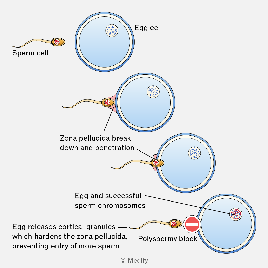
Fertilisation is the process in which a male gamete (sperm) and a female gamete (egg) form a zygote. This can be broken down into the following steps:
- Acrosome reaction – the sperm reaches the egg and binds to its zona pellucida. The acrosome releases digestive enzymes, breaking down the zona pellucida, allowing the sperm to reach and fuse with the egg cell membrane.
- Cortical reaction – the egg releases cortical granules into the zona pellucida (the layer that surrounds the membrane), which hardens and prevents polyspermy (entry of multiple sperm).
- Fusion of nuclei – the sperm’s nucleus enters the egg cytoplasm, and the sperm and egg nuclei fuse, forming a diploid zygote with 46 chromosomes (in humans).