Forensic biology (6.1, 6.2, 6.3, 6.4)
On this page
Establishing the time of death is a key role of forensics when investigating an unexplained death. The predictable physical and chemical changes which occur in a mammalian body post mortem are used to predict the time a person died.
Key factors observed when establishing time of death are:
- body temperature
- degree of rigor mortis
- stage of decomposition
- entomological (insect-based) evidence and succession.
Body temperature is a useful predictor of time of death within the first 24 hours
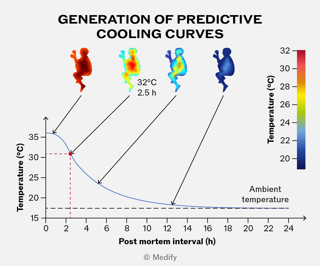
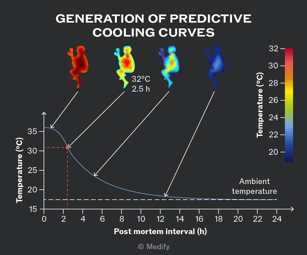
Cooling curves are generally sigmoid, with a stable temperature for the first 30 minutes to an hour.
Many factors influence the shape of a predictive cooling curve. Extensive studies go into producing curves specific to different conditions.
Rigor mortis is the stiffening of muscles and joints in the body after death.
When a mammal dies, the muscles initially relax, then stiffen, then relax again. The pattern of rigor across the body is predictable, with small muscles stiffening before larger ones (face and fingers before arms and legs).
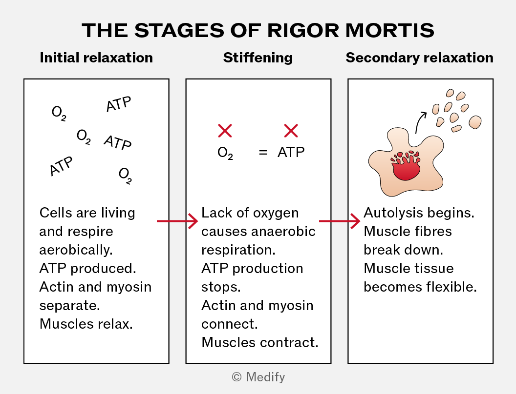
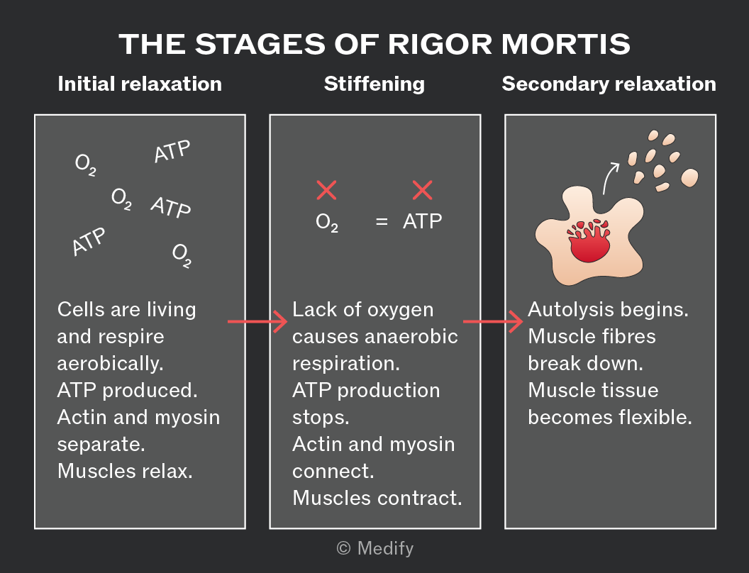
The timelines associated with rigour mortis can be influenced by other factors like exercise prior to death and ambient conditions. These are considered when predicting time of death.
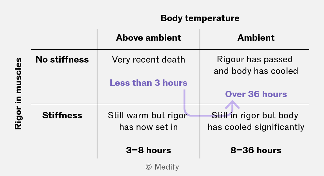
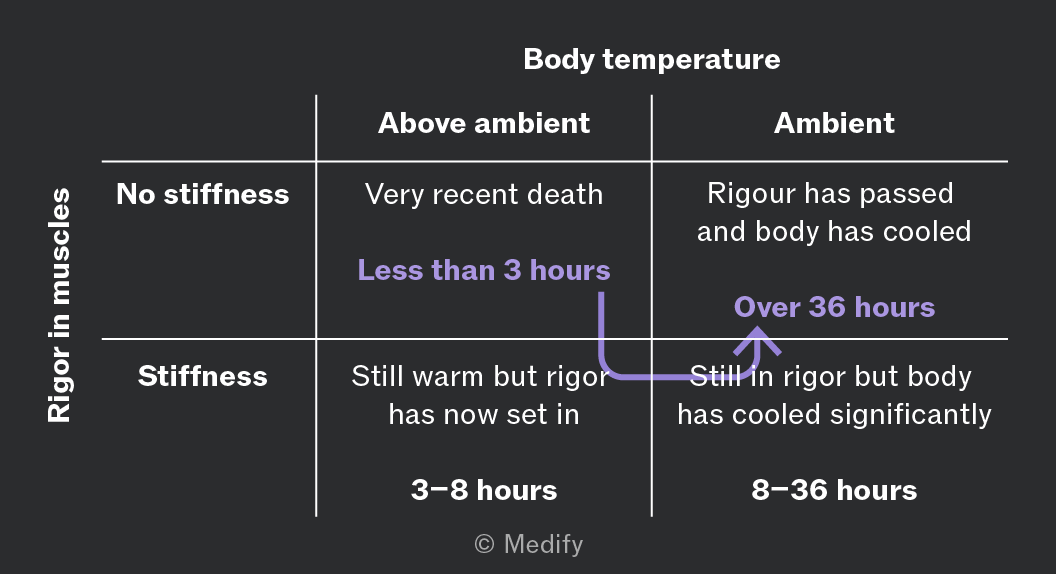
The cooling curve and the degree of rigor mortis can be combined to provide time-of-death windows.
Decomposition (also known as putrefaction) in humans begins with autolysis, where the body’s own enzymes break down its cells.
The subsequent stages of decomposition can be observed over time:
- Initially, there is a greenish discolouration on the lower abdomen caused by sulfhaemoglobin in the blood.
- Discolouration spreads across the body, darkening to reddish-green, then to purplish-black.
- Blistering and bloating then occur due to gas production. The body begins to smell.
- The body deflates, and the putrefaction fluids drain away.
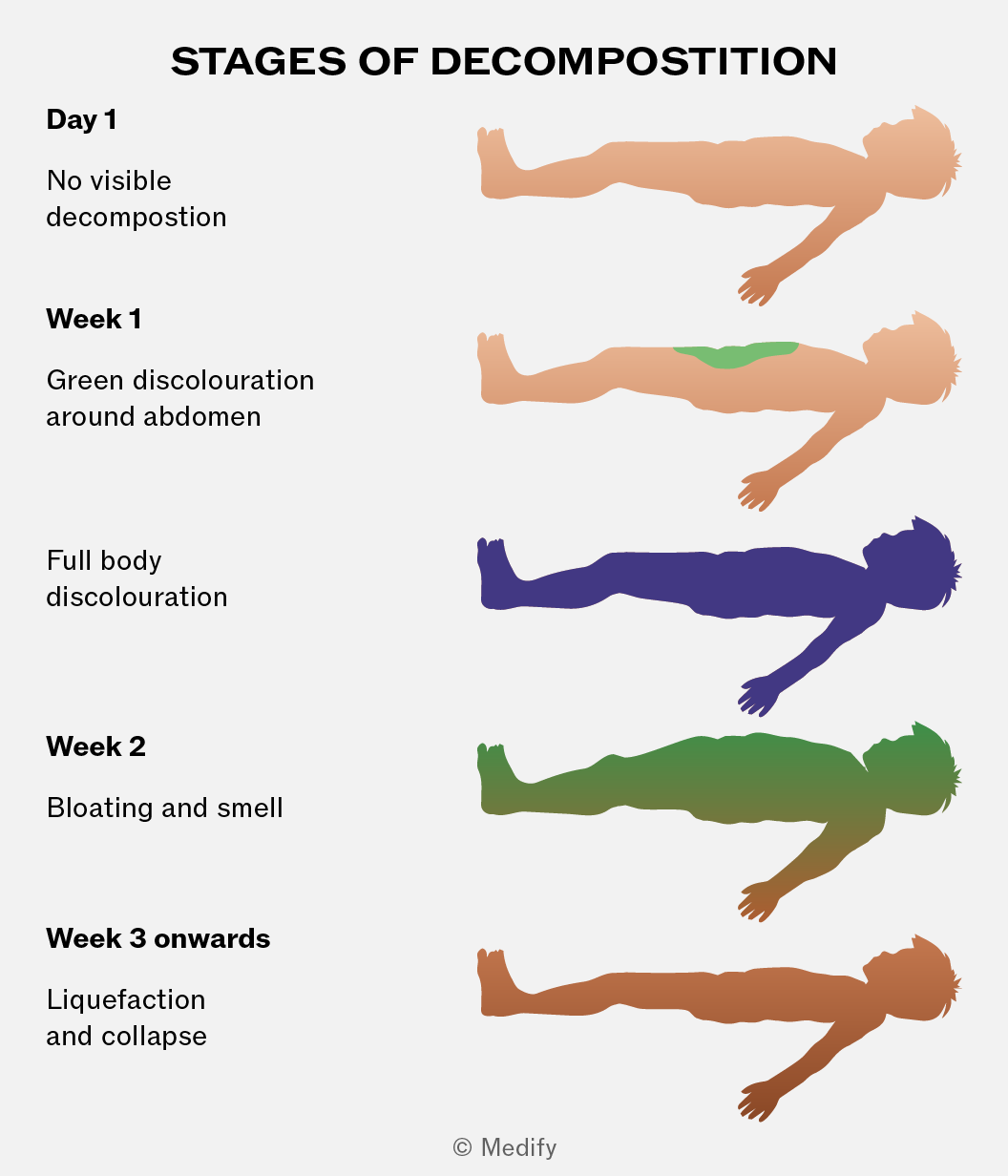
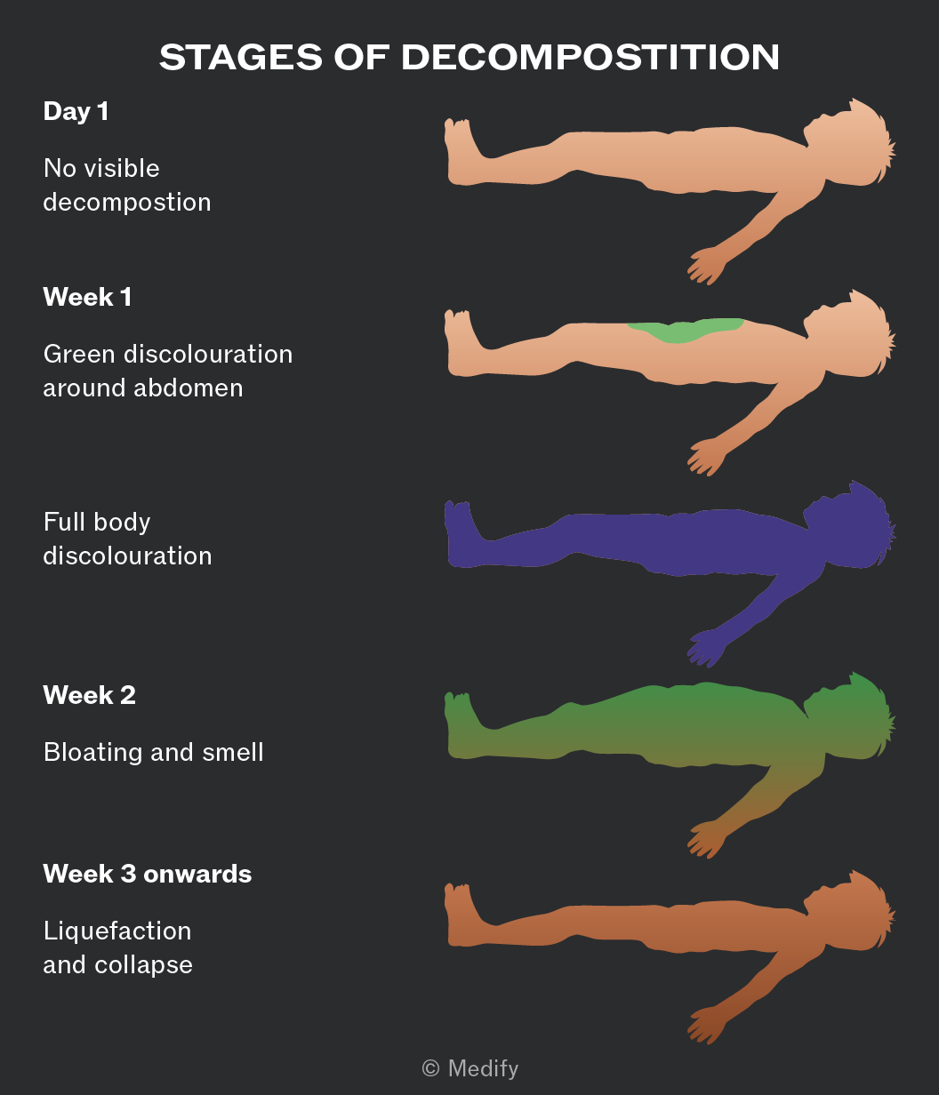
The actual timescale associated with each stage is highly dependent on environmental conditions. In general, hotter, moister conditions and external body damage lead to faster decomposition.
Forensic entomology uses the study of insects to predict time of death. In entomological research, the decaying body is viewed as a habitat in which insect colonies can establish themselves.
The two main fields of research in forensic entomology are documenting insect life cycles and understanding species succession in a variety of conditions.
Data does not exist to support analysis of the full range of insects present in nature, in all conceivable conditions. The short life cycles of insects allow data to be gathered specifically for a case, under the exact conditions in which a corpse has been found.
Flies lay eggs in body openings shortly after death. The maggot mass and stage of development of the larvae or pupae at the point a body is discovered can be used to determine how long ago the eggs were laid.
Extensive data exist for common fly species, such as the bluebottle, across multiple environmental conditions. This encompasses the stage of development as well as the predicted size within each stage.
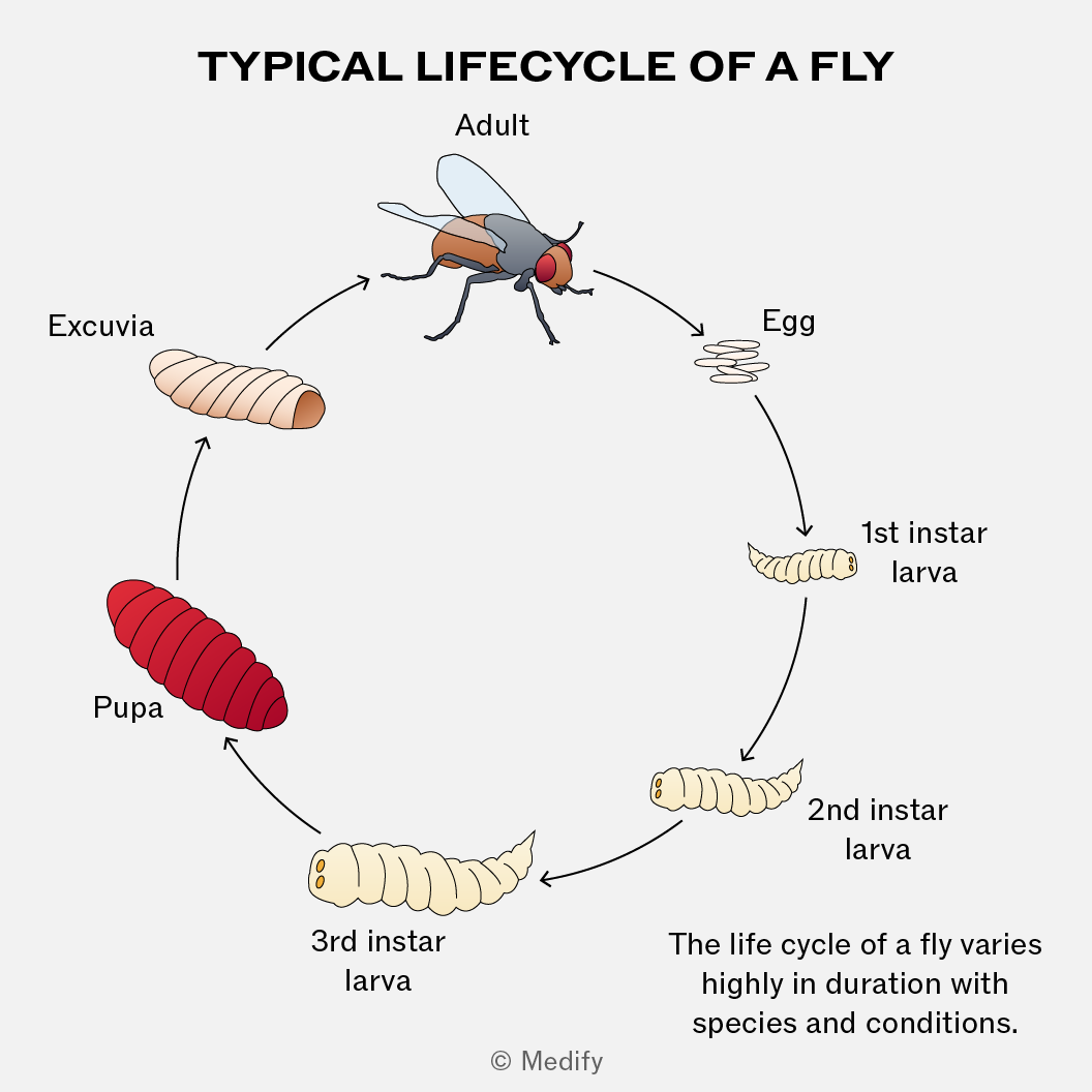
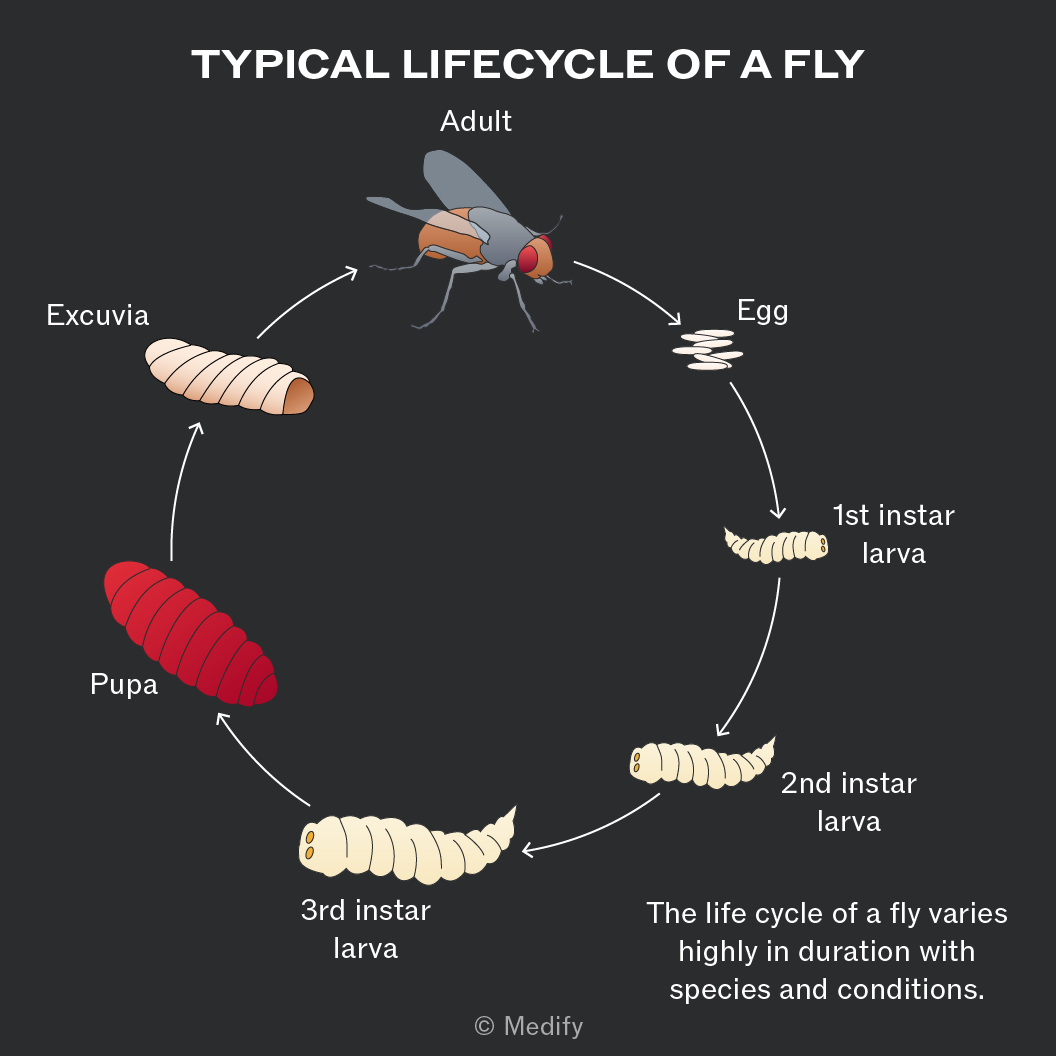
For corpses found under unusual conditions, or for rarer fly species, live samples are removed from the corpse and used to conduct controlled studies to obtain the required information.
Species succession refers to the order in which different insect species will colonise a decaying corpse.
Insect succession is cumulative; the arrival of a new species does not equate to the departure of the original species.
Some species thrive on corpses at the later stages of decay, so a certain time lapse post mortem can be expected before the eggs from that species are laid.
Succession can also be used to give evidence that a corpse has been moved, as different species are associated with different locations.
Decomposition of bodies and other organic matter is facilitated by fungi and bacteria. The predictability of the type and number of these decomposers on a corpse during decay is low, limiting their usefulness in forensic analysis.
Dead organic matter is used as a food source for fungi and bacteria. These reproduce rapidly as decomposition progresses.
A combination of aerobic and anaerobic respiration in the decomposers converts organic matter into carbon dioxide. The carbon dioxide can then be converted back into organic matter through photosynthesis, completing the carbon cycle.
DNA profiling (also called DNA fingerprinting) analyses non-coding, variable regions of DNA to compare individuals.
DNA profiling most commonly uses short tandem repeats (STRs), also known as microsatellites, which consist of short repeated DNA sequences. The number of repeats at each STR locus varies between individuals, producing a unique DNA profile (except in identical twins).
DNA profiling follows these steps:
- Sample collection – a biological sample containing DNA is obtained, such as blood, saliva, hair roots or plant tissue.
- DNA extraction – DNA is isolated from the cells in the sample.
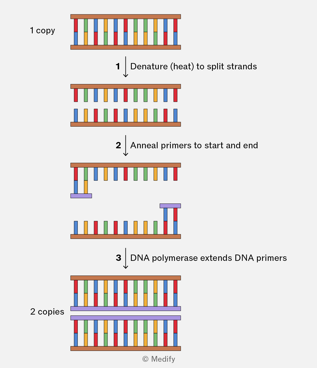
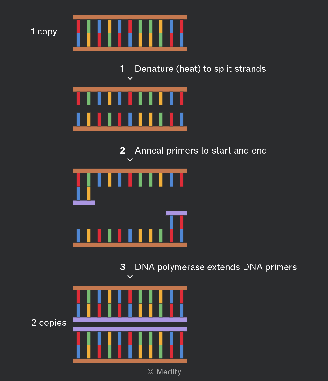
- PCR – the short tandem repeat regions (STR) are amplified using PCR to produce many copies for analysis.
- Electrophoresis – the amplified DNA fragments are separated according to their length (smaller fragments move further), producing a pattern of bands.
- Profile comparison – resulting DNA profiles are compared between samples. Matching banding patterns indicate that the DNA is likely to have originated from the same individual.
Uses of DNA profiling include:
- Matching suspects to crime scene samples.
- Identifying victims in disasters.
- Distinguishing between similar-looking organisms (rare species, plant cultivars).
- Conservation – preventing inbreeding by checking relatedness and tracking illegal wildlife trade (timber, ivory).
Relatives share a predictable proportion of STR patterns:
- Parents and offspring share approximately of their STR alleles / bands.
- Siblings share many STR alleles / bands, but usually have different overall DNA profiles.
DNA profiling can be used to confirm parentage, breeding lineages, or inheritance disputes. In plants, profiling is used to confirm breeding crosses and verify cultivar purity. In animals, it is used to manage captive breeding programmes (zoos) and maintain genetic diversity.
PCR (Polymerase Chain Reaction) is a technique used to rapidly amplify specific fragments of DNA.
The reaction mixture contains:
- Template DNA – the DNA containing the target sequence to be copied.
- Primers – short single-stranded DNA sequences that bind to the ends of the target region.
- Free DNA nucleotides – used to build new DNA strands.
- DNA polymerase – a heat-stable enzyme that synthesises new DNA strands.
- Buffer – maintains the optimum and conditions for the enzyme.
The PCR Cycle (repeated 25–35×):
- Denaturation (~95): hydrogen bonds between complementary DNA strands break.
- Annealing (~50–65): primers bind (anneal) to complementary sequences on the template (target) DNA.
- Extension (~72): DNA polymerase adds complementary nucleotides to the primers creating new DNA strands.
Each cycle approximately doubles the amount of target DNA resulting in exponential amplification.
After n cycles, the number of DNA molecules ≈ 2n (assuming 100 efficiency).