
Responses to infection (6.7, 6.8, 6.9)
On this page
Animals have a number of non-specific defences that help prevent infection:
- Inflammation: increases blood flow and capillary permeability allowing immune cells and antimicrobial substances to reach infected tissue.
- Lysozyme action: breaks down bacterial cell walls.
- Interferon: prevents viruses replicating / spreading.
- Phagocytosis: pathogens are engulfed and digested by immune cells.
Phagocytes are a type of white blood cell that play a key role in the non-specific immune response by engulfing and destroying pathogens.
Neutrophils are the most abundant type of phagocyte. They rapidly respond to infections and die after engulfing pathogens.
Macrophages are larger, longer-lived phagocytes that develop from monocytes (a type of white blood cell). Macrophages act as antigen-presenting cells (APCs) to activate specific immune responses.
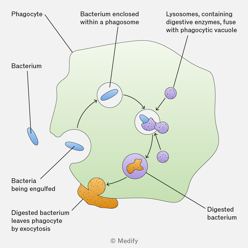
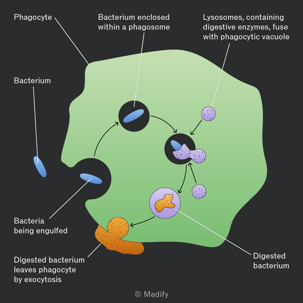
Phagocytosis is the process by which phagocytes (e.g., neutrophils, macrophages) engulf and destroy pathogens.
Phagocytosis involves the following steps:
- Chemotaxis – phagocytes use chemical signals to move toward pathogens.
- Recognition and attachment – receptors on the phagocyte membrane bind to antigens on the pathogen surface.
- Engulfment – the pathogen is enclosed in a phagosome (a membrane-bound vesicle).
- Digestion – lysosomes fuse with the phagosome to form a phagolysosome, releasing enzymes to break down the pathogen.
- Antigen presentation – macrophages display the pathogen’s antigens on their surface membrane to activate the immune response.
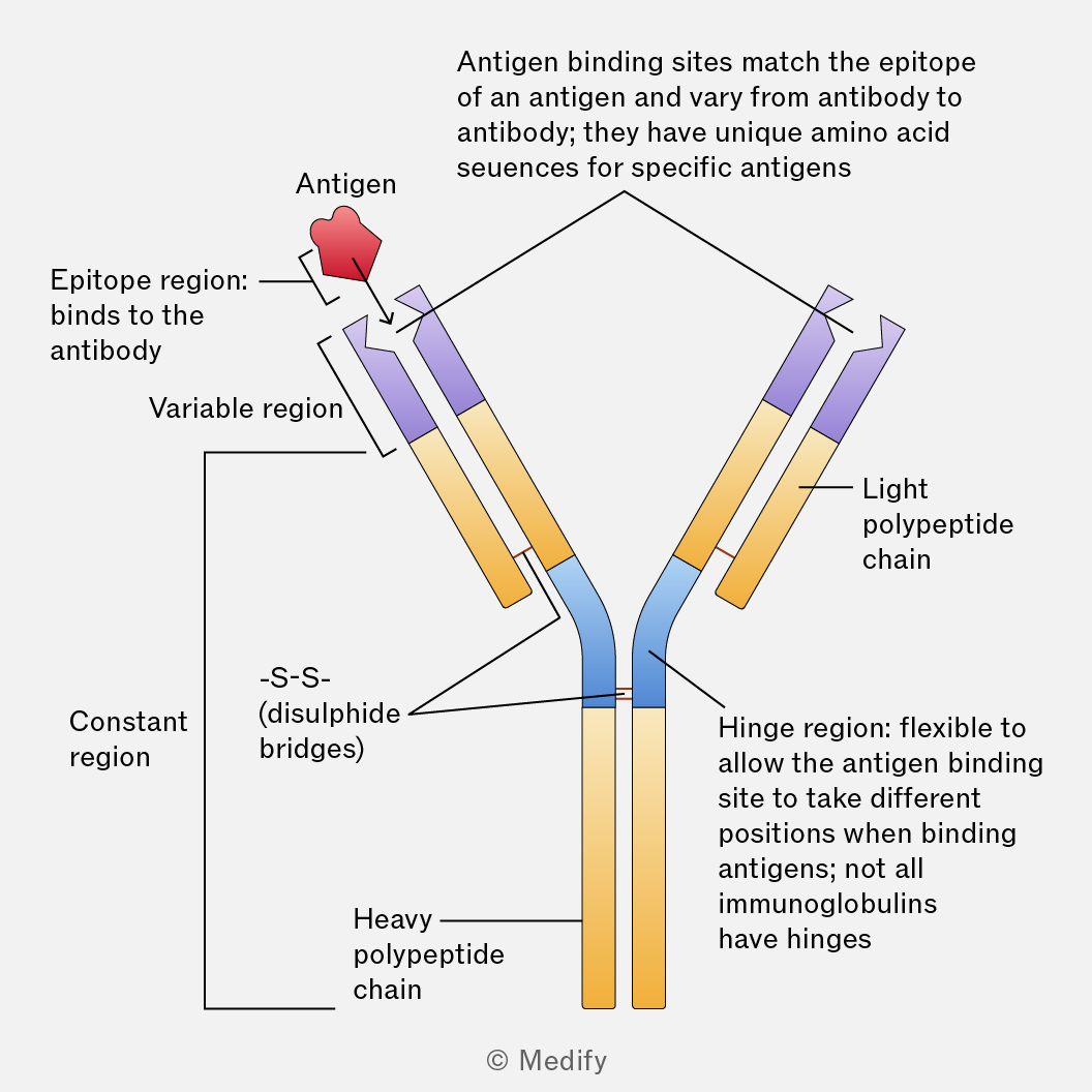
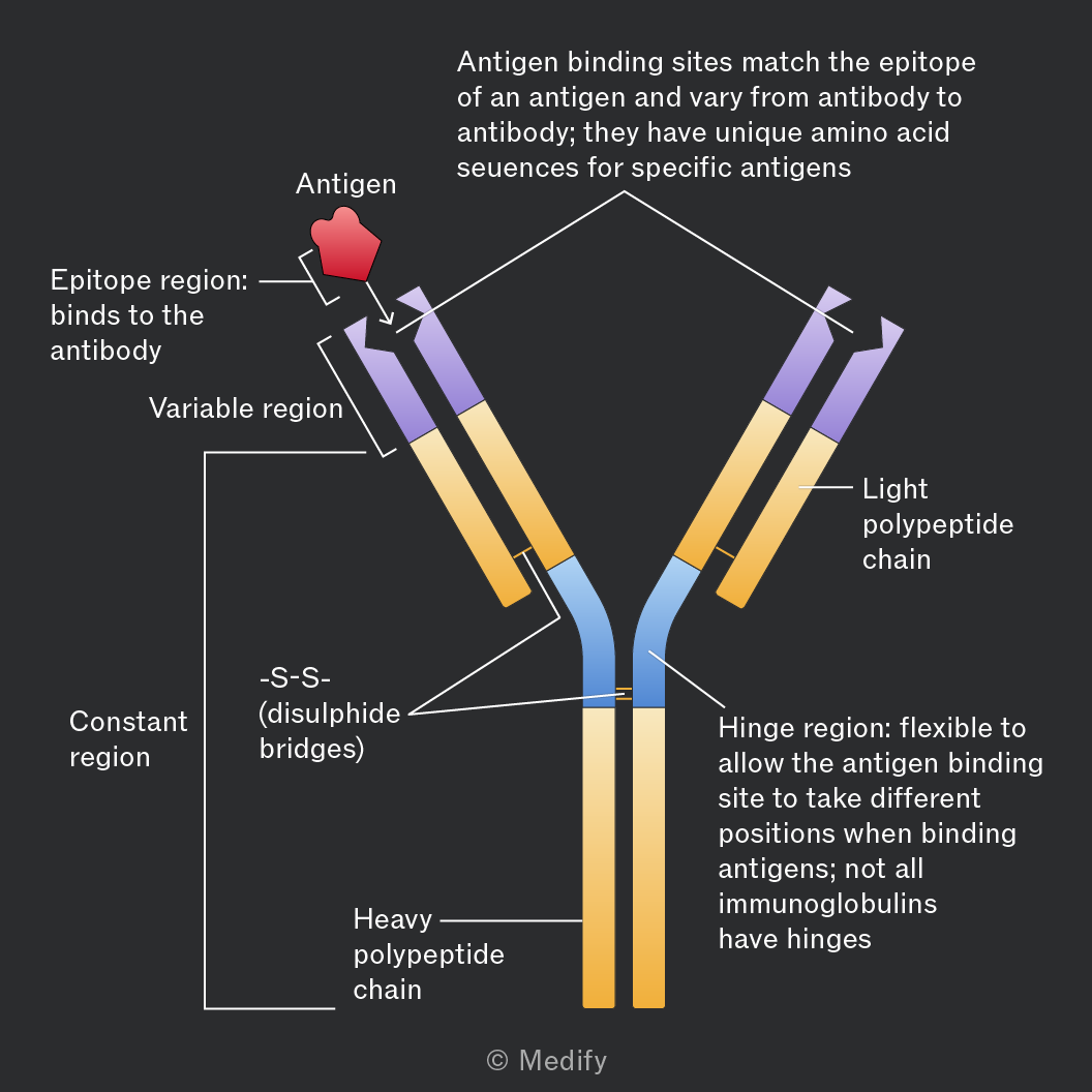
An antibody is a Y-shaped globular protein made of four polypeptide chains (two heavy and two light), held by disulfide bonds.
The variable region at the tips form two antigen-binding sites; they have a specific tertiary structure that is complementary to a specific antigen, meaning specific binding will occur at these sites.
The constant region is the same in all antibodies of that class and interacts with other components of the immune system, helping to recruit immune cells.
The hinge region provides flexibility, allowing the antibody to bind to antigens at different angles.
Antibodies bind to antigens on pathogens (or to toxins). The binding is specific, like a lock and key and forms an antigen–antibody complex.
Once the antigen-antibody complex is formed then there are several different ways in which the antibody itself or the immune system will destroy the pathogen.
An antigen is a molecule, usually a protein or polysaccharide, that triggers an immune response by stimulating the production of antibodies or activating immune cells.
Antigens are usually found on the surface of pathogens (e.g., bacteria, viruses) but can also be on toxins or foreign substances.
Each antigen has a specific shape that is recognised by complementary antibodies or T-cell receptors.
This recognition enables the immune system to distinguish between self (the body’s own cells) and non-self (foreign invaders).
Some pathogens (e.g., viruses and bacteria) change their antigens over time through mutations or genetic recombination.
This allows them to evade immune recognition, because previously produced antibodies and memory cells may no longer recognise the altered antigens. This makes it harder for the immune system to respond effectively.
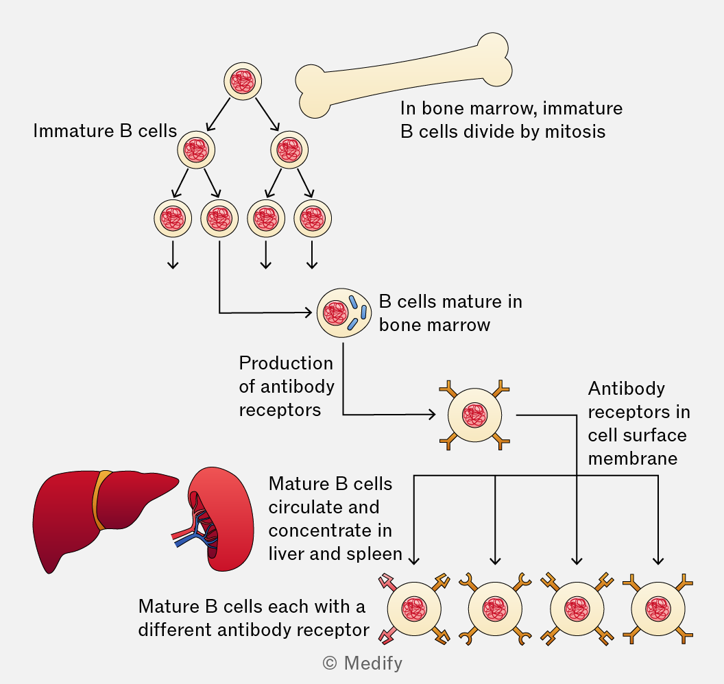
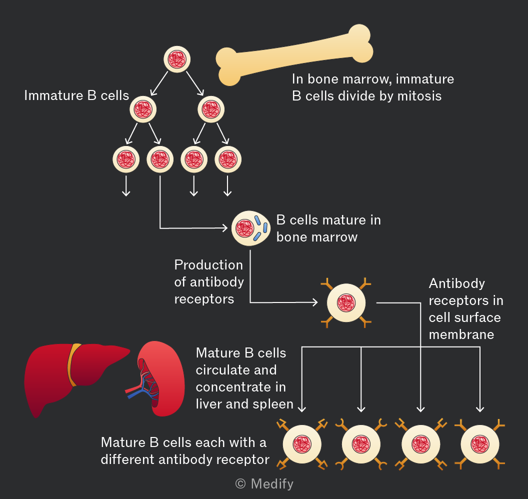
B lymphocytes are a type of lymphocyte that mature in the bone marrow and play a key role in the specific (humoral) immune response. They are responsible for targeting pathogens in body fluids such as blood and lymph.
When a B cell detects a specific antigen, it becomes activated and divides to form plasma cells (B effector cells), which produce antibodies that bind to and neutralise the pathogen.
Some B cells become memory cells, which remain in the body and provide long-term immunity by responding quickly if the same antigen is encountered again.
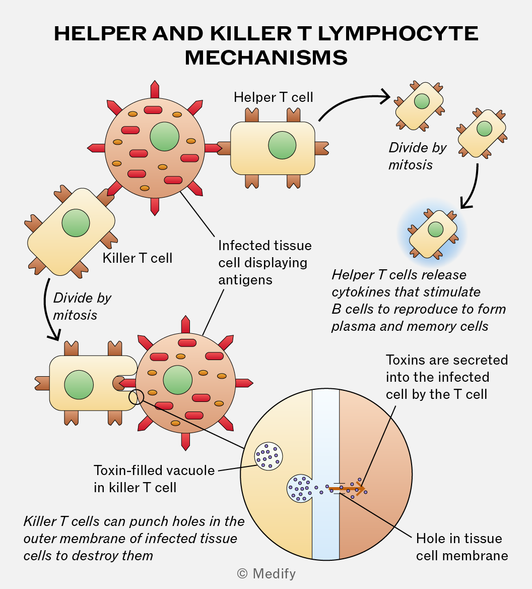
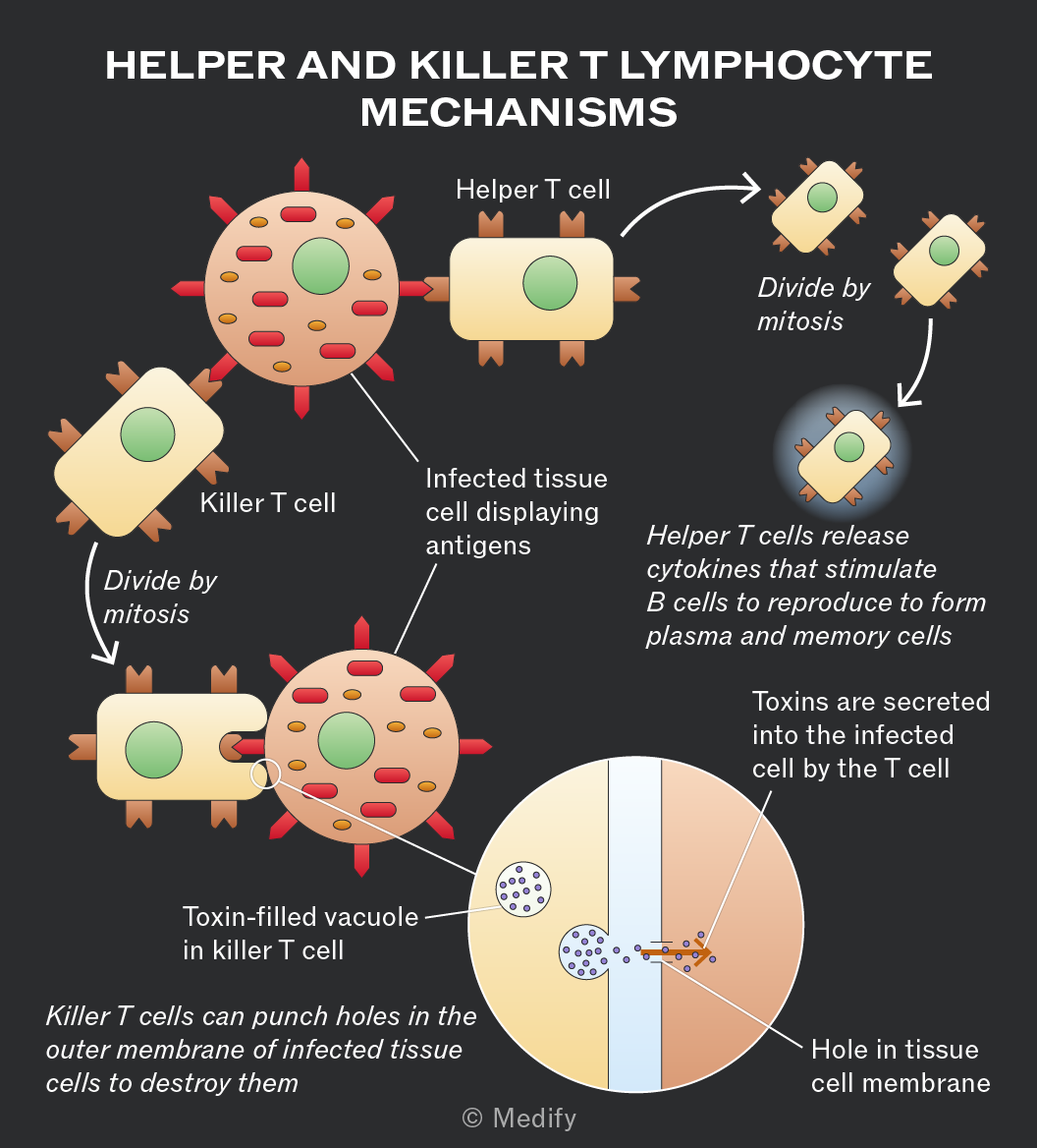
There are several types of T lymphocytes:
- T helper cells (Th): release cytokines to activate B cells, phagocytes, and other T cells
- T killer (cytotoxic) cells (Tc): destroy infected or abnormal cells by releasing substances that cause cell lysis.
- T memory cells: provide long-term immunity by responding quickly to reinfection.