
Plant and animal responses (5.1.5)
On this page
Biotic factors (living) and abiotic factors (non-living) are external stimuli that influence plant survival and growth. Plants have evolved several types of responses to these factors, allowing them to adapt to changing environmental conditions.
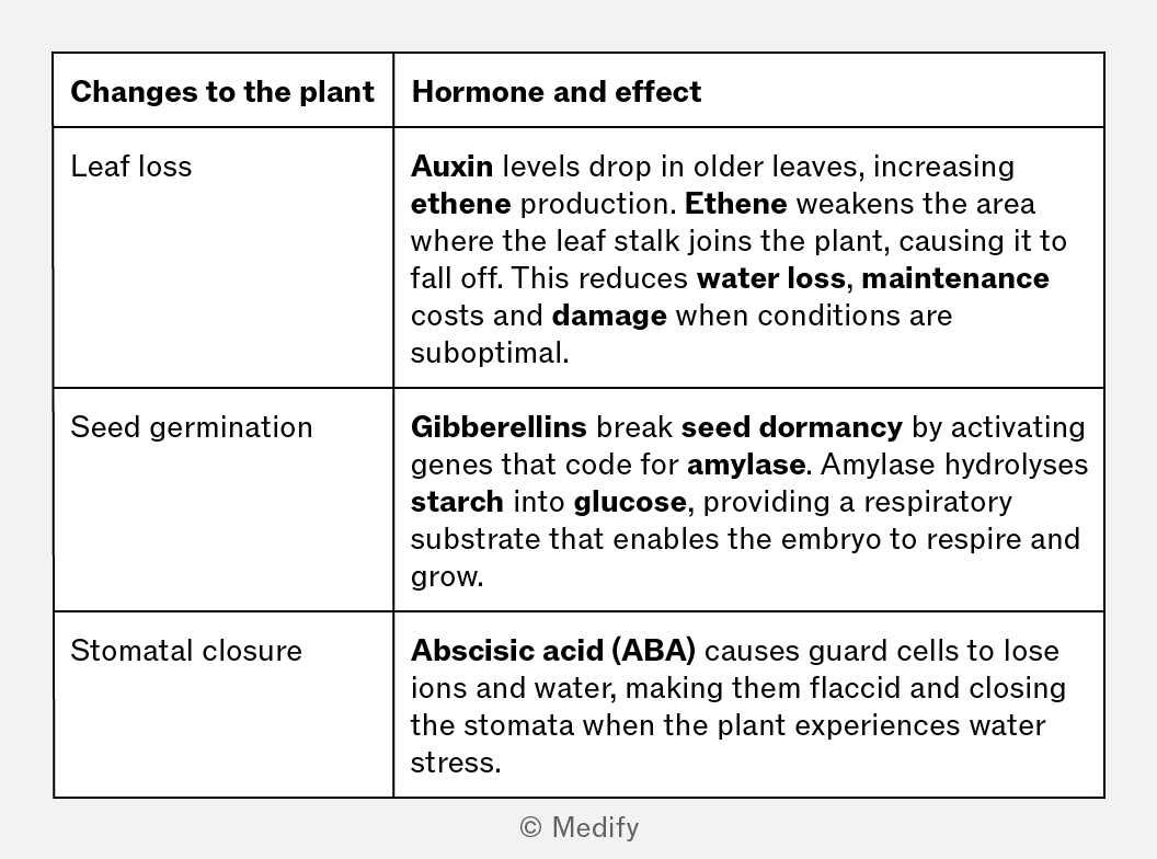
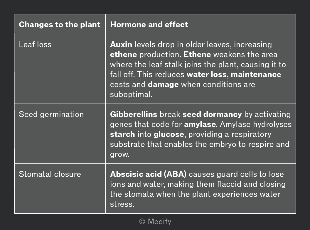
Plants responses to abiotic stress triggered by changes in day length, temperature and water availability are controlled by hormones and include:
- Leaf loss – deciduous plants shed leaves in autumn when maintenance costs exceed photosynthetic output.
- Freezing prevention – some plants produce molecules that prevent ice crystal formation in cells or mitigate damage if freezing occurs.
- Stomatal closure – stomata close when water is scarce to reduce transpiration and open in favourable conditions for gas exchange.
Plants have several defences that enable them to respond to their environment, including chemical defences such as:
- Alkaloids (e.g., caffeine, nicotine, morphine) – bitter–tasting chemicals found in many plants. They make the plant unpalatable or toxic to herbivores. They can also prevent seed germination in competitor plants.
- Pheromones - chemicals that affect the physiology or behaviour of other organisms.
Plants must respond to changing environmental conditions to optimise growth.
Tropisms are directional growth responses to an environmental stimulus:- A positive tropic response is when a plant grows towards a stimulus.
- A negative tropic response is when a plant grows away from a stimulus.
There are many types of tropism each resulting from a different environmental stimulus:
- Light triggers phototropism; shoots grow towards light.
- Gravity triggers geotropism; roots grow downward.
- Chemicals trigger chemotropism; pollen tubes grow through the style, towards the ovule.
- Climbing plants gain support using thigmotropism, wrapping their shoots around supports.
Responses that are not directional in relation to a stimulus are called nastic responses.
Some plants respond to touch by folding or curling their leaves as a form of defense. This response is called thigmonasty.
Plant hormones are transported throughout the plant in the xylem and phloem. Their movement may occur by diffusion, active transport between cells, and mass flow within transport tissues.
There are many types of plant hormones, each with a specific effect.
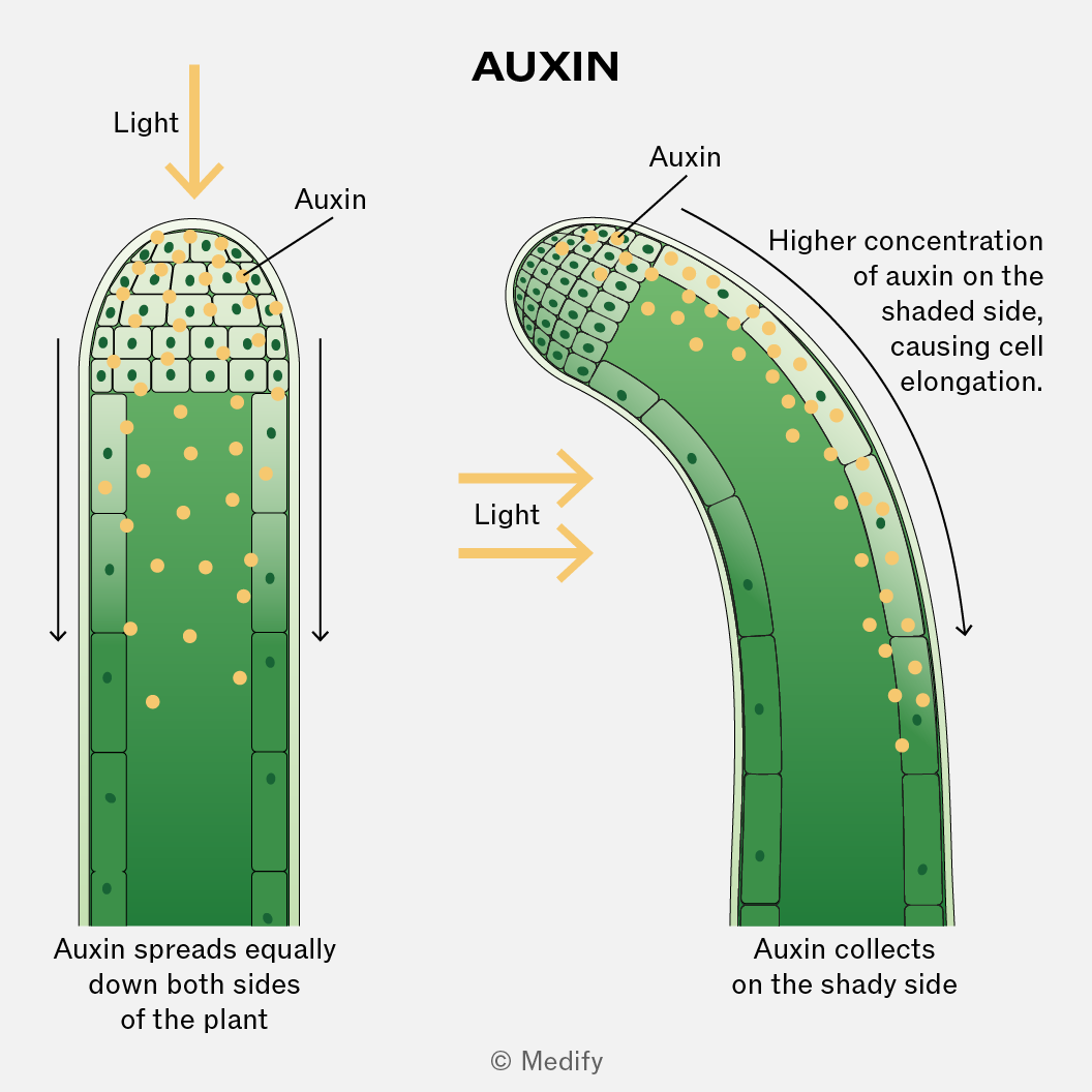
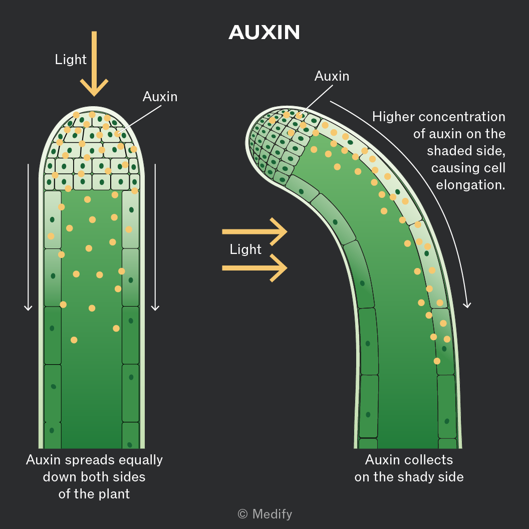
Auxins are plant hormones (e.g., IAA) that play many roles. They are involved in regulating plant growth and are responsible for phototropism, where plant shoots grow towards a light source. Auxins are produced mainly in the shoot tip and move down the shoot.
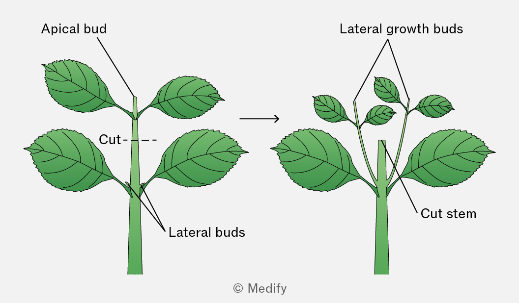
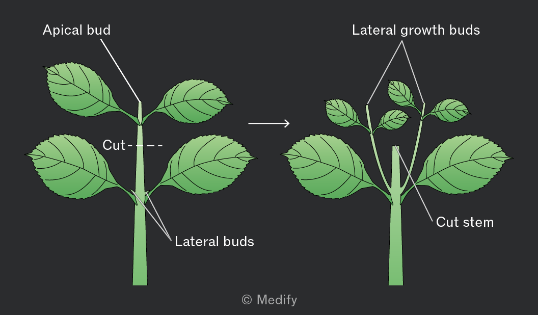
Apical dominance is the inhibition of lateral bud growth by auxins produced in the apical bud (shoot tip).
Removing the apical bud lowers auxin concentration, allowing lateral buds (side shoots) to grow.
Applying auxin paste to the cut tip restores inhibition, confirming that auxin controls apical dominance.
Higher auxin concentrations produce stronger apical dominance.
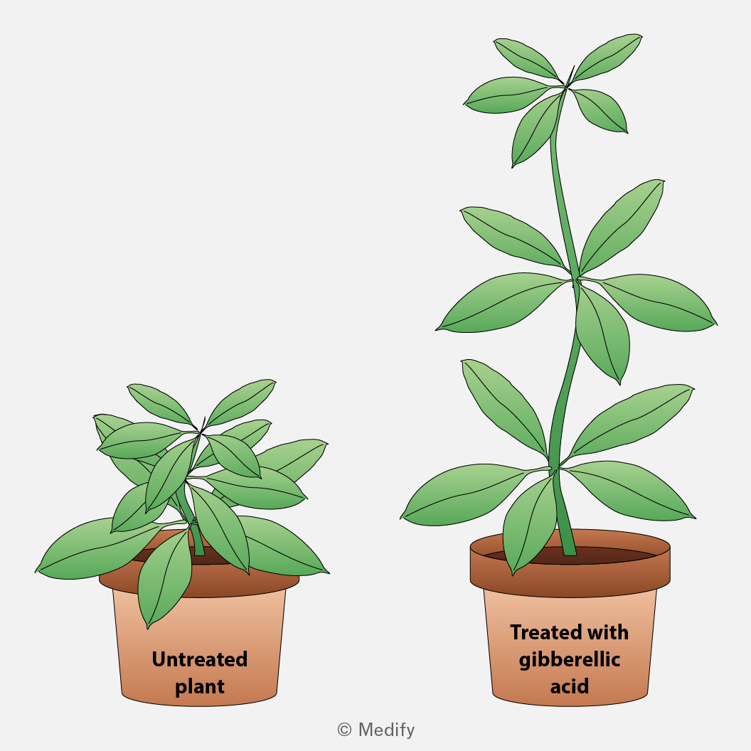
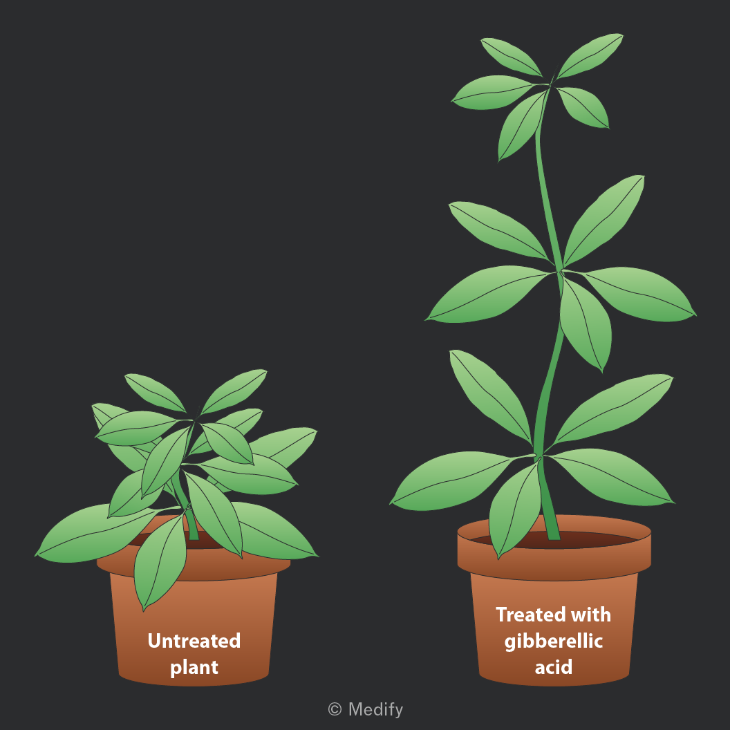
Gibberellins stimulate stem elongation, so plants with higher gibberellin concentrations generally develop longer stems between leaf nodes.
Applying gibberellic acid (a type of gibberellin) to the plant’s stem increases growth. Experiments show that gibberellins promote stem elongation by stimulating both cell division and cell elongation.
Gibberellins are important in seed germination and are produced by the plant embryo. They stimulate the production of enzymes, such as amylase, which hydrolyse starch reserves in the seed into sugars that can be used in respiration to generate ATP for growth.
Seeds unable to produce gibberellins, or in which gibberellin action is inhibited, may fail to germinate. Applying gibberellins artificially can restore germination.
Plants contain meristems, which are regions of actively dividing, undifferentiated cells that can differentiate into many specialised cell types. Meristems are found at the tips of roots and shoots and in the vascular cambium. They allow plants to continue growing throughout their lives.
Plant hormones such as auxins and gibberellins regulate growth and differentiation in the meristematic regions.
- Apical meristems are located at the tips of roots and shoots and enable primary growth (increase in length).
- Lateral meristems, such as the vascular cambium, enable secondary growth (increase in width).
The effects of auxin presence or absence on plant growth can be tested by removing the tips of plant shoots and placing agar blocks containing auxin at varying positions on the shoots.
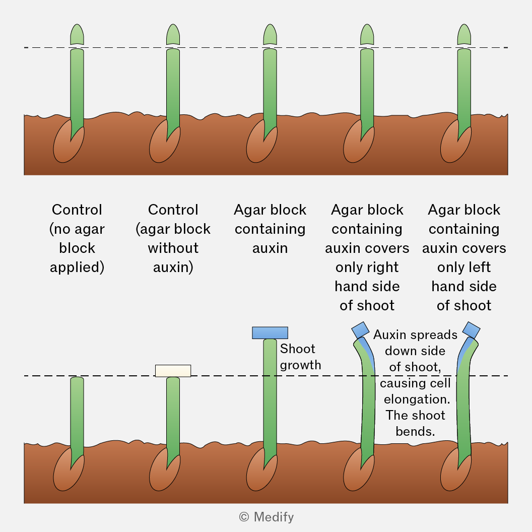
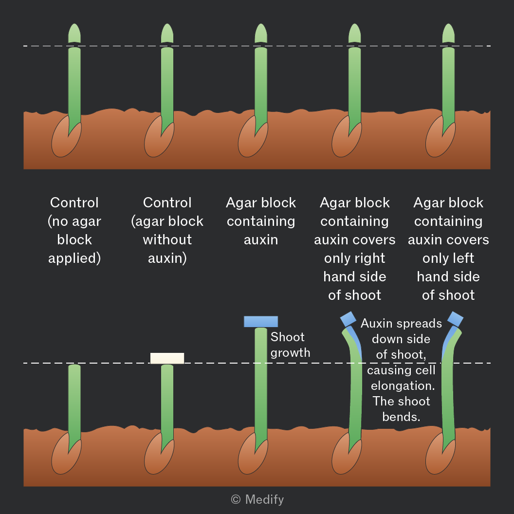
The concentration of auxins affects the extent of cell elongation and plant growth.
Serial dilution can be used to make agar blocks containing different concentrations of auxins such as IAA (indoleacetic acid). These blocks can be placed on shoot tips to investigate the effect of IAA concentration on plant growth.
Positive phototropism is a growth response caused by the unequal distribution of auxin in a shoot.
When light shines on one side of a shoot, auxin moves laterally to the shaded side. This causes greater cell elongation on the shaded side, so the shoot bends towards the light.
When light is evenly distributed, auxin is distributed evenly, causing the shoot to grow straight.
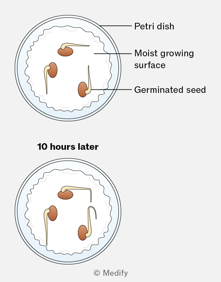
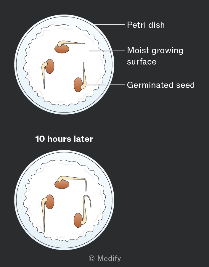
Plant growth is affected by gravity; shoots are negatively geotropic, and roots are positively geotropic. This ensures that roots always grow down into the soil.
This effect can be investigated by altering the orientation of roots and shoots as they grow (either in darkness or non-directional light).
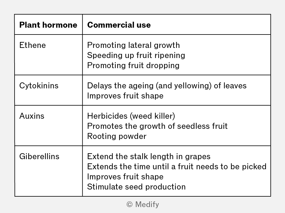
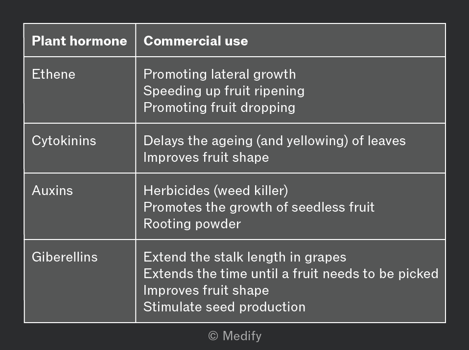
Plant hormones can be used in a variety of commercial contexts to aid industries such as farming and food production.
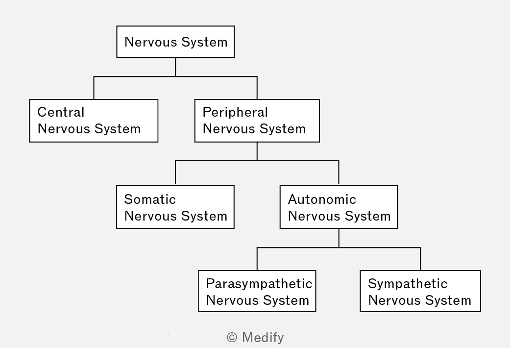
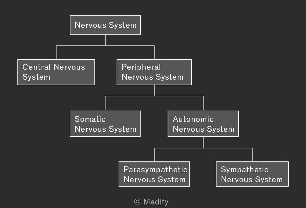
The mammalian nervous system coordinates responses to internal and external stimuli. It is organised in the following way:
- The Central Nervous System (CNS) consists of the brain and spinal cord. It is responsible for processing, integrating, and coordinating responses.
- The Peripheral Nervous System (PNS) is made up of all neurones outside the CNS (sensory and motor neurones). It connects the CNS to the rest of the body and can be divided into the sensory (afferent) and motor (efferent) pathways.
The peripheral nervous system is further organised in the following way:
- The somatic nervous system (voluntary).
- The autonomic nervous system (ANS; involuntary), which can be further split into sympathetic and parasympathetic nervous systems.
The somatic nervous system manages the voluntary control of skeletal muscles. This involves myelinated neurones for rapid response such as lifting your arm or walking.
The autonomic nervous system (ANS) manages the involuntary control of smooth muscle, cardiac muscle, and glands.
The ANS can be divided into two systems:
- Sympathetic system: “fight or flight”; uses noradrenaline; increases heart rate, dilates pupils.
- Parasympathetic system: “rest and digest”; uses acetylcholine; slows heart rate, promotes digestion.
The human brain is located in the skull and controls behaviour, perception, and homeostasis. The brain is surrounded by protective membranes called the meninges.
The brain receives and processes signals from nervous and endocrine systems, coordinating a response. It is made up of distinct regions with specialised functions.
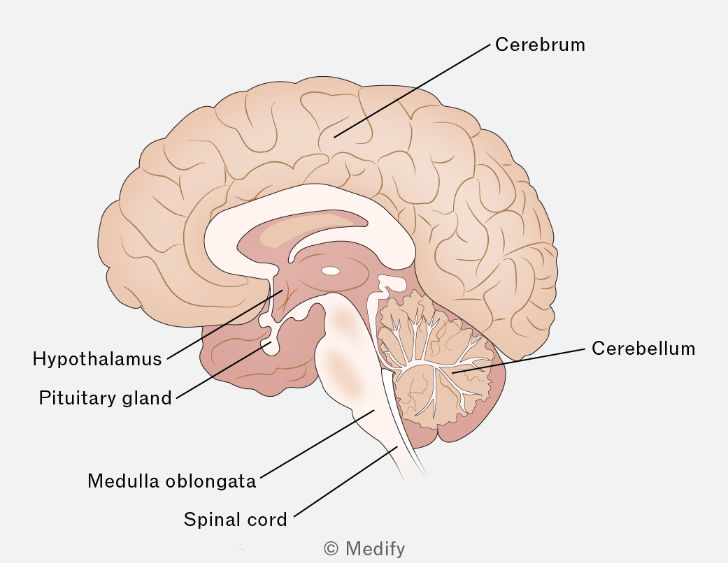
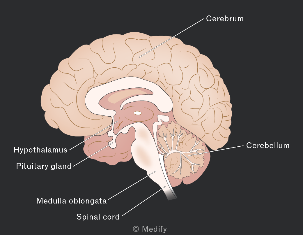
The cerebrum is the largest region of the brain and is divided into two hemispheres; each hemisphere is responsible for the opposite half of the body. It receives sensory information and coordinates responses.
- The outer layer of the cerebrum is known as the cerebral cortex. The prefrontal cortex is involved in decision-making and reasoning.
- The sensory and motor areas are sized in proportion to the number of receptors and motor endings.
- The sensory area receives and passes on information to association areas to be analysed and acted upon.
- Movement is controlled by the motor cortex.
The cerebellum is located near the brainstem and is responsible for controlling and coordinating muscle movement.
It receives information about balance and muscle tone and then relays the information to the motor control areas.
The medulla oblongata is located at the base of the brainstem.
The medulla oblongata is part of the autonomic nervous system (involuntary processes) and is involved in reflexes such as control of heart rate, breathing, peristalsis, and coughing.
The role of the hypothalamus is to maintain homeostasis. It controls complex behaviour patterns, monitors blood plasma concentration and produces hormones.
It is the control centre for both the sympathetic and parasympathetic nervous systems.
The pituitary gland is connected to the base of the hypothalamus.
The pituitary gland releases hormones that control other endocrine glands.
It is made up of an anterior and posterior section that produce, store and release hormones.
A reflex is an involuntary response to a sensory stimulus (e.g., blinking).
Reflexive actions are faster than actions done under conscious thought and increase the chances of survival.
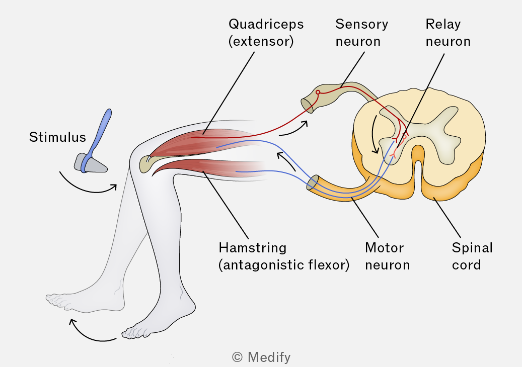
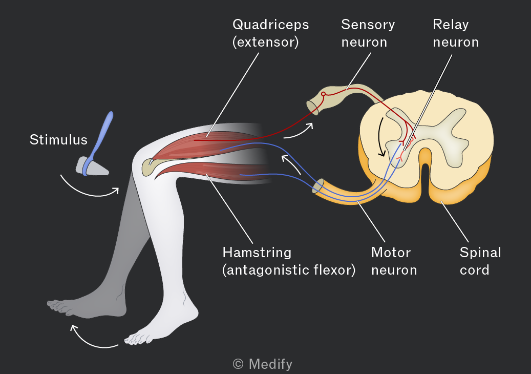
Nerve pathways responsible for reflexes are called reflex arcs.
The mechanism of action in a reflex arc is:
- Stimulus detected by a receptor (e.g., pain receptor in the skin).
- A sensory neurone transmits an impulse to the CNS.
- A relay neurone in the CNS processes the signal and links to a motor neurone.
- The motor neurone carries an impulse to an effector (e.g., muscle or gland).
- The effector produces a response (e.g., muscle contraction).
The knee–jerk reflex is a spinal reflex that involves only the spinal cord, not the brain. It is responsible for the unconscious maintenance of balance and posture.
If the tendon located below the knee is tapped then the extensor in the thigh contracts. The relay neurone inhibits the motor neurone of the hamstring (the antagonistic flexor) and the leg kicks.
Large multicellular organisms use two main systems to respond to internal and external stimuli:
- Hormonal responses in plants and animals involve chemical messengers and are usually slower but longer-lasting.
- Nervous responses in animals involve electrical impulses and produce rapid, short-term responses.
The ‘fight or flight’ response in mammals is coordinated by the endocrine and nervous systems.
The autonomic nervous system detects a threat, and the hypothalamus:
- Activates the sympathetic nervous system, which sends impulses to smooth muscle and the adrenal glands, to release adrenaline and noradrenaline into the blood
- Stimulates the pituitary gland, which secretes ACTH, causing the release of many longer-acting stress hormones from the adrenal cortex.
Physiological responses include pupil dilation, increased heart rate and increased blood glucose levels.
Hormones play a role in cell signalling during the ‘fight or flight’ response (e.g., to trigger glycogenolysis in liver cells).
Adrenaline acts as a first messenger, meaning it binds to a receptor on a target cell’s surface membrane, causing the activation of adenyl cyclase (an enzyme) within the cell.
Adenyl cyclase triggers the conversion of ATP to cyclic AMP, a second messenger that activates enzymes that convert glycogen to glucose. Glucose is then released into the blood to be used in respiration.
Cardiac muscle is myogenic, meaning it can initiate its own contractions, but heart rate is controlled by the medulla oblongata in the brain. It is connected to the sinoatrial node (SAN) by motor neurons.
- Impulses carried by the sympathetic nerves release noradrenaline at the SAN, increasing heart rate.
- Impulses carried by the parasympathetic nerve (vagus nerve) release acetylcholine at the SAN, decreasing heart rate.
Pressure, stretch and chemical receptors detect stimuli and activate one or other of these pathways to alter heart rate.
During exercise, stretch receptors in muscles detect limb movement and send impulses to the medulla oblongata. The resultant increase in heart rate facilitates increased oxygen delivery to muscles.
Baroreceptors in the aorta and carotid arteries detect changes in blood pressure and reduce heart rate when pressure is too high or increase it when pressure is too low.
Chemoreceptors monitor the of the blood. These receptors are found in the brain, carotid arteries and aorta.
During exercise, cells respire and create carbon dioxide, which forms carbonic acid and reduces the level of the blood. The medulla oblongata coordinates an increase in heart rate to increase the speed with which blood flows to the lungs for gas exchange.
There are several different muscle types:
- Skeletal muscle: these muscles control movement.
- Cardiac muscle: specialised muscle only found in the heart.
- Smooth muscle: involved in involuntary processes, found in the bladder, blood vessels, and digestive tract.
Each type of muscle has a different structure:
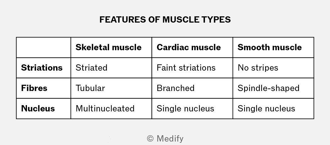
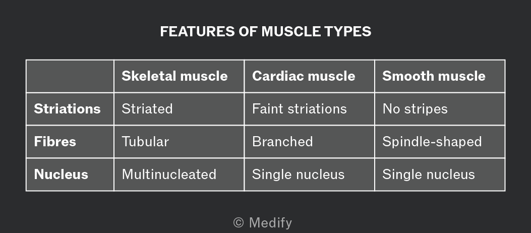
Skeletal muscle is made up of muscle fibres, which are long, multinucleated cells. Each muscle fibre is surrounded by a membrane known as the sarcolemma. T-tubules carry action potentials from the surface of the muscle cell to its interior. They are tiny tube-like extensions of the sarcolemma.
The cytoplasm in a muscle cell is specialised and called the sarcoplasm. It contains many mitochondria and a specialised endoplasmic reticulum called sarcoplasmic reticulum.
Muscle fibres contain myofibrils, contractile threads with repeating units (sarcomeres). These are protein-based organelles specialised for contraction. Myofibrils are laid down in parallel and combined, are very strong.
Myofibrils contain two types of protein filaments:
- Actin is a thinner filament, made up of two twisted strands. Actin has binding sites for myosin, which are blocked by tropomyosin and held in place by troponin.
- Myosin filaments are thicker, long, rod-shaped fibres with hinged projections to the sides. Each head has one binding site for actin and one for ATP.
When viewed under a microscope, skeletal muscle has bands:
- A band: dark, myosin (with overlapping actin).
- I band: light, actin only.
- H zone: central region, myosin only.
- Z lines: mark the boundaries of each sarcomere. The distance between two adjacent Z lines is one sarcomere.
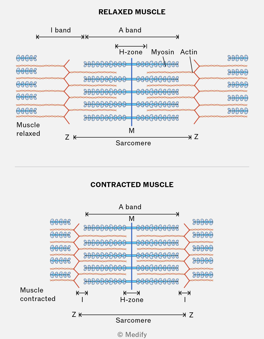
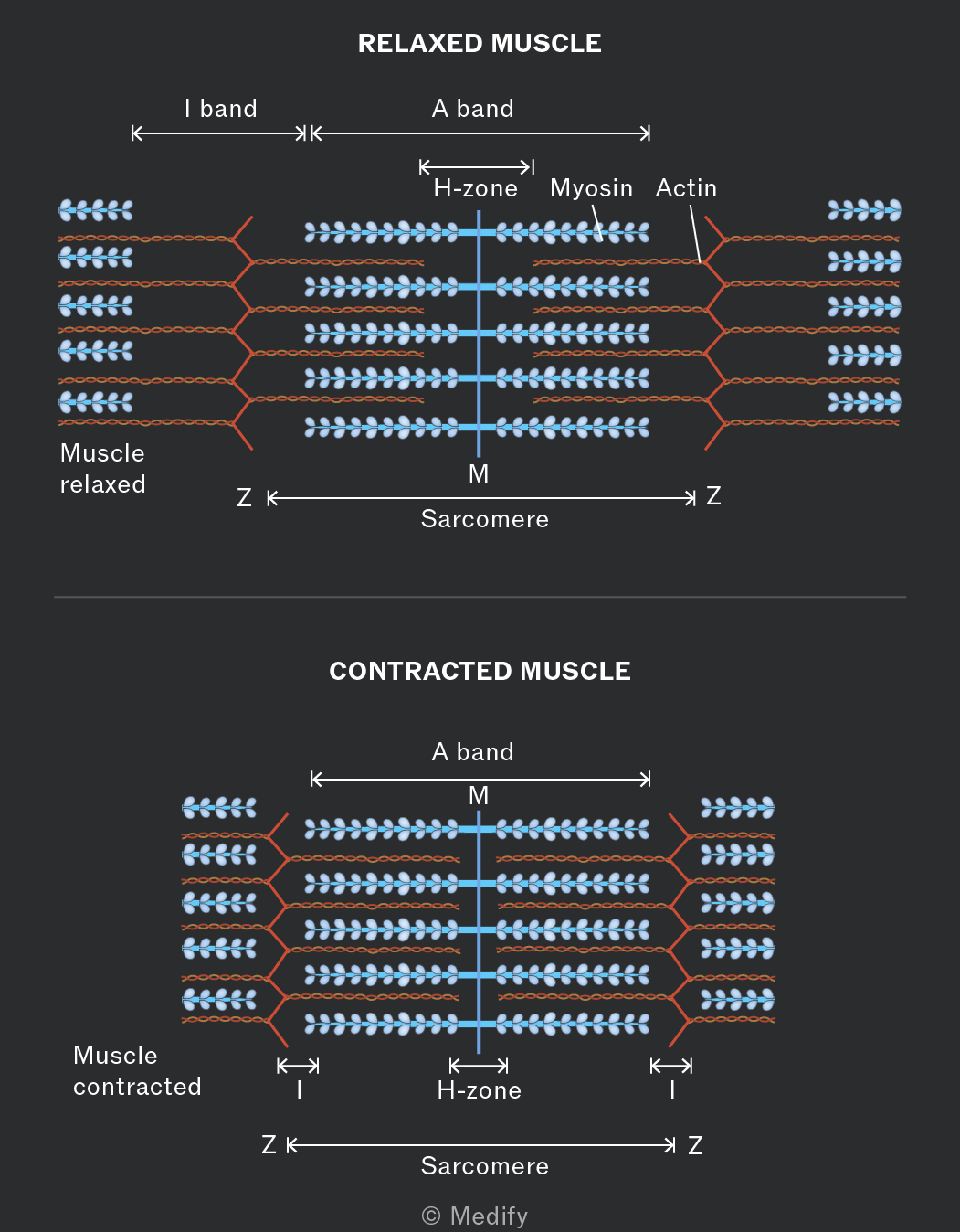
The sliding filament model explains how muscle fibres contract:
- Myosin filaments pull actin filaments towards the centre of the sarcomere, sliding past one another.
- The I band and H zone become narrower.
- The A band remains the same.
- The Z lines move closer together so the sarcomere shortens.
As many sarcomeres shorten simultaneously, the muscle fibre contracts, causing the whole muscle to contract and produce movement.
Skeletal muscle contraction occurs when actin and myosin filaments slide past one another, shortening the sarcomere:
- Calcium ions () bind to troponin, causing tropomyosin to move and expose the myosin-binding sites on actin.
- Myosin heads bind to actin, forming cross-bridges.
- ATP is hydrolysed providing the energy for the movement of the myosin head which pulls the actin filament towards the centre of the sarcomere.
- The myosin detaches, the heads move back and attach further along the actin filament. This requires ATP.
This cycle occurs rapidly, occurring many times per second.
A neuromuscular junction (NMJ) connects a neurone to a muscle fibre.
There are multiple neuromuscular junctions along a muscle to ensure rapid, strong and efficient contraction. A motor unit is all the muscle fibres associated with one motor neurone.
An action potential reaches the presynaptic bulb / knob and calcium ion channels open.
Vesicles fuse with the presynaptic membrane and release acetylcholine into the synapse by exocytosis. The acetylcholine binds to the sarcolemma causing sodium ion channels to open, leading to depolarisation.
Acetylcholine is broken down in the junction to give muscles a rest period.
Depolarisation spreads through the sarcolemma via T-tubules that touch the sarcoplasmic reticulum. The sarcoplasmic reticulum stores ions but when depolarisation occurs the channels open and ions move into the sarcoplasm.
binds to troponin which moves tropomyosin, allowing an actin-myosin cross bridge to form. Myosin then pulls actin along. ADP on myosin is replaced with ATP and the myosin head detaches from actin.
ions stimulate ATPase and myosin reattaches further along the actin filament. This process will continue for however long the muscle is stimulated for.
The hydrolysis of ATP releases energy.
Muscle contraction requires large amounts of ATP, which is used to move myosin heads and actively reabsorb back into the sarcoplasmic reticulum.
ATP is regenerated by:
- Aerobic respiration (longer, low intensity); lots of mitochondria are involved and require oxygen.
- Anaerobic respiration (short-term, high intensity); muscles use oxygen faster than it is delivered, leading to pyruvate being broken down into lactic acid, causing fatigue.
- Creatine phosphate (short-term, vigorous); provides a supply of Pi, for rapidly generating ATP but is used up quickly.