Cells (3.2)›All cells arise from other cells (3.2.2)

All cells arise from other cells (3.2.2)

An overview of all cells arise from other cells (3.2.2) from AQA A level Biology including: the cell cycle, the stages of mitosis and cancer
5 min

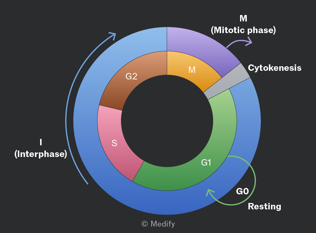
Not all eukaryotic cells retain the ability to divide, but those that can undergo the cell cycle, a series of stages involving growth and division. The outcome is two genetically identical daughter cells.

The cycle consists of interphase, followed by mitosis and cytokinesis (cytoplasmic division).
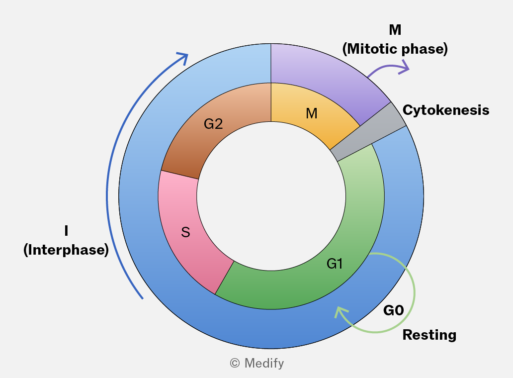
The image depicts a circular diagram representing the cell cycle. The outer circle is labeled with two main phases: 'I (Interphase)' on the left in blue and 'M (Mitotic phase)' on the right in purple, with arrows indicating the direction of the cycle. The inner circle is divided into segments labeled 'G1' in green, 'S' in pink, 'G2' in brown, 'M' in yellow, and a small gray segment labeled 'Cytokinesis.' An additional green arrow curves into 'G1' from a segment labeled 'G0 Resting.'
Add to favourites

Interphase is the stage during which the cell grows and prepares for division. It occupies approximately 90 of the cell cycle and consists of three phases:

  • G1 (first growth)
  • S (DNA synthesis)
  • G2 (second growth).
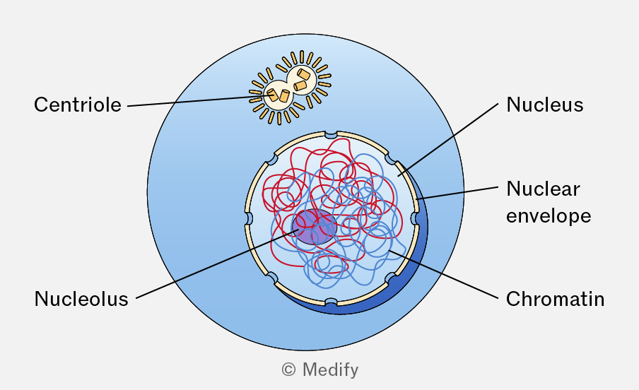
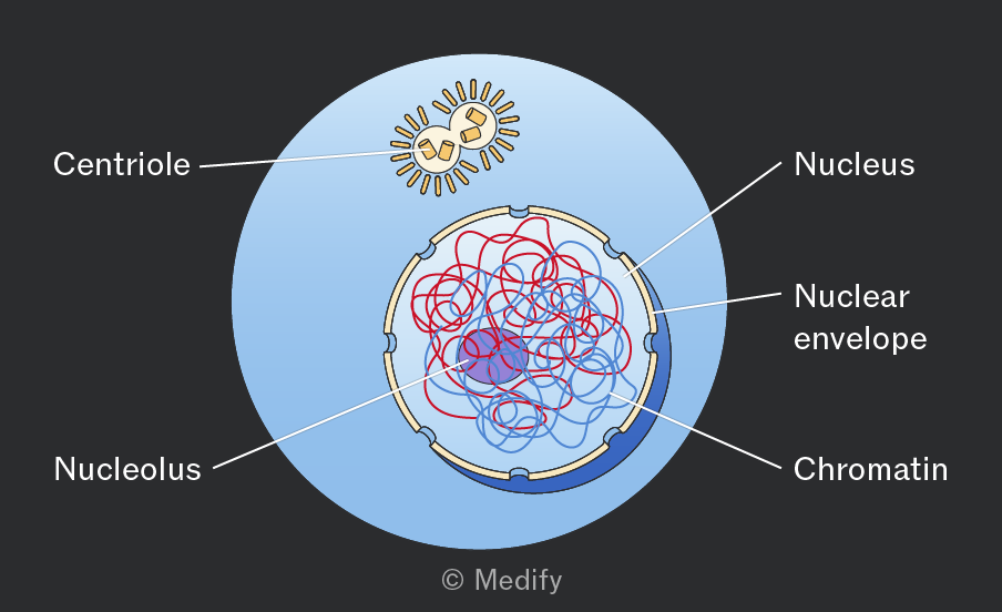
A diagram of a cell displaying various structures labeled with text. The outer blue circle is labeled 'Nucleus'. Within this circle are two concentric circles forming the 'Nuclear envelope'. Inside the nuclear envelope, there is a dense, purple area labeled 'Nucleolus'. Surrounding the nucleolus is a network of red lines labeled 'Chromatin'. Above the nucleus is a structure labeled 'Centriole', depicted as a sun-like shape with a central circle and radiating lines. The background is light blue. The image is copyrighted by Medify.
Add to favourites

Mitosis is the nuclear division process during which a eukaryotic cell separates its duplicated chromosomes into two identical nuclei. It consists of prophase, metaphase, anaphase and telophase and is followed by cytokinesis.

Cytokinesis is the division of the cytoplasm following nuclear division, resulting in the formation of two daughter cells.
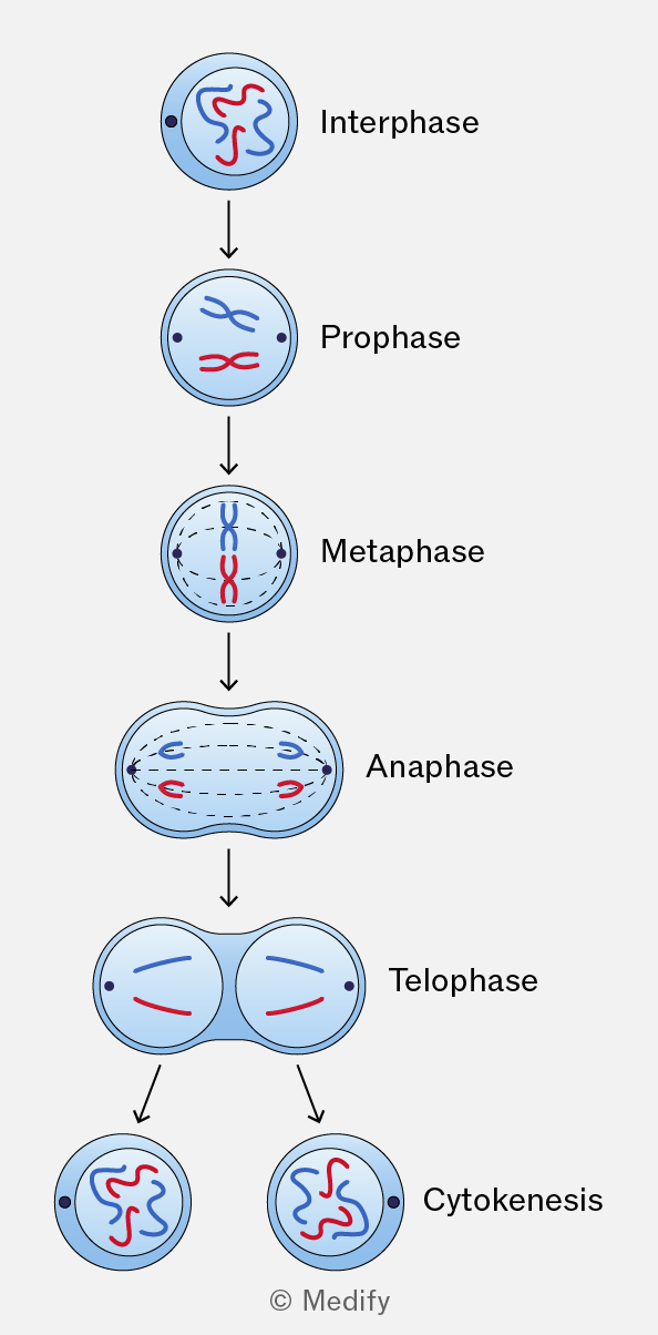
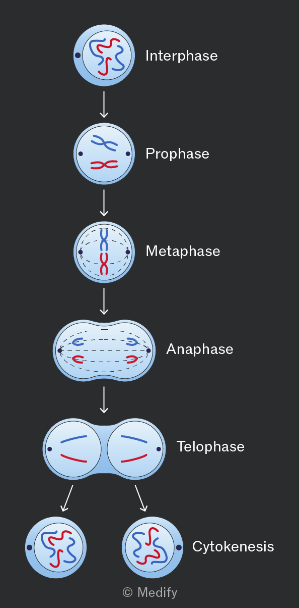
Diagram showing stages of cell division: Interphase with tangled chromatin, Prophase with condensed chromosomes, Metaphase with chromosomes aligned at the center, Anaphase with separated chromosomes moving to poles, Telophase with two distinct nuclei forming, and Cytokinesis with two separate cells. Each stage is labeled accordingly.
Add to favourites

Prophase is the first stage of mitosis. Chromatin condenses into chromosomes, each consisting of two sister chromatids joined at the centromere.

The nuclear envelope breaks down and the nucleolus disappears.

Centrosomes (centrioles) move to opposite poles of the cell, forming spindle fibres that guide chromosome movement.
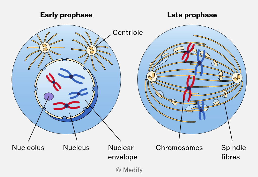
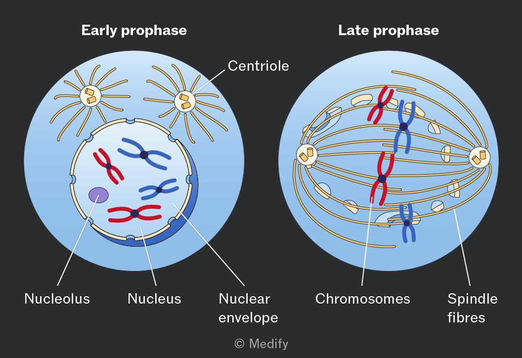
The image shows two diagrams labeled 'Early prophase' and 'Late prophase'. In the 'Early prophase' diagram, there are two centrioles with spindle fibers extending from them, positioned outside a nucleus. Inside the nucleus, labeled structures include the nucleolus, and several chromosomes depicted as pairs of connected blue and red lines. The nuclear envelope surrounds the nucleus. The 'Late prophase' diagram shows spindle fibers extending from centrioles towards the center where chromosomes, depicted as red and blue paired lines, are more condensed and aligned. Labels indicate the chromosomes and spindle fibers, with the centrioles positioned at opposite poles.
Add to favourites

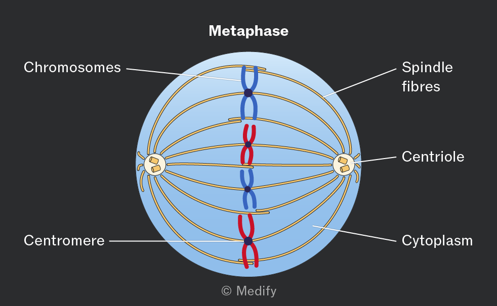
During metaphase, chromosomes align at the cell equator, also called the metaphase plate.

Spindle fibres from opposite poles attach to each centromere.
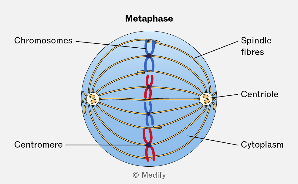
Diagram labeled 'Metaphase' showing a cell with a light blue circular background. In the center, blue and red structures labeled 'Chromosomes' are aligned vertically. Each chromosome has a central black dot labeled 'Centromere.' Gold lines extend from the centromeres to structures labeled 'Centriole' at each end of the cell. These lines are labeled 'Spindle fibres.' The entire cell is labeled 'Cytoplasm.'
Add to favourites

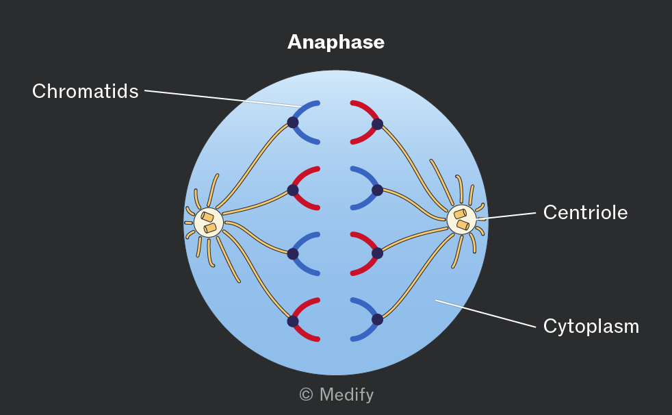
During the anaphase, the centromeres divide, allowing sister chromatids to separate.

The spindle fibres shorten, moving the chromatids towards opposite poles of the cell.
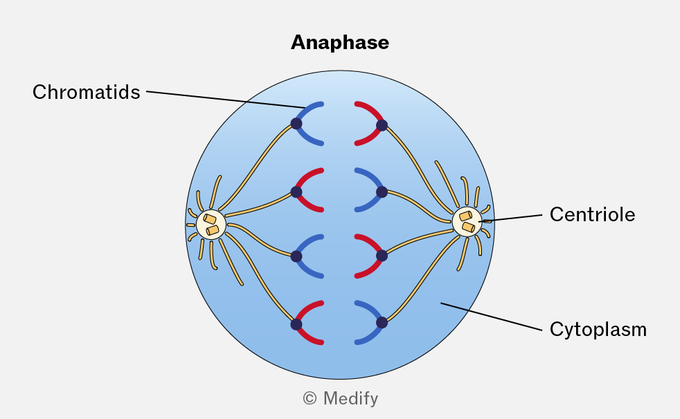
The image illustrates the anaphase stage of cell division. Inside a blue circle representing the cytoplasm, two groups of chromatids, depicted as red and blue curved lines, are being pulled apart toward opposite sides. Each group is connected to a centriole, represented as a yellow structure with radiating lines, located on either side of the circle. Labels indicate 'Chromatids' pointing to the red and blue structures, 'Centriole' pointing to the yellow structure, and 'Cytoplasm' pointing to the blue circle. The word 'Anaphase' is written at the top of the image.
Add to favourites

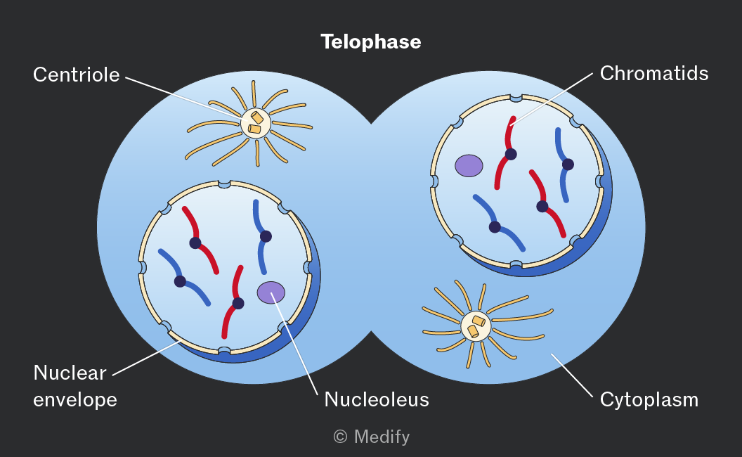
During telophase, the chromatids are at the cell’s poles and are now chromosomes. The nuclear envelope reforms around each set of chromosomes, creating two nuclei.

The chromosomes uncoil back into chromatin, and the nucleolus is formed.

The mitotic spindle breaks down as the cell prepares for cytokinesis.
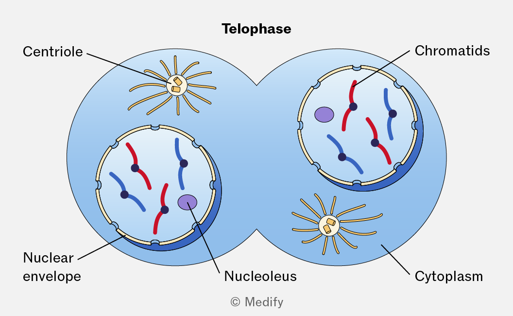
The image illustrates the telophase stage of cell division, showing two circular cells connected in the center. Each cell contains a labeled centriole with radiating spindle fibers. Inside each cell, a nuclear envelope encloses chromatids, depicted as red and blue curved lines, some connected by black dots. Labels include 'Centriole', 'Chromatids', 'Nuclear envelope', 'Nucleolus', and 'Cytoplasm'. The title 'Telophase' is centered at the top of the image. The copyright notice '© Medify' is at the bottom center.
Add to favourites

Viruses are non-living, so they cannot undergo cell division.

Viruses attach to a host cell and inject their genetic material (DNA or RNA) into it. The host cell then synthesises the viral components and assembles new viruses.

Add to favourites

Binary fission is the method of cell division (asexual reproduction) in prokaryotic cells (e.g., bacteria).

  1. Replication of circular DNA and plasmids.
  2. The cell increases in size.
  3. The circular DNA molecules move to opposite ends of the cell.
  4. The cytoplasm divides.
  5. A new cell wall and cell membrane form between the two circular DNA molecules.

Two genetically identical daughter cells are produced, containing one copy of the circular DNA and a variable number of plasmids.
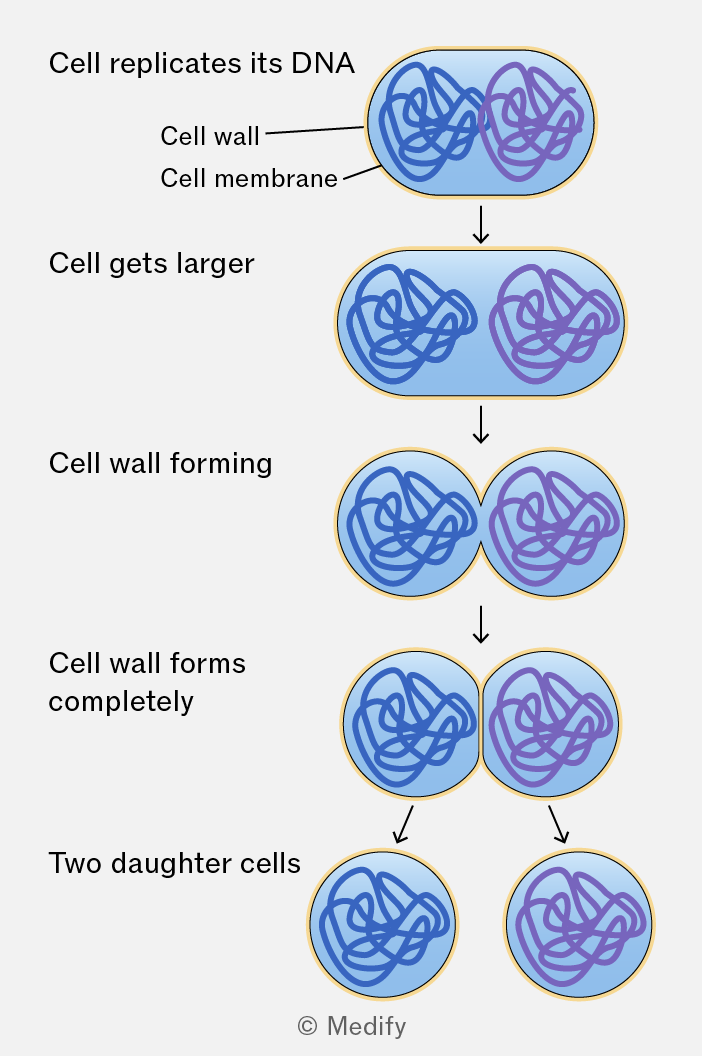
A diagram showing the process of cell division with five stages. 1. 'Cell replicates its DNA': A single cell with two sets of DNA inside, labeled 'Cell wall' and 'Cell membrane'. 2. 'Cell gets larger': The cell enlarges with two distinct DNA regions. 3. 'Cell wall forming': The cell begins to constrict in the middle. 4. 'Cell wall forms completely': The constriction completes, and the cell is nearly divided. 5. 'Two daughter cells': Two separate cells, each with its own set of DNA. Arrows indicate progression from one stage to the next. © Medify.
Add to favourites

If mitotic control mechanisms fail, cells may divide uncontrollably, forming a tumour (a mass of abnormal cells). Tumours may be benign (harmless) or malignant.

Benign tumours grow slowly, remain contained and are unlikely to be life-threatening.
Malignant tumours are cancerous, grow rapidly and can invade surrounding tissues and spread to other parts of the body, making them more likely to be life-threatening.

Add to favourites

Many cancer treatments work by controlling or stopping cell division.

Examples include:

  • Chemotherapy – uses drugs to prevent mitosis.
  • Radiotherapy – uses radiation to damage DNA and stop cell division.

These treatments also affect normal dividing cells, causing side effects such as hair loss, nausea, and a weakened immune system.

Add to favourites

There are multiple types of gene mutations. In substitution (point) mutations, one DNA base is replaced by another during DNA replication. There are three different types of substitution mutation:

  • Silent: the base sequence changes, but the same amino acid is coded for, so the protein is unchanged.
  • Missense: the base substitution changes the codon, resulting in a different amino acid being incorporated into the protein. This may alter the protein’s structure and function.
  • Nonsense: the base substitution creates a stop codon, causing translation to end prematurely and producing a shortened, usually non-functional protein.
Add to favourites

In insertion / deletion (indel) mutations, one or more bases are added or removed. Unless the number of bases inserted or deleted is a multiple of three, this causes a frameshift mutation altering the triplet reading frame.

As a result, amino acid sequence from the mutation onwards is changed, often producing a protein with an altered tertiary structure that is unable to function normally.

Add to favourites