Cell recognition and the immune system (3.2.4)
On this page
Cell identification allows the immune system to distinguish self from non-self, preventing attacks on the body’s own cells (autoimmune diseases) while targeting pathogens.
Body cells possess specific self-antigens on their cell-surface membranes. These allow immune cells to recognise the body’s own cells meaning healthy body cells are not normally attacked.
Pathogens have different antigens and are recognised as non-self triggering an immune response against them.
HIV (human immunodeficiency virus) is a retrovirus that infects and destroys helper T cells (also called T helper cells).
This weakens the immune system by reducing the number of helper T cells. If left untreated, HIV infection can progress to AIDS (acquired immune deficiency syndrome). People with AIDS are highly susceptible to opportunistic infections and certain cancers, as their immune system is no longer able to respond effectively.
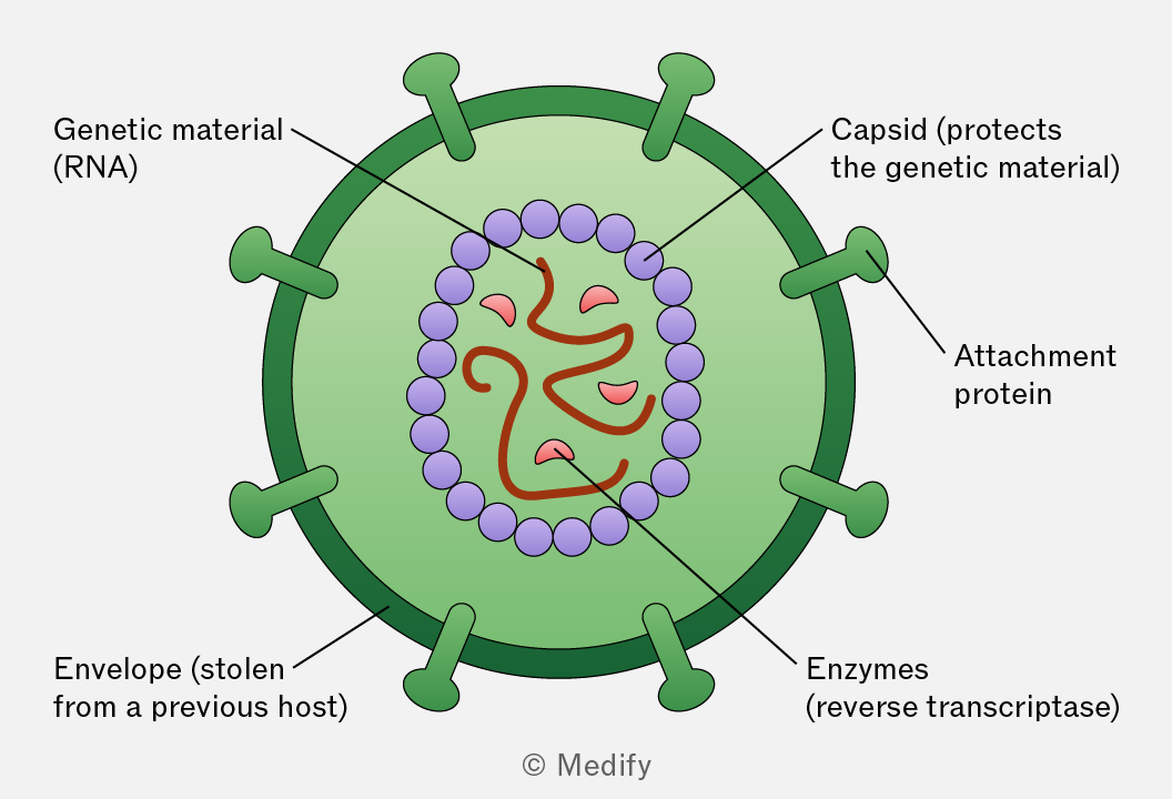
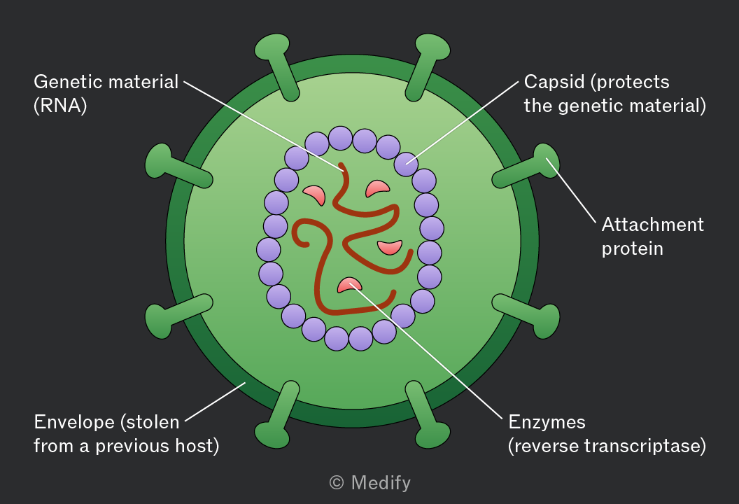
HIV is a retrovirus with an outer envelope derived from a previous host cell, containing attachment proteins (e.g., gp120) that bind to CD4 receptors on helper T cells.
Inside, a protein capsid protects two RNA strands and the enzyme reverse transcriptase, which converts viral RNA into DNA in the host cell.
This allows HIV to hijack the host cell’s machinery and replicate.
Antibiotics are drugs that kill or inhibit the growth of bacteria by targeting structures or processes that are unique to bacterial cells, such as:
- Cell wall synthesis (e.g. penicillin).
- Ribosomes (used for protein synthesis).
- Metabolic pathways unique to bacteria.
Viruses, such as HIV, are acellular (not made of cells). Viruses have no cell wall, ribosomes and no independent metabolism. Viruses rely completely on host cells to replicate; none of the target structures present in bacteria are found within viruses or host cells, making antibiotics ineffective against viral infections.
Phagocytes are a type of white blood cell that play a key role in the non-specific immune response by engulfing and destroying pathogens.
Neutrophils are the most abundant type of phagocyte. They rapidly respond to infections and die after engulfing pathogens.
Macrophages are larger, longer-lived phagocytes that develop from monocytes (a type of white blood cell). Macrophages act as antigen-presenting cells (APCs) to activate specific immune responses.
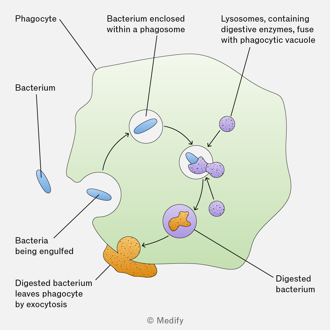
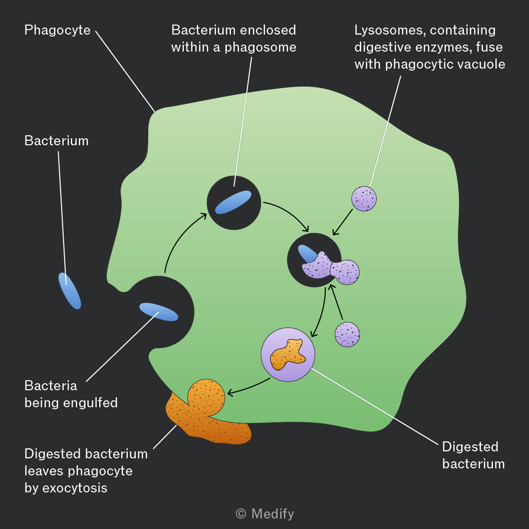
Phagocytosis is the process by which phagocytes (e.g., neutrophils, macrophages) engulf and destroy pathogens.
Phagocytosis involves the following steps:
- Chemotaxis – phagocytes use chemical signals to move toward pathogens.
- Recognition and attachment – receptors on the phagocyte membrane bind to antigens on the pathogen surface.
- Engulfment – the pathogen is enclosed in a phagosome (a membrane-bound vesicle).
- Digestion – lysosomes fuse with the phagosome to form a phagolysosome, releasing enzymes to break down the pathogen.
- Antigen presentation – macrophages display the pathogen’s antigens on their surface membrane to activate the immune response.
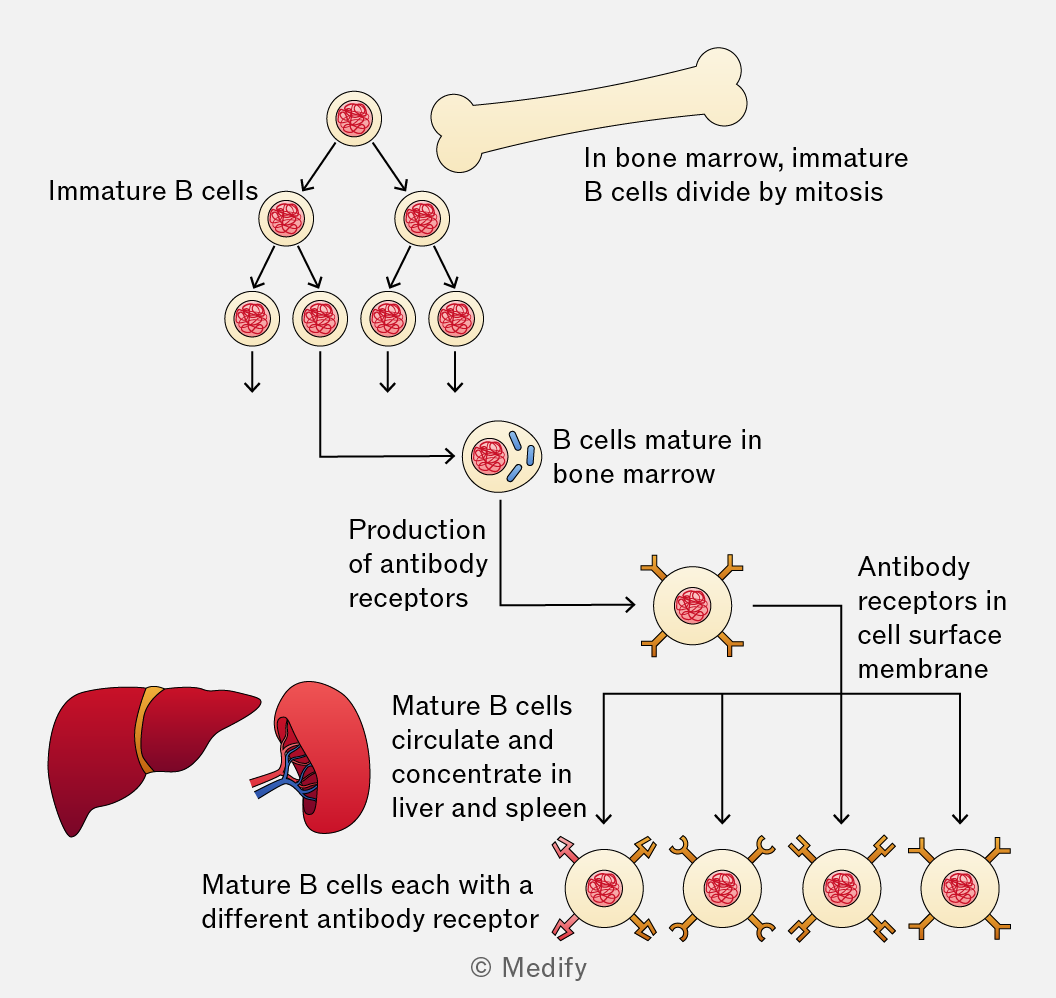
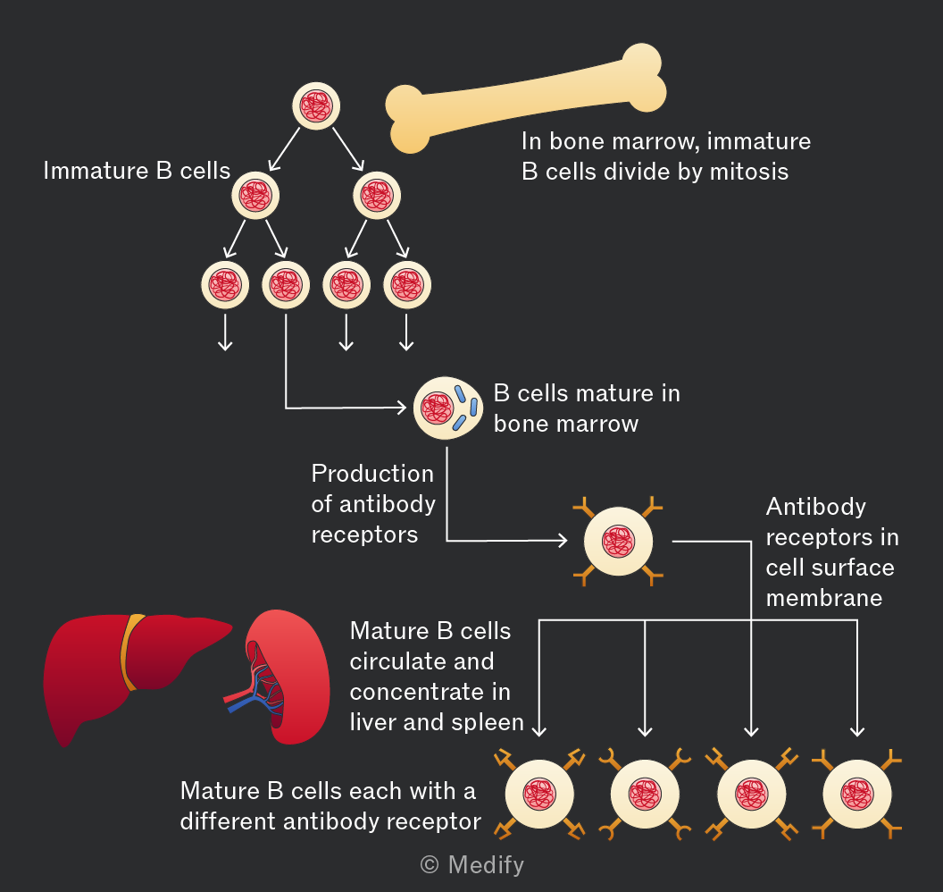
B lymphocytes are a type of lymphocyte that mature in the bone marrow and play a key role in the specific (humoral) immune response. They are responsible for targeting pathogens in body fluids such as blood and lymph.
When a B cell detects a specific antigen, it becomes activated and divides to form plasma cells (B effector cells), which produce antibodies that bind to and neutralise the pathogen.
Some B cells become memory cells, which remain in the body and provide long-term immunity by responding quickly if the same antigen is encountered again.
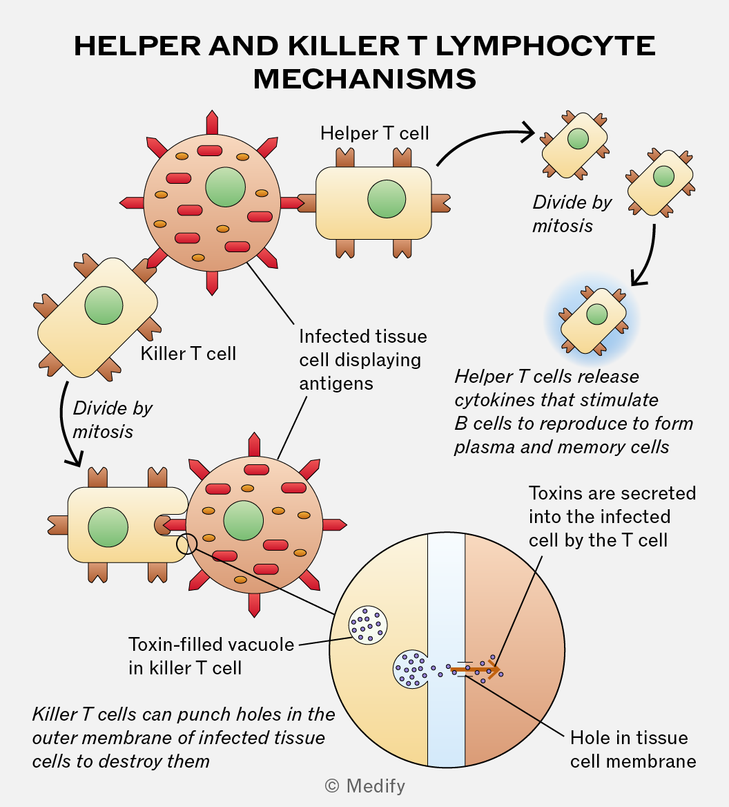
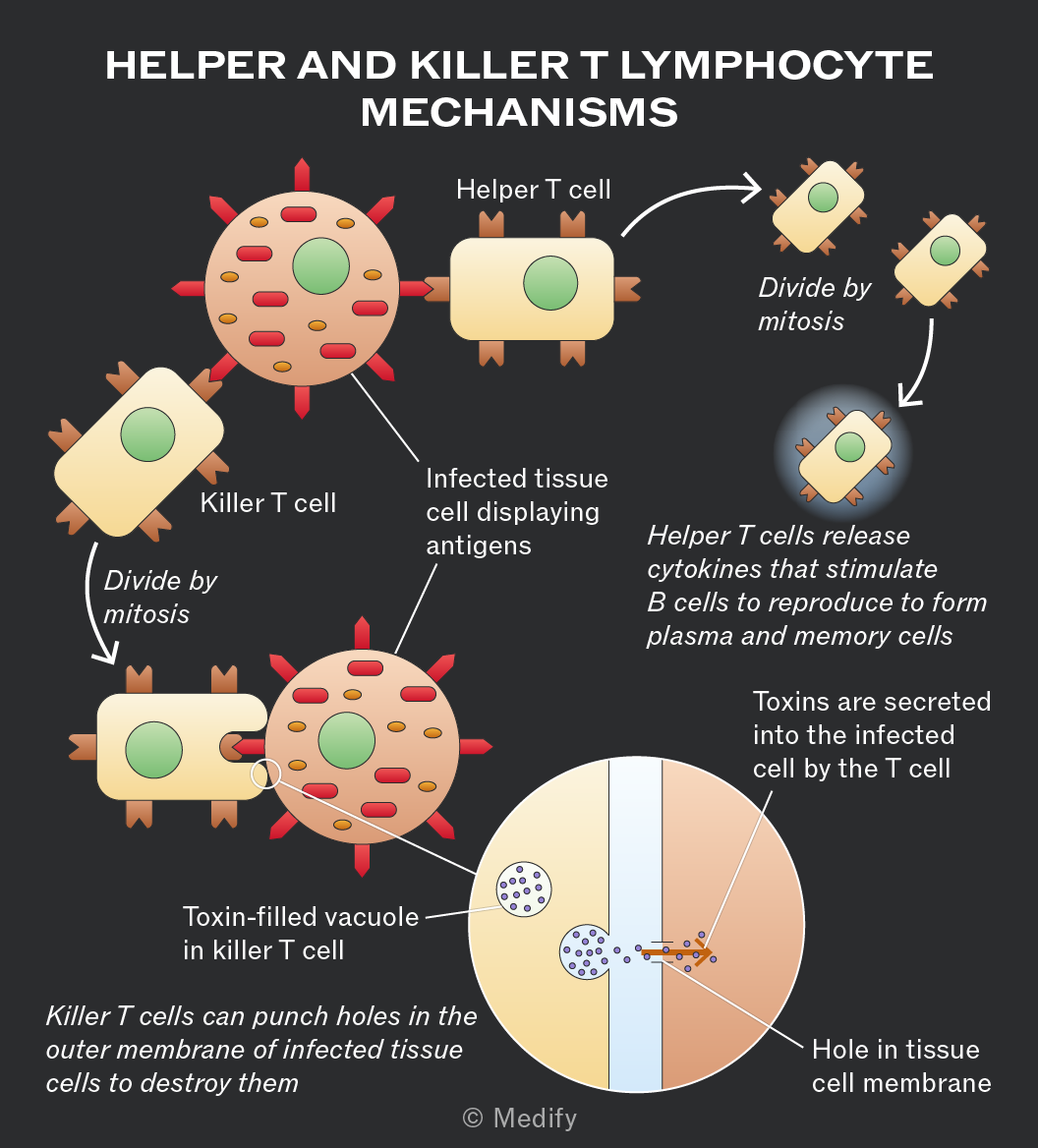
There are several types of T lymphocytes:
- T helper cells (Th): release cytokines to activate B cells, phagocytes, and other T cells such as cytotoxic T cells.
- T killer (cytotoxic) cells (Tc): destroy infected or abnormal cells by releasing substances that cause cell lysis.
Clonal selection occurs when an antigen binds to a B cell with a complementary receptor on its cell surface.
The B cell presents the antigen on its surface, where it is recognised by a helper T cell with a complementary receptor. The helper T cell activates the selected B cell, ensuring that only the B cell specific to that antigen responds.
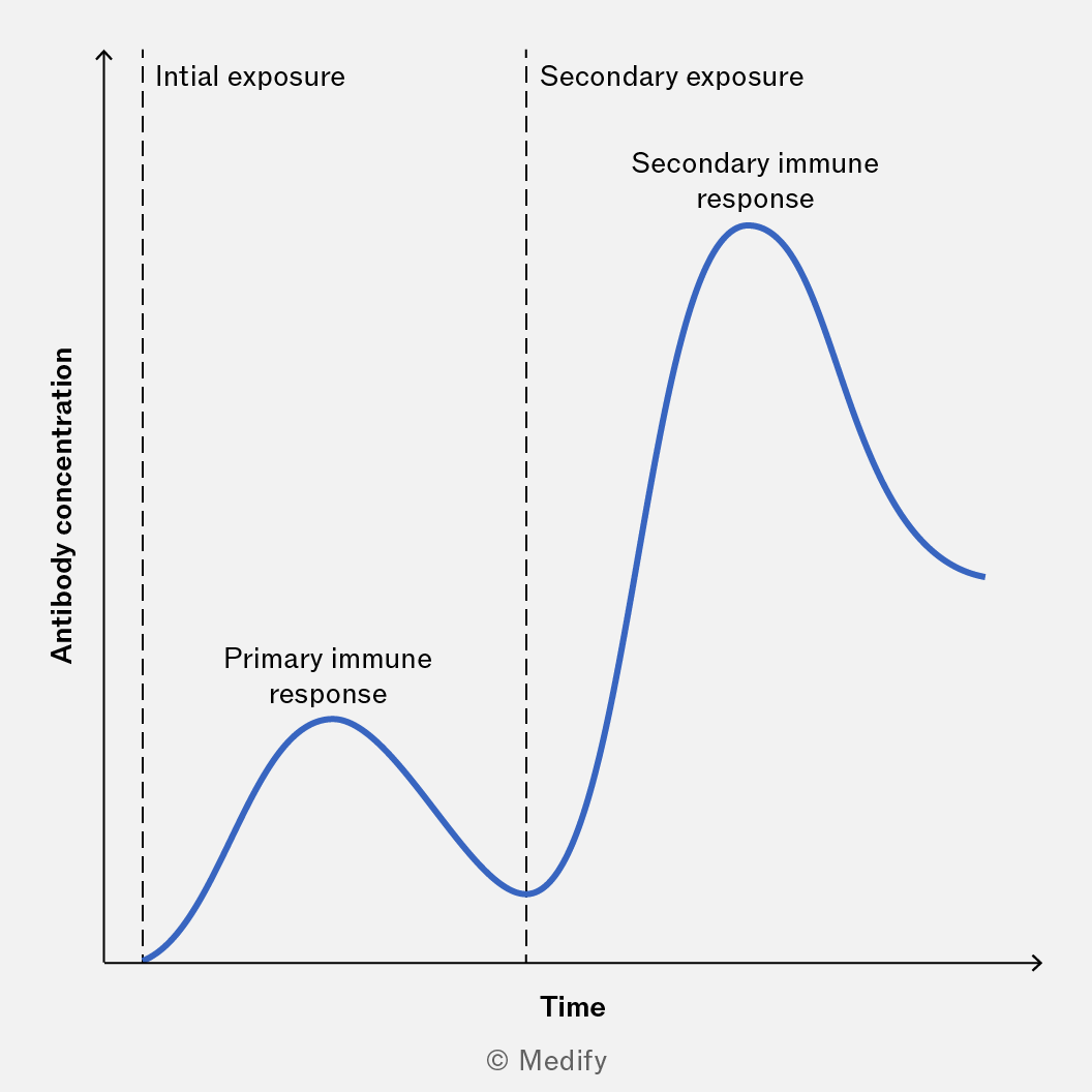
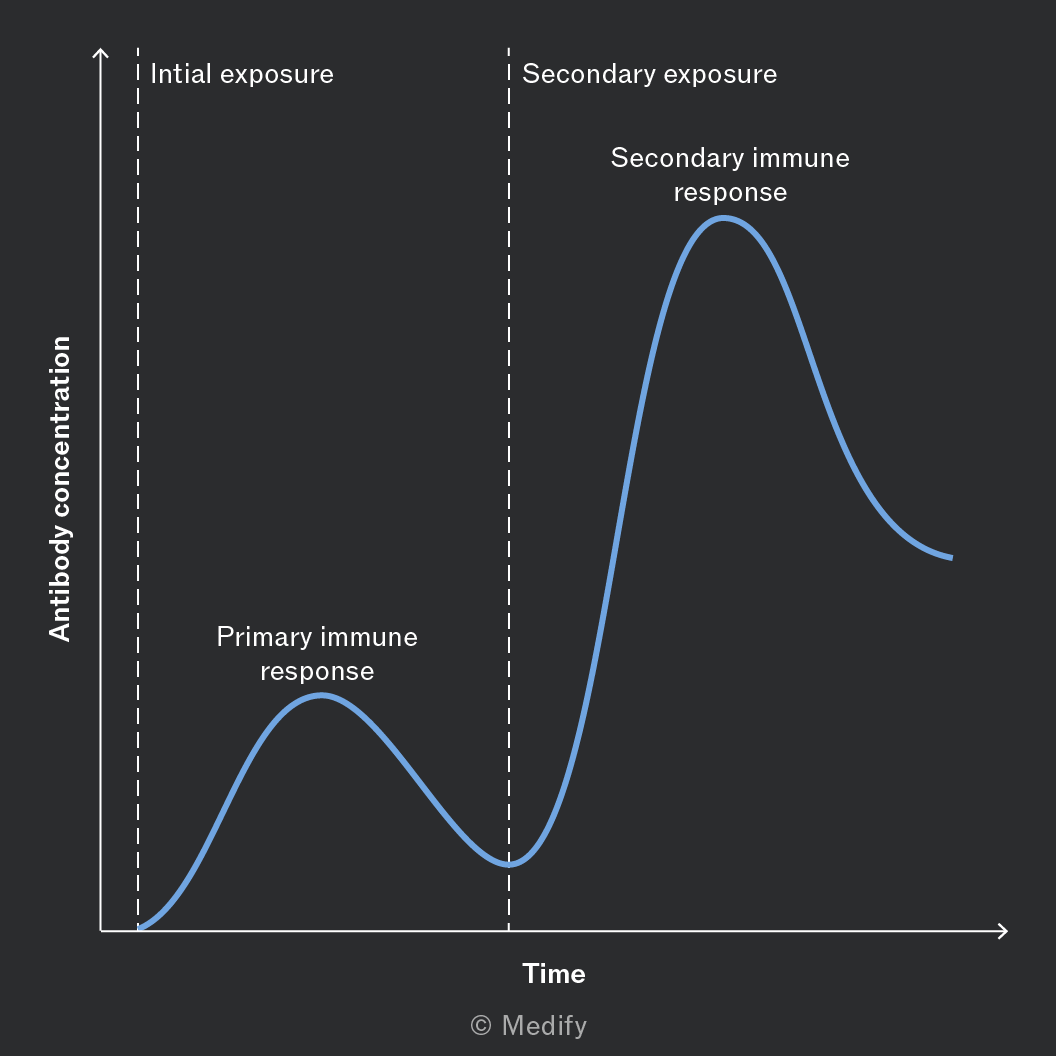
During the primary immune response, plasma cells are produced from activated B cells.
They make and release antibodies specific to the antigen which help destroy the pathogen.
This response is slow, so symptoms may appear before the pathogen is removed. Memory cells are also made during this time.
During the secondary immune response, memory B cells quickly recognise the pathogen’s antigen.
They rapidly divide and form plasma cells. These plasma cells produce antibodies faster and in larger quantities than during the primary immune response.
The pathogen is usually destroyed before symptoms develop which is why people are often immune after the first infection or following vaccination.
An antibody is a Y-shaped globular protein made of four polypeptide chains (two heavy and two light), held by disulfide bonds.
The variable region at the tips form two antigen-binding sites; they have a specific tertiary structure that is complementary to a specific antigen, meaning specific binding will occur at these sites.
The constant region is the same in all antibodies of that class and interacts with other components of the immune system, helping to recruit immune cells.
The hinge region provides flexibility, allowing the antibody to bind to antigens at different angles.
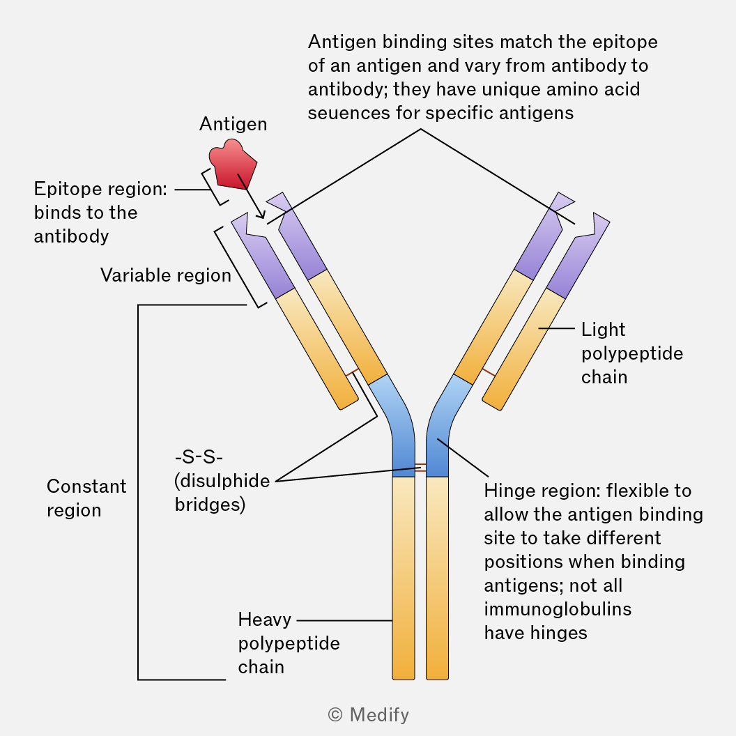
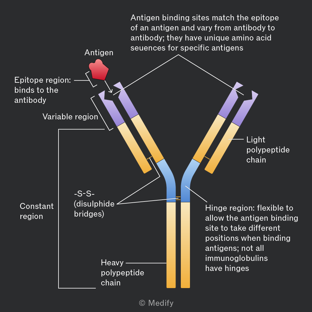
Antibodies bind to antigens on pathogens (or to toxins). The binding is specific, like a lock and key and forms an antigen–antibody complex.
Once the antigen-antibody complex is formed then there are several different ways in which the antibody itself or the immune system will destroy the pathogen.
An antigen is a molecule, usually a protein or polysaccharide, that triggers an immune response by stimulating the production of antibodies or activating immune cells.
Antigens are usually found on the surface of pathogens (e.g., bacteria, viruses) but can also be on toxins or foreign substances.
Each antigen has a specific shape that is recognised by complementary antibodies or T-cell receptors.
This recognition enables the immune system to distinguish between self (the body’s own cells) and non-self (foreign invaders).
Some pathogens (e.g., viruses and bacteria) change their antigens over time through mutations or genetic recombination.
This allows them to evade immune recognition, because previously produced antibodies and memory cells may no longer recognise the altered antigens. This makes it harder for the immune system to respond effectively.
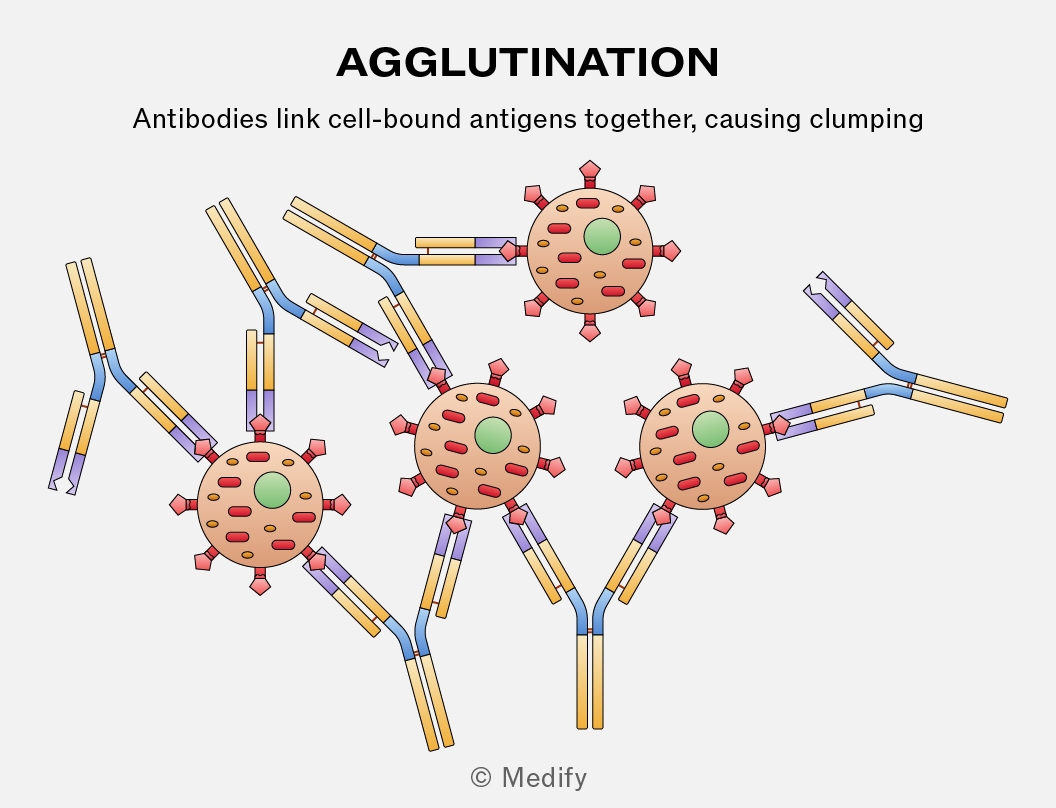
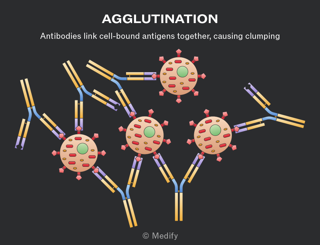
The role of agglutins is to clump pathogens together. They bind to antigens on the surface of multiple pathogens, forming antigen–antibody complexes.
The clumped pathogens are more easily recognised and engulfed by phagocytes.
There are two types of immunity:
- Active immunity: the body’s own immune system makes antibodies and memory cells following exposure to an antigen (e.g., vaccinations or after infection).
- Passive immunity: ready-made antibodies are transferred to an individual, so no memory cells are made (e.g., breast milk being ingested by infants or antibody injection such as antivenom).
A vaccine contains antigens from a pathogen (e.g., a dead, weakened or part of a pathogen), stimulating the immune system without causing disease.
The antigens in the vaccine are recognised by the immune system. Antigen-presenting cells process the antigens and present them to T helper cells which activate B cells. The activated B cells undergo mitosis and differentiate to form:
- Plasma cells: produce antibodies (short term).
- Memory cells: remain in the body (long term).
This results in active immunity, because the body’s own immune system makes the antibodies and memory cells.
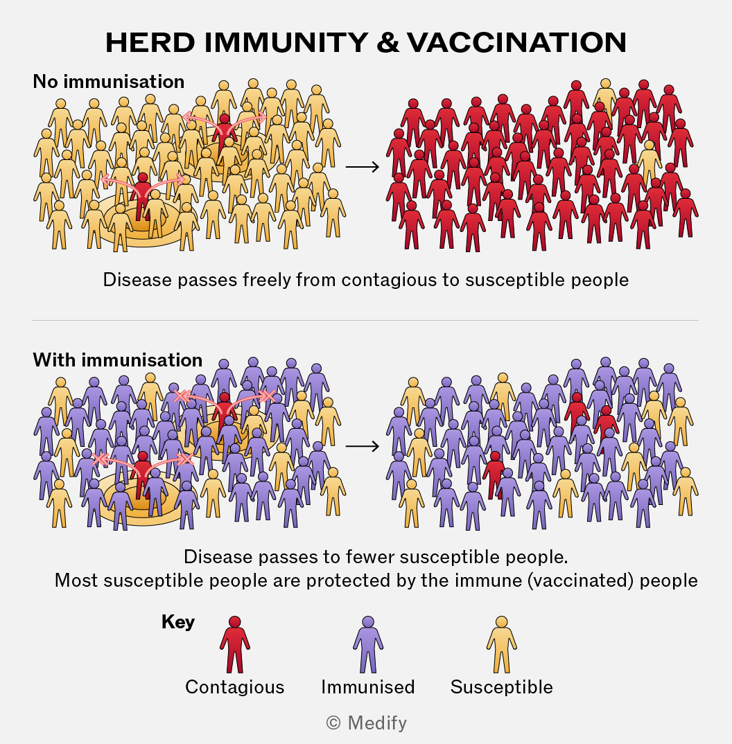
Herd immunity occurs when a large proportion of a population is immune to a specific infectious disease, making it more difficult for the disease to spread.
Pathogens spread through contact between infected and susceptible individuals but if most people are immune (through vaccination), the pathogen has fewer people to infect. As a result, unvaccinated or vulnerable individuals are indirectly protected because they are less likely to come into contact with an infected person.
There are social and ethical issues associated with the production and use of vaccines, including:
- Animal testing: vaccines are tested on animals before human use, raising concerns about animal welfare.
- Informed consent: people must understand the risks before being vaccinated. In some countries, this can be difficult to ensure.
- Access and fairness: some people or countries may not have equal access to vaccines, especially during shortages.
- Herd immunity decisions: choosing not to vaccinate can put others at risk, especially those who cannot be vaccinated for medical reasons.
Plasma cells release large amounts of identical antibodies that are specific to one antigen. These antibodies bind to antigens helping to neutralise pathogens and toxins, and also help phagocytes recognise and destroy pathogens.
Monoclonal antibodies are laboratory-produced antibodies that are identical and specific to one antigen. They can be used to deliver drugs directly to target cells (e.g., cancer cells).
A therapeutic drug (e.g., chemotherapy agent) is attached to a monoclonal antibody which binds to antigens on the target cells. This reduces damage to healthy cells, increasing treatment precision and reducing side effects.
The use of monoclonal antibodies in medicine raises some ethical and social concerns:
- Use of animals: mice are often used in the development and testing of antibodies, raising ethical concerns about animal welfare.
- Cost and access: these treatments can be expensive, so not everyone may have equal access to them.
- Side effects: some monoclonal antibody treatments have serious side effects, so patients must be fully informed of the risks before treatment.
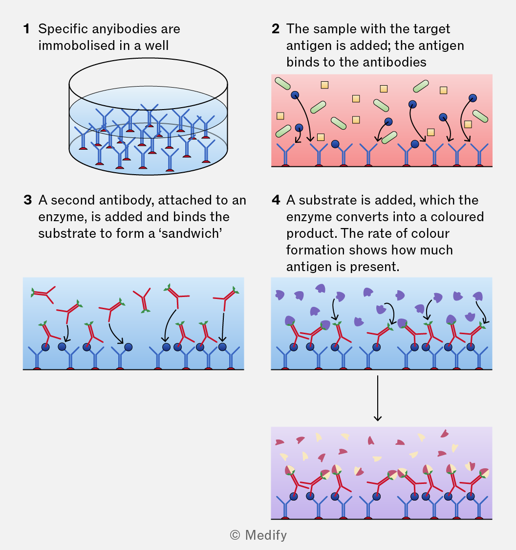
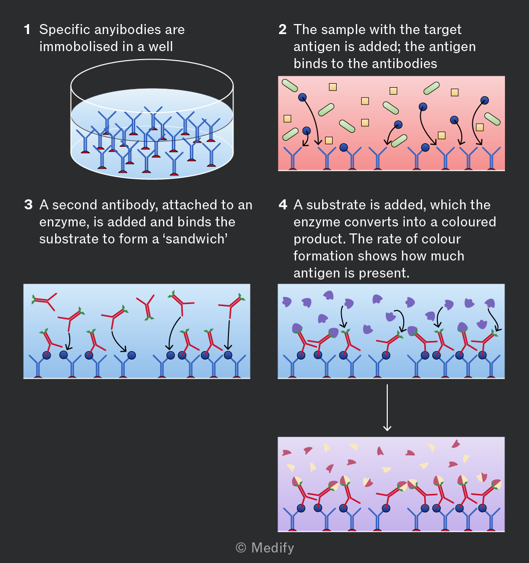
ELISA (enzyme-linked immunosorbent assay) is a test used to detect and measure the presence of a specific antigen or antibody protein in a sample.
Monoclonal antibodies bind specifically to the target antigen. An enzyme-linked antibody is then added, followed by a substrate. If the target antigen is present, the enzyme catalyses a reaction that produces a colour change.
The intensity of the colour is proportional to the amount of antigen (or antibody) present in the sample.
ELISA tests are highly specific because the antibodies only bind to their complementary antigen. They can be used to test for HIV, TB, hepatitis as well as to detect hormones and other proteins.