Gas exchange (3.3.2)
On this page
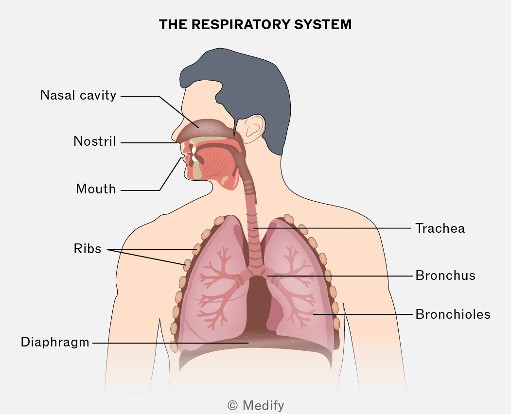
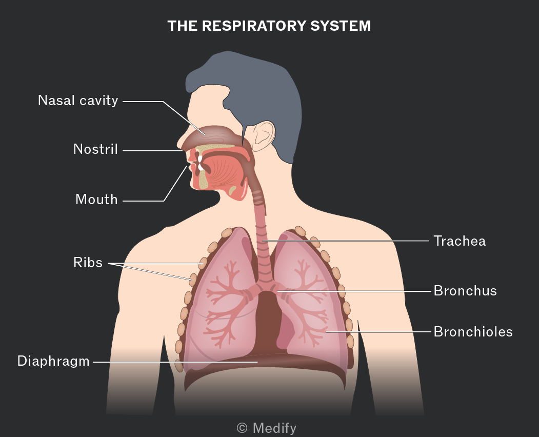
The trachea (windpipe) connects the mouth to the bronchi. The trachea is lined with incomplete rings of strong, flexible cartilage which maintains its shape and prevents collapse. The trachea has elastic fibres, which allow it to stretch and recoil during ventilation.
The cells lining the trachea are mostly ciliated epithelial cells, with goblet cells in between. Goblet cells produce mucus, which trap dirt and microorganisms. The ciliated epithelial cells waft and move mucus away from the lungs and towards the throat, where it is swallowed.
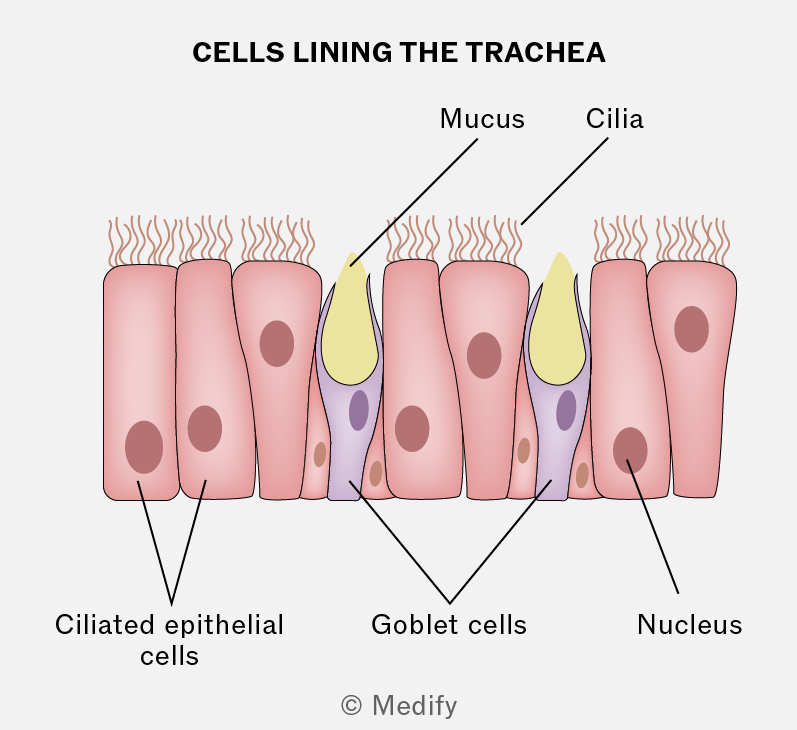
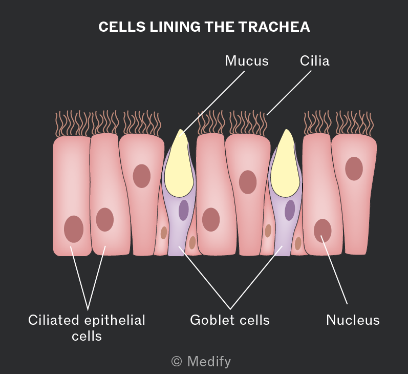
The trachea branches into two smaller bronchi, which lead to each lung. The bronchi have a similar function to the trachea and also contain supporting rings of cartilage. The bronchi then branch into bronchioles.
The smaller bronchioles do not have cartilage rings and contain smooth muscle. The smooth muscle can contract and relax, controlling the air reaching the lungs. Bronchioles are lined with a thin layer of flattened epithelial cells. Due to the thin layer of cells, a small amount of gas exchange can happen in the bronchioles.
The branches of the small bronchioles end at the alveoli. The alveoli are tiny air sacs in the lungs where the majority of gas exchange takes place.
The alveoli have many adaptations:
- Lined with elastic tissue, allowing them to stretch and recoil during ventilation.
- Large surface area for increased rate of diffusion.
- Good ventilation and blood supply maintain a steep concentration gradient, which increases the rate of diffusion.
- Surfactant makes the surface of the alveoli moist, which aids diffusion.
- Each alveoli is lined with flattened epithelial cells, and the capillaries are lined with a thin layer of endothelial cells. This makes the distance required for diffusion short, increasing the rate of gas exchange.
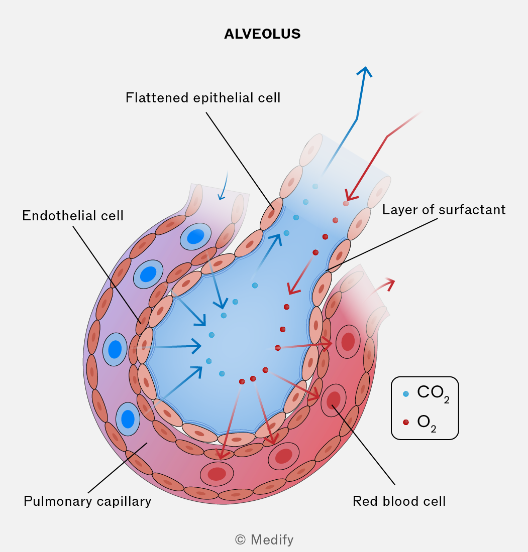
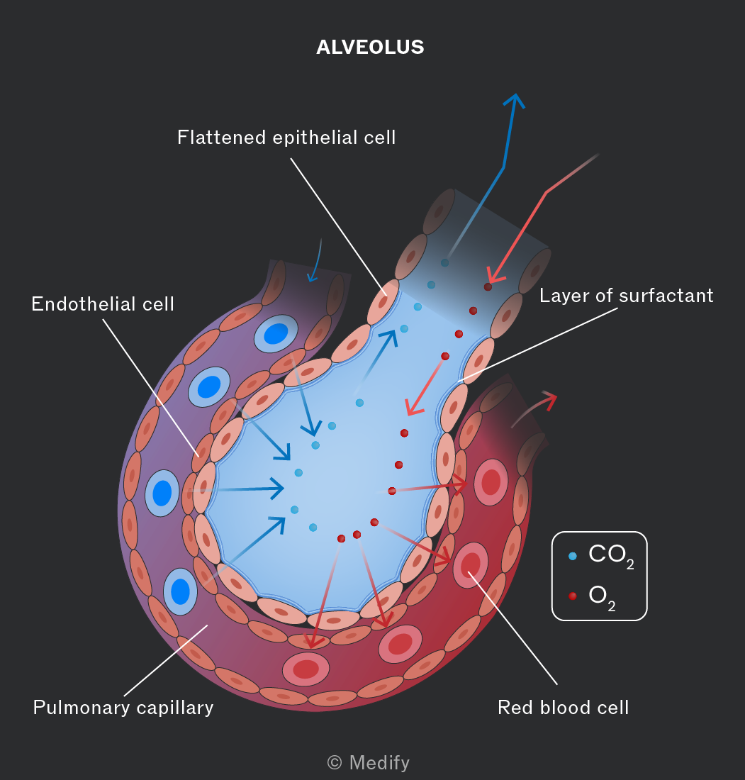
Lung diseases, such as chronic obstructive pulmonary disease (COPD), can reduce gas exchange owing to reduced alveolar surface area.
COPD also reduces the elasticity of tissues in the lungs, reducing their ability to stretch and recoil, making ventilation more difficult. This reduces the ability to maintain the concentration gradient, reducing gas exchange.
Certain risk factors increase the likelihood of developing COPD:
- smoking
- air pollution, (e.g., )
- frequent chest infections
- occupational exposure to hazardous substances
- genetics.
The respiratory system carries out the process of ventilation:
- Ribs: bones which surround and protect the lungs and heart and move during ventilation.
- Diaphragm: a dome-shaped sheet of muscle which contracts and relaxes to facilitate breathing.
- External intercostal muscles: contract during inhalation and move the ribs up and outwards.
- Internal intercostal muscles: contract during expiration.
- Pleural membranes: surround the lungs and make pleural fluid.
- Pleural fluid: reduces friction during ventilation when the pressure in the lungs changes.
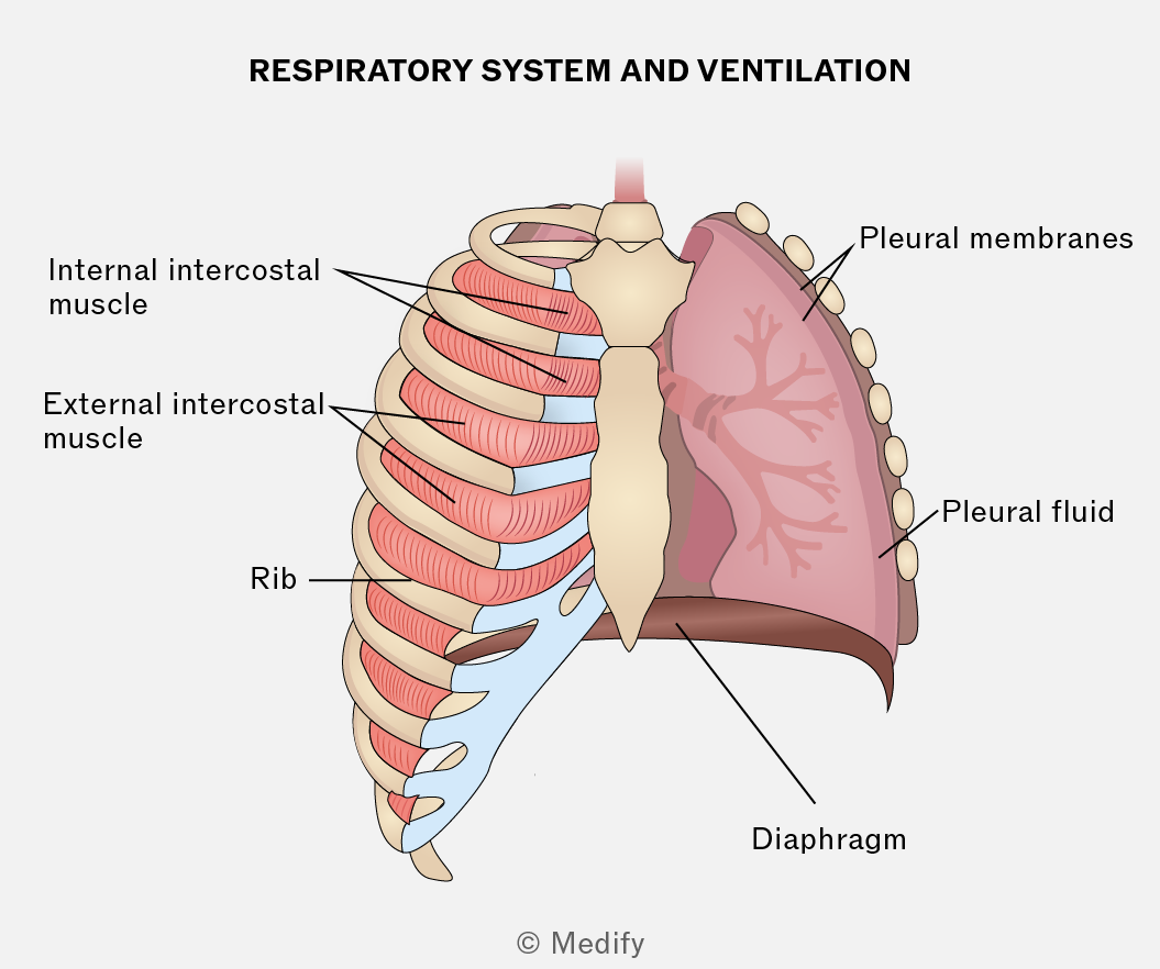
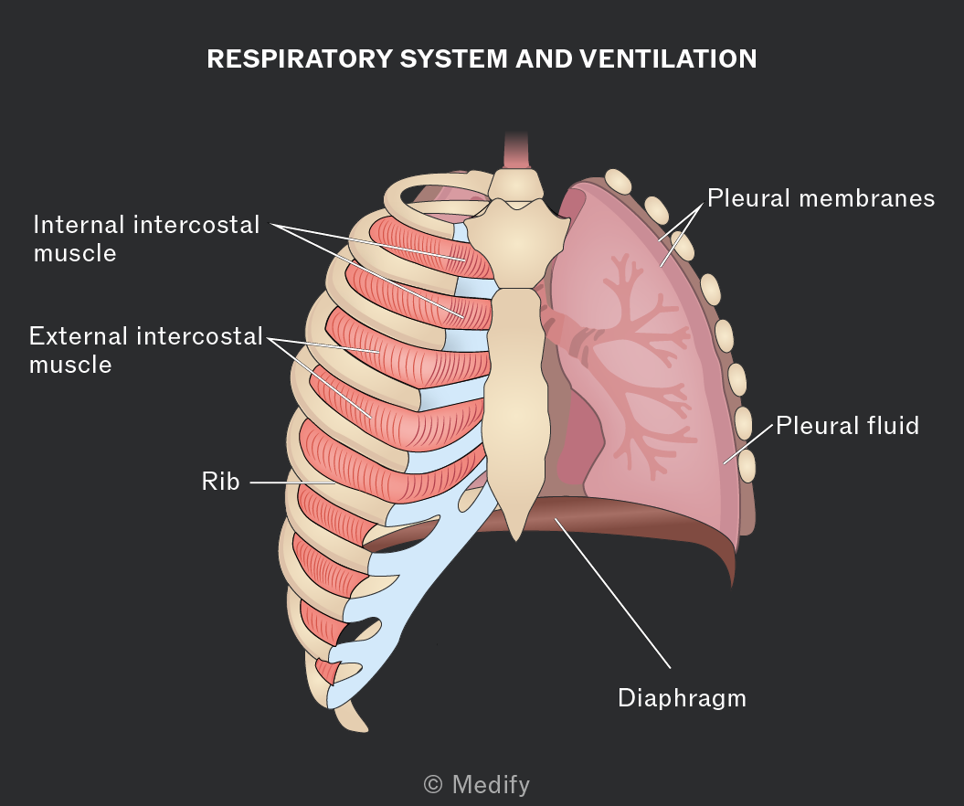
During inspiration:
- The diaphragm contracts and flattens.
- The external intercostal muscles contract and pull the rib cage up and out.
- The volume in the thorax increases, and the pressure decreases.
- Air moves into the lungs.
During expiration:
- The diaphragm relaxes and moves upward.
- The external intercostal muscles relax and the rib cage moves down and in.
- The internal intercostal muscles contract, also causing the rib cage to move down and in.
- The volume in the thorax decreases, and the pressure increases.
- Air moves out of the lungs.
When a person is breathing at rest, most of the movement of the rib cage is due to elastic recoil of the tissues. The internal intercostal muscles play a greater role during forced expiration.
Insects require a gas exchange system because they:
- Do not have oxygen–carrying pigments, such as haemoglobin, to transport oxygen.
- Have exoskeletons made of chitin, which are impermeable so gases are not able to diffuse through.
- Do not have a large enough SA:V ratio to rely on diffusion alone.
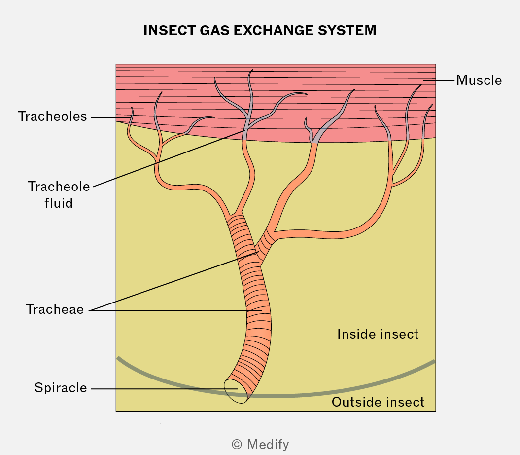
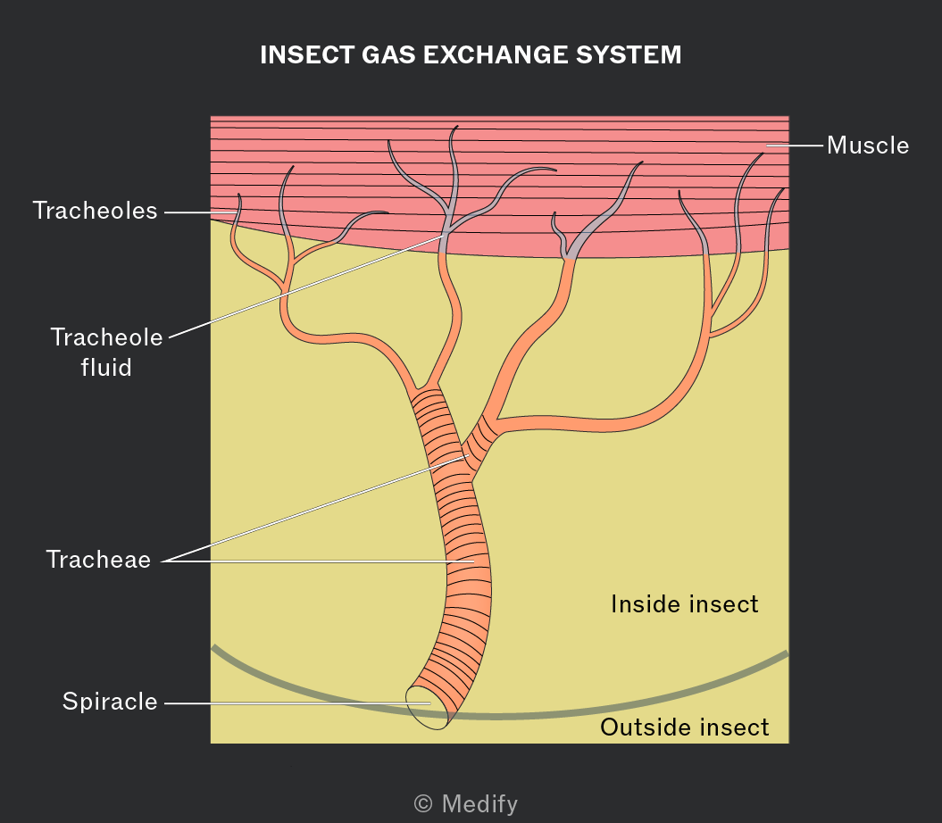
Insects have sphincters around the external openings of the tracheae which can be opened and closed:
- To prevent water loss from the tracheal fluid evaporation.
- In response to oxygen and carbon dioxide levels.
Some insects use mechanical ventilation, moving their thorax or abdomen to move air through the tracheal system. Air sacs or enlarged tracheae can further increase ventilation efficiency.
Anaerobic respiration can occur in insects during periods of intense activity. This produces lactate which lowers the water potential of the muscles causing water to move from the tracheoles into the muscles.
Less water in the tracheoles increases the space available for air, increasing air movement. This increases the rate of diffusion of gases but can also lead to greater water loss through evaporation in the tracheoles.
Insects have various adaptations to reduce water loss:
- Exoskeletons made of chitin are impermeable to water.
- A small surface area to volume ratio reduces the area where water can be lost.
- Spiracles have sphincters, which can be closed to reduce water loss from the tracheoles. However, this also prevents gas exchange required for respiration.
Fish have low SA:V ratios, so require specialised exchange surfaces to absorb oxygen and excrete carbon dioxide.
Being aquatic, fish do not need to prevent water loss from their exchange surfaces.
Water is more viscous than air and has a lower oxygen content. Due to its viscosity, moving water mechanically in and out of the body would take too much energy. Fish have developed gills as their specialised exchange surfaces.
Gills have the following structures:
- Operculum: flap covering and protecting the gills.
- Opercular valve: controls the opening and closing of the operculum.
- Gill arch: support the gill filaments.
- Gill lamellae: thin structures where exchange of oxygen and carbon dioxide occurs.
- Afferent blood vessel: supplies blood to the gills.
- Efferent blood vessel: carries the blood from the gills to the body.
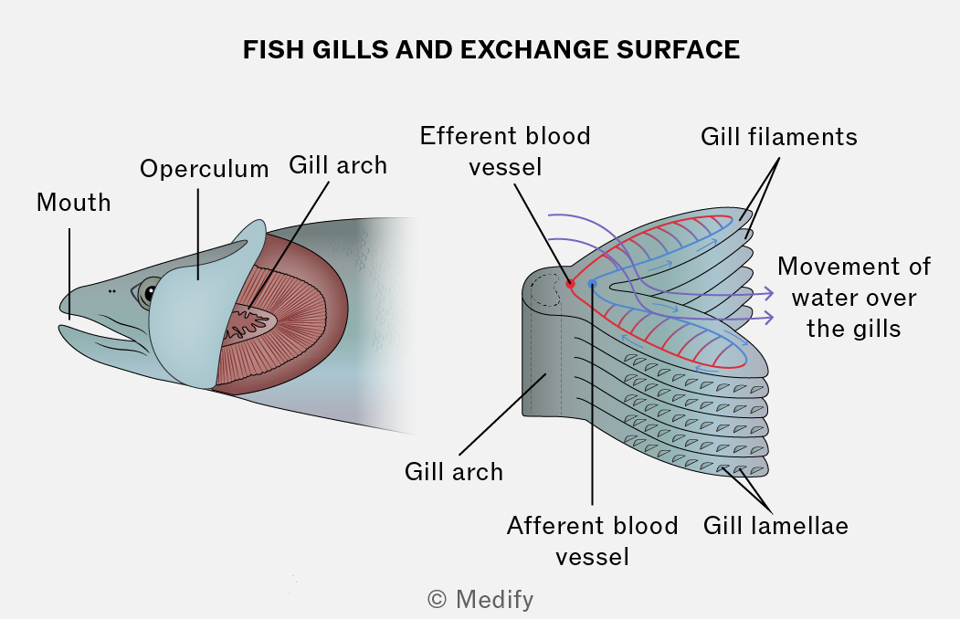
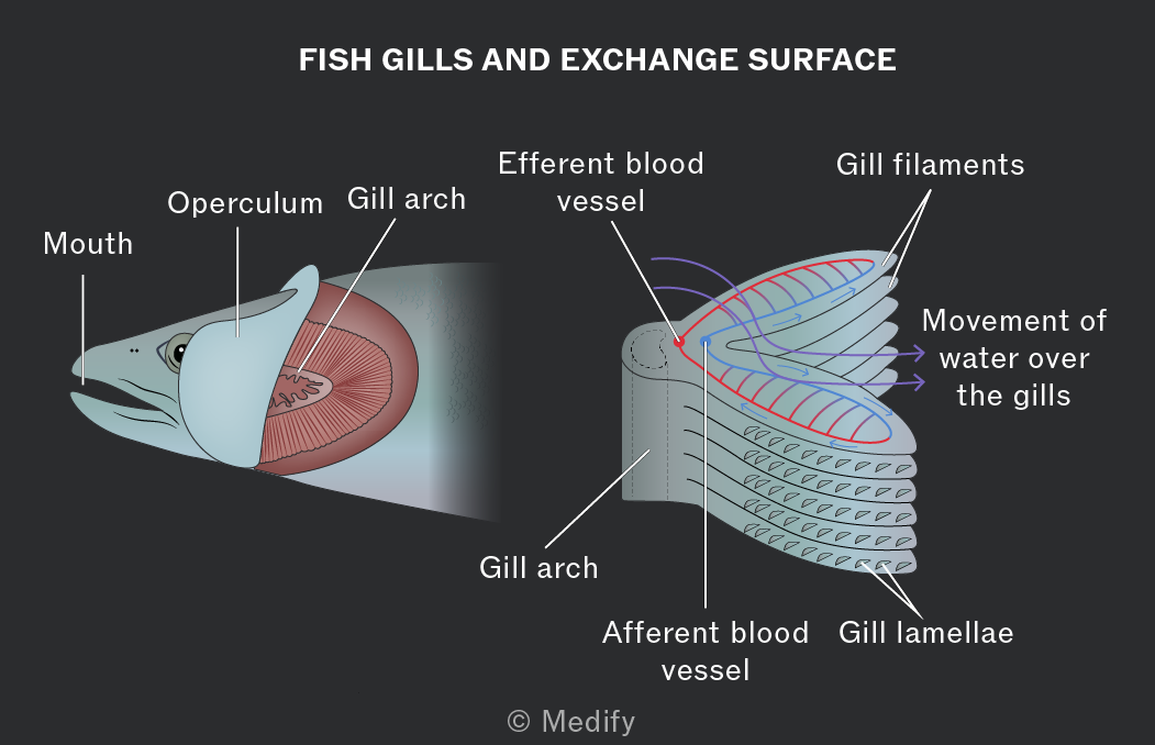
The gills are adapted for diffusion:
- Large surface area.
- Good blood flow to maintain the diffusion gradient.
- The layers in the gill are thin, leading to a short diffusion distance.
- Water flows in one direction over the gills. This leads to a countercurrent system between the water and blood flow in the gills, which increases the rate of diffusion.
Many fish have to keep moving for water to flow over their gills.
Some fish species are adapted to use their buccal cavity and operculum to mechanically move water over their gills when not swimming:
- The mouth opens with the opercular valve closed. This increases the volume of the buccal cavity.
- This lowers the pressure, and water moves in and over the gills.
- The mouth closes and the opercular valve opens. This decreases the volume of the buccal cavity.
- This increases the pressure and water moves out through the opercular valve.
In plants, gas exchange mostly occurs in the leaves. The gases being exchanged with the environment will depend on which processes the plant is carrying out at that time.
Plants require carbon dioxide for photosynthesis, this is also produced during respiration.
Plants produce oxygen during photosynthesis as a waste product, this is also used during respiration.
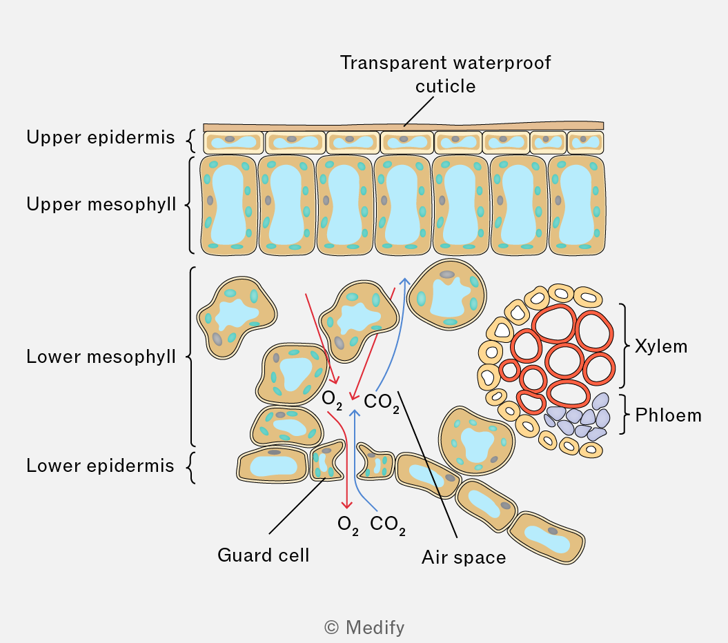
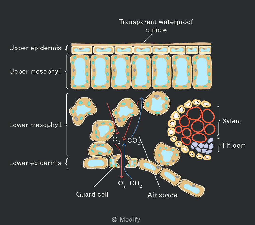
Plant leaves are adapted for gas exchange:
- Leaves are normally thin which reduces diffusion distance.
- Large surface area of mesophyll cells to increase diffusion.
- Air spaces in the spongy mesophyll for gases to easily move and diffuse through the leaf.
Stomata are pores mainly on the underside of leaves that allow carbon dioxide to enter and oxygen to leave the plant.
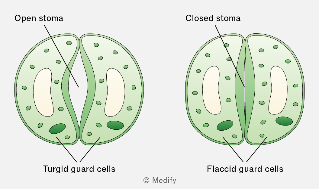
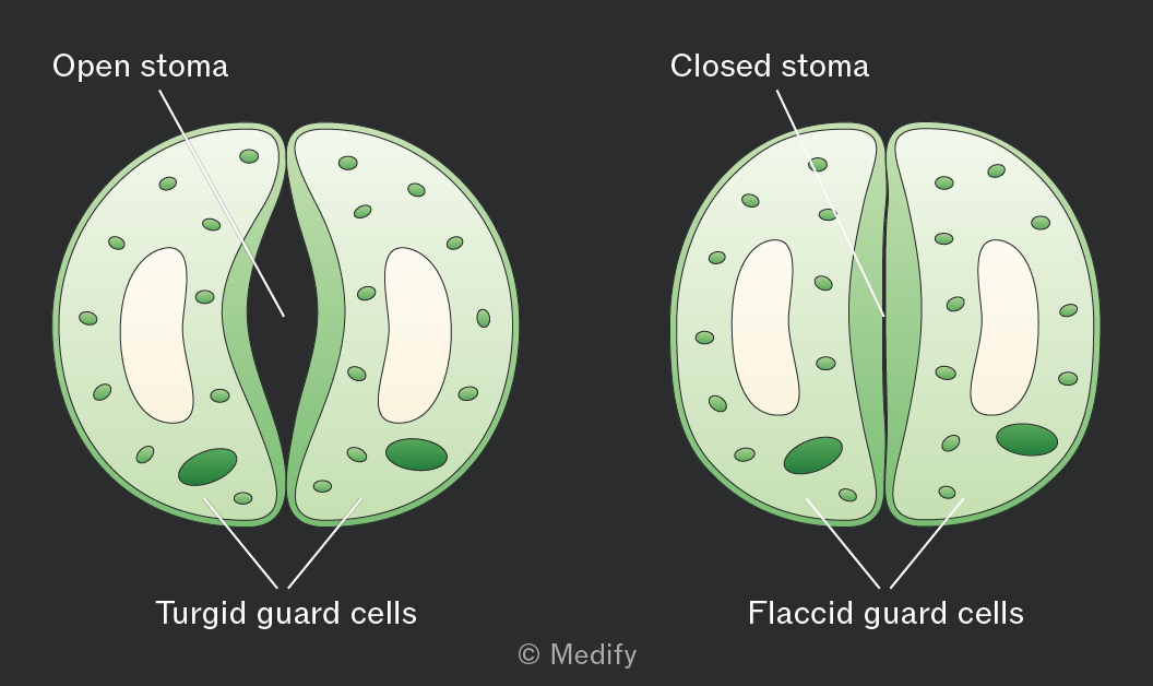
Each stomatal pore is surrounded by kidney-shaped guard cells with a thicker inner wall and thinner outer wall.
When guard cells take in water and become turgid, they swell and bend, opening the stomata. This happens during the day when photosynthesis occurs in the presence of sunlight.
When guard cells lose water and become flaccid, the stomata close. This happens at night when photosynthesis cannot occur.
Xerophytes are plants that live in dry environments, such as deserts or sand dunes. They require adaptations to reduce water loss and conserve water. Examples of xerophytes include cacti and marram grass.
Xerophytes have several adaptations to reduce water loss.
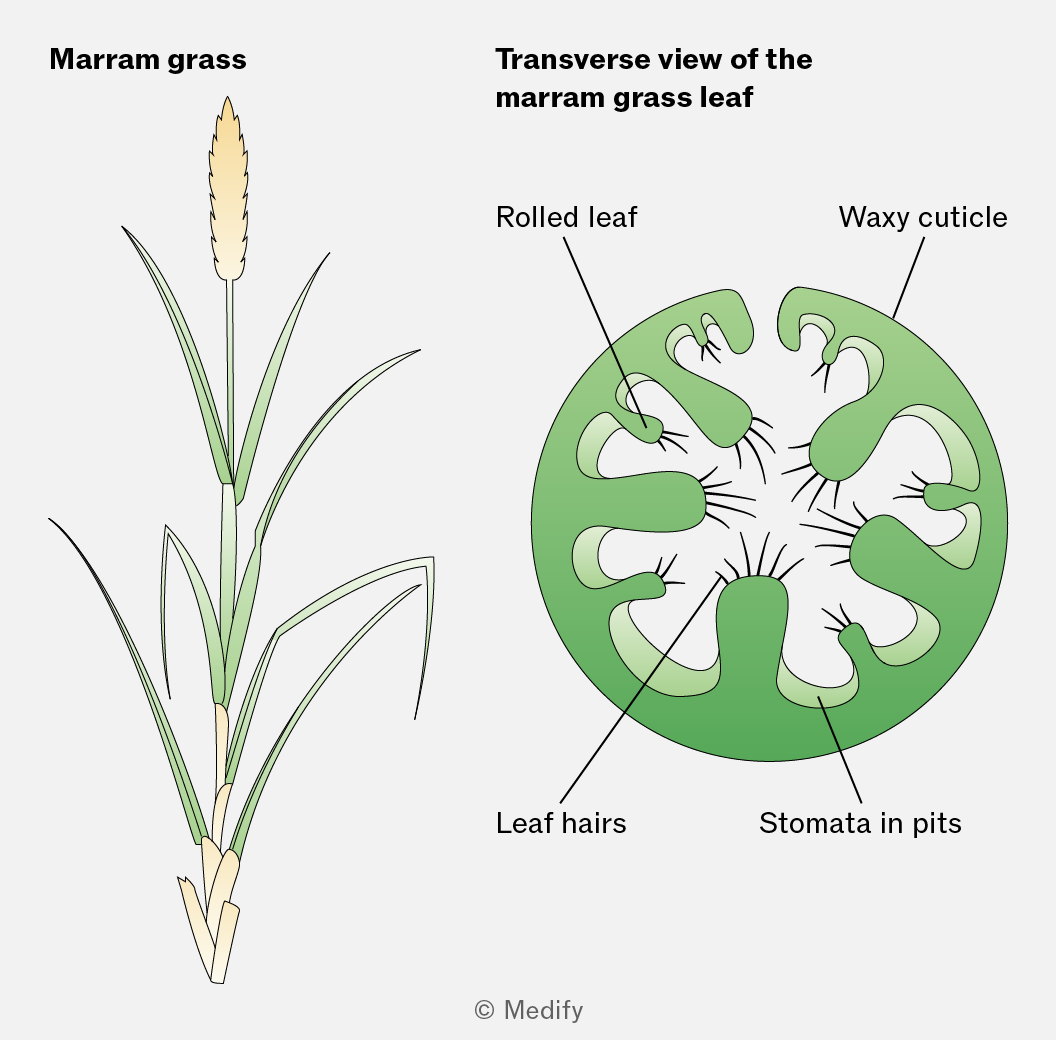
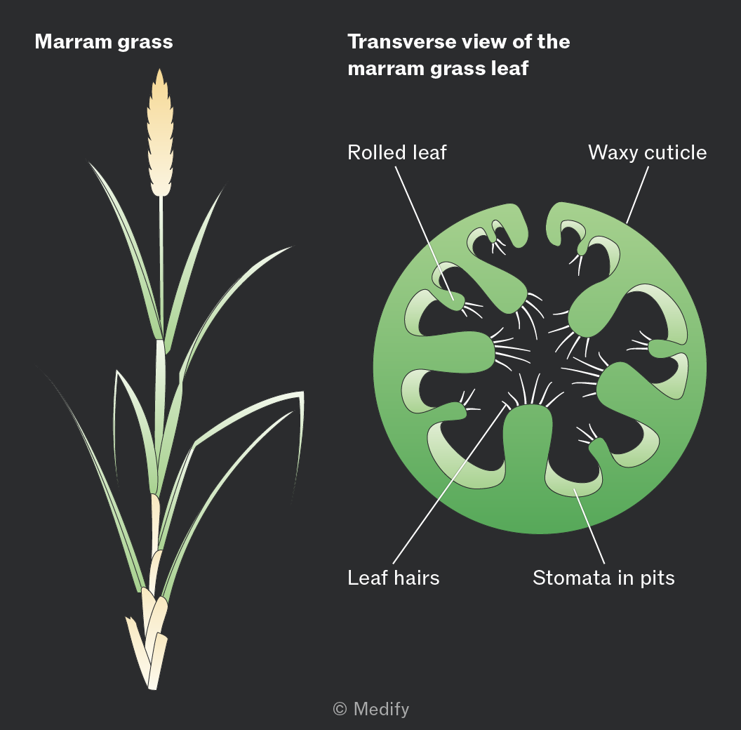
Marram grass has sunken stomata, leaf hairs, and rolled leaves, which help reduce transpiration by:
- reducing air movement and trapping water vapour, increasing humidity around the stomata. This lowers the water potential gradient between the leaf and the surrounding air, reducing water loss.
- reducing the surface area exposed for transpiration.
Xerophytes have several adaptations to reduce water loss.
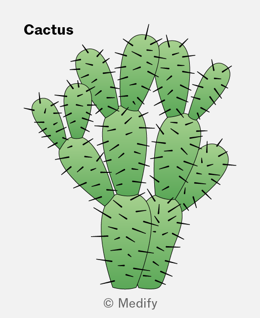
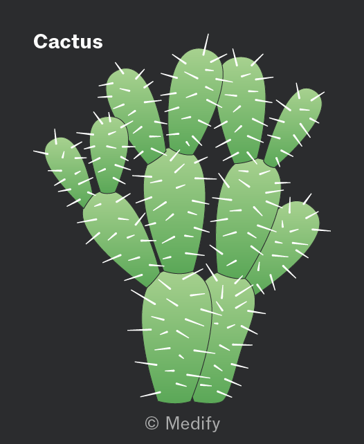
Cacti reduce water loss by:
- Having spines instead of leaves, reducing their surface area:volume ratio and therefore reducing transpiration.
- Closing their stomata during the hottest part of the day.
- Having a thick, waxy cuticle, which is waterproof. Its thickness increases the diffusion distance, while the wax layer reduces evaporation from the leaf surface.