Genetic information, variation and relationships between organisms (3.4)›DNA and protein synthesis (3.4.2)

DNA and protein synthesis (3.4.2)

An overview of DNA and protein synthesis (3.4.2) from AQA A level Biology including: the genome, proteome, transcription and translation
6 min

The genome is the complete set of DNA in a cell or organism, including all of its genes. It contains the instructions for building and maintaining that organism.

The proteome is the full set of proteins that a cell or organism can produce.

The genome is mostly constant, while the proteome varies between cells and over time, depending on which genes are expressed.
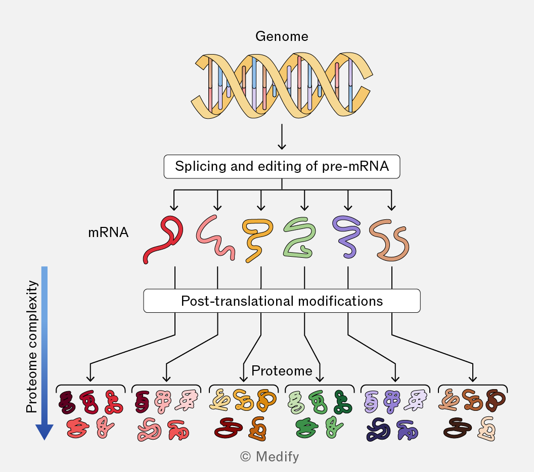
The image is a flowchart illustrating the process from genome to proteome complexity. At the top, there is a DNA double helix labeled 'Genome.' Below it, an arrow points to a box labeled 'Splicing and editing of pre-mRNA.' Five arrows extend downward from this box to five different colored squiggly shapes labeled 'mRNA.' Each mRNA shape has its own arrow leading to a box labeled 'Post-translational modifications.' From this box, several arrows point downward to various groups of colored and shaped forms representing the 'Proteome.' There is a vertical blue arrow on the left side labeled 'Proteome complexity,' pointing downward. The image is labeled © Medify at the bottom.
Add to favourites

DNA contains the ‘instructions’ for making polypeptides, but is contained in the nucleus of cells. The ribosomes, which assemble polypeptides, are in the cytoplasm.

Protein synthesis, therefore, happens in two stages:

  • Transcription – messenger RNA (mRNA) is made from the DNA template. The mRNA takes the instructions from the nucleus to the ribosomes.
  • Translation – ribosomes join amino acids together in the order specified by the sequence of bases in the mRNA, forming a polypeptide.
Add to favourites

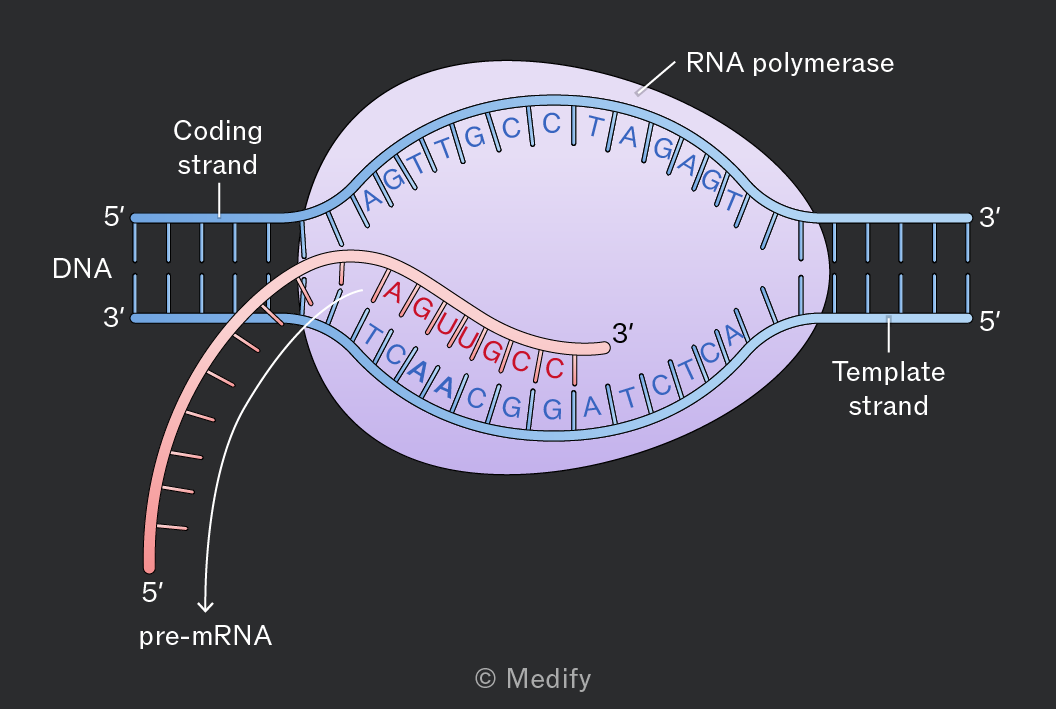
During transcription, the DNA sequence of a gene is copied into pre-messenger RNA (pre-mRNA):

  1. At the start codon of a gene, an enzyme unzips the DNA double helix.
  2. Free RNA nucleotides match up with their complementary base pairs on the exposed DNA strand.
  3. RNA polymerase joins the RNA nucleotides in condensation reactions, forming phosphodiester bonds.
  4. DNA rejoins behind RNA polymerase to limit the number of exposed bases.
  5. Transcription stops at a terminator sequence, and the pre-mRNA molecule leaves the nucleus through a nuclear pore.
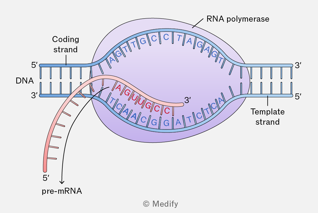
The image illustrates the process of transcription, focusing on RNA polymerase and DNA strands. The DNA is shown with two strands: the coding strand labeled 5' to 3' and the template strand labeled 3' to 5'. RNA polymerase is depicted as a purple oval encompassing part of the DNA. The template strand inside the RNA polymerase is paired with complementary RNA nucleotides forming pre-mRNA, labeled from 5' to 3'. The DNA sequence in the template strand is shown as TCAACGGTAGCTC, and the corresponding pre-mRNA sequence is AGUUGCCUCGAG. The coding strand sequence is AGTTGCGATCAGT. An arrow indicates the direction of pre-mRNA synthesis, leading away from RNA polymerase. Labels include 'RNA polymerase,' 'Coding strand,' 'Template strand,' and 'pre-mRNA.'
Add to favourites

In eukaryotes, the RNA molecule created during transcription contains both introns and exons. This is pre-mRNA. The sequences of introns are removed, and exons are joined together in a process called splicing which forms mature mRNA.

Most prokaryotic DNA does not contain introns, so transcription directly results in mRNA, which can be used to synthesise polypeptides.

Add to favourites

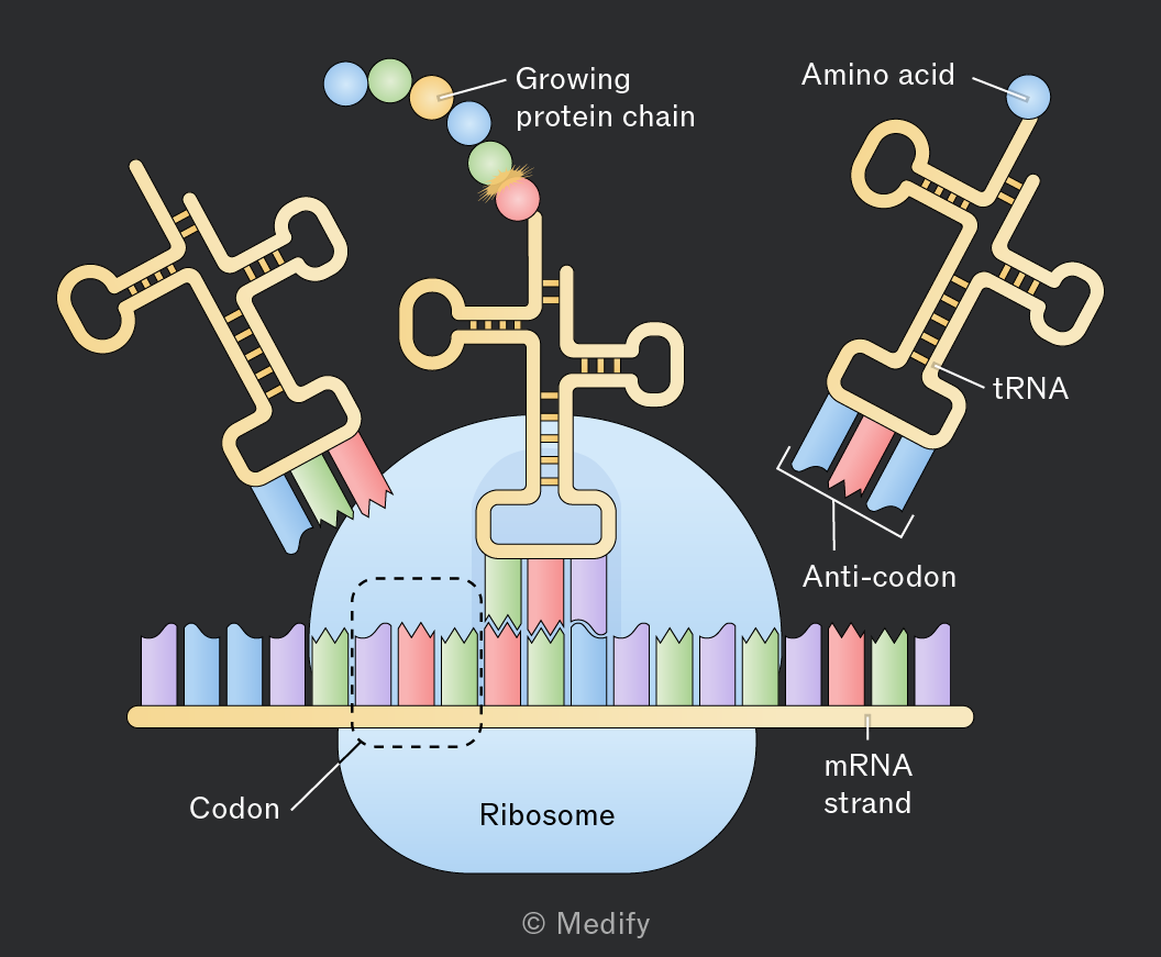
During translation, ribosomes join amino acids together in the order specified by the sequence of bases in the mRNA codons, forming a polypeptide and determining how it will fold and function.

Each codon specifies a particular amino acid, which is delivered to the ribosome by a transfer RNA (tRNA) molecule.

ATP provides energy for an enzyme to join amino acids together by peptide bonds.
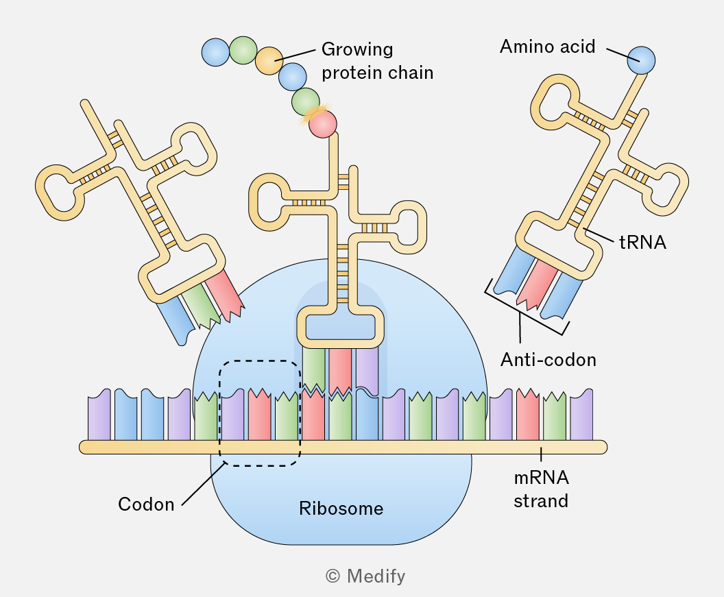
The diagram illustrates the process of protein synthesis. At the center is a ribosome depicted as a blue structure interacting with an mRNA strand shown as a sequence of colored bars labeled 'mRNA strand'. Above the ribosome, a 'Growing protein chain' is shown as a series of colored spheres. On the right, a tRNA molecule labeled 'tRNA' is depicted with an 'Amino acid' attached at one end and an 'Anti-codon' at the other. The tRNA is shown interacting with the mRNA strand. On the left, another tRNA is shown entering the ribosome with a different anti-codon. The mRNA strand has colored segments labeled 'Codon', indicating the interaction with anti-codons on the tRNA. The diagram is attributed to © Medify.
Add to favourites

Transfer RNA, tRNA is made of RNA nucleotides. It is a small, single-stranded molecule that folds into a clover leaf shape due to complementary base pairing within its single strand.

Each tRNA has an anticodon region that pairs with a complementary codon on the mRNA.

At the other end of the molecule from the anticodon, tRNA molecules have an amino acid attachment site. Each tRNA can only bind to one specific amino acid which is determined by its anti-codon.
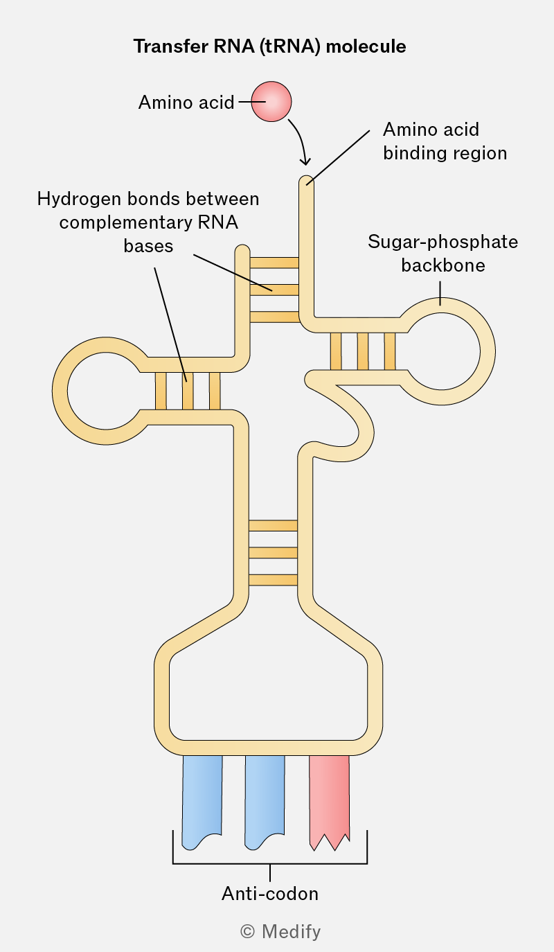
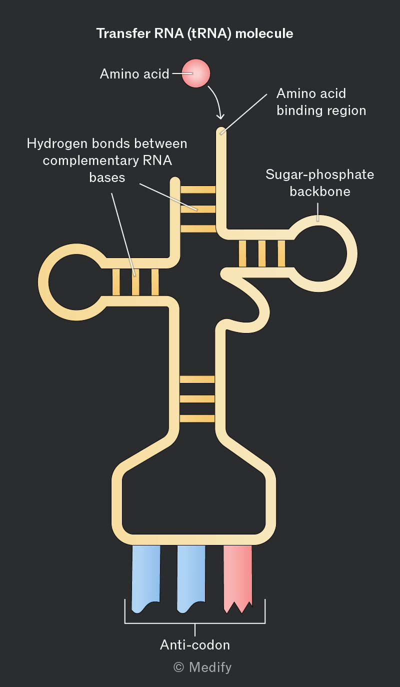
Diagram of a Transfer RNA (tRNA) molecule. The structure includes an amino acid depicted as a red sphere connected to the tRNA at the top, labeled 'Amino acid'. An arrow points to the 'Amino acid binding region' on the tRNA. The backbone of the tRNA is labeled 'Sugar-phosphate backbone'. There are regions with parallel lines labeled 'Hydrogen bonds between complementary RNA bases'. At the bottom, there are three colored segments, two blue and one red, labeled collectively as 'Anti-codon'.
Add to favourites

Translation is a multistep process involving mRNA, ribosomes, tRNA and amino acids:

  1. A ribosome binds to the start codon of an mRNA molecule.
  2. A tRNA molecule with the complementary start anticodon binds to the mRNA and the ribosome, bringing with it an amino acid.
  3. A tRNA molecule with an anticodon complementary to the next codon along on the mRNA molecule and an amino acid binds next.
  4. An enzyme catalyses the formation of a peptide bond, joining the two amino acids together, requiring ATP for energy.
  5. The ribosome moves along the length of the mRNA molecule, repeating the above process until reaching a stop codon. There is no complementary tRNA anticodon for the stop codon, so the polypeptide chain is released.

Multiple ribosomes can simultaneously synthesise multiple polypeptide chains from a single mRNA molecule by following each other along the mRNA.
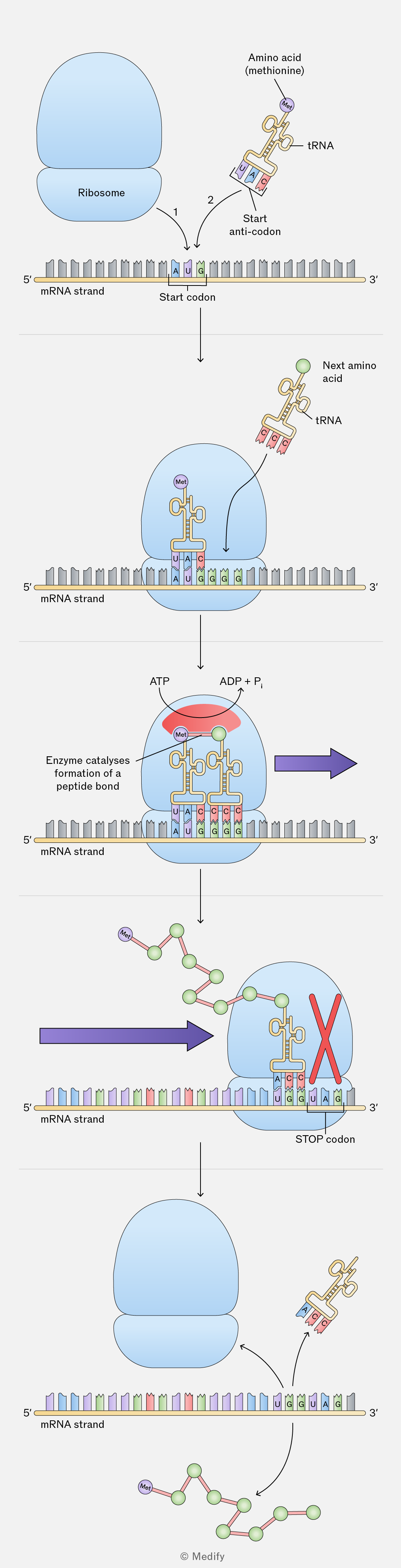
Five-panel diagram of protein synthesis on an mRNA strand. 

Top panel: A large blue ribosome is shown above a horizontal strand labeled "mRNA strand," with 5′ at the left end and 3′ at the right end. Three boxed bases on the mRNA are labeled "A U G," and below them is the text "Start codon." To the right is a tRNA drawing labeled "tRNA," carrying a small circle labeled "Met" with the text "Amino acid (methionine)." The three-base tRNA sequence is labeled "Start anti-codon" and shown as "U A C." Curved arrows labeled "1" and "2" point down toward the start codon.

Second panel: The ribosome now surrounds the mRNA. The mRNA strand is again labeled "mRNA strand" with 5′ on the left and 3′ on the right. Visible boxed bases on the mRNA include "A U G G G G." A tRNA with "U A C" is paired to the "A U G" codon inside the ribosome, carrying a small circle labeled "Met." The letter "P" appears beside this tRNA. Another tRNA approaching from the upper right is labeled "tRNA" and carries a green sphere with the text "Next amino acid."

Third panel: The ribosome contains two tRNAs over the mRNA. The mRNA strand is labeled "mRNA strand" with 5′ left and 3′ right. Visible boxed mRNA bases are "A U G G G G." The left tRNA carries "Met" and has anticodon "U A C." The right tRNA has anticodon "C C C." A short linkage connects the methionine and the green sphere of the next amino acid. Text at the left reads "Enzyme catalyses formation of a peptide bond." Above the ribosome are the labels "ATP" and "ADP + Pi" with curved arrows. A large purple arrow points to the right.

Fourth panel: A growing peptide chain of connected green spheres with one purple-labeled sphere marked "Met" extends from the ribosome. The ribosome sits over the mRNA near a labeled "STOP codon." The mRNA strand is labeled "mRNA strand" with 5′ at left and 3′ at right. Near the stop codon, the visible boxed bases read "U G G U A G." A tRNA with anticodon "A C C" is paired to "U G G." A large red X is drawn over the position aligned with "U A G," indicating no tRNA binding. A large purple arrow points to the right.

Bottom panel: The ribosome has separated into two blue subunits above the mRNA strand, which is labeled "mRNA strand" with 5′ at left and 3′ at right. Near the right side, visible boxed bases still include "U G G U A G." A released tRNA with anticodon "A C C" is shown to the upper right. A completed peptide chain of green spheres, beginning with a purple-labeled "Met," is shown below the mRNA with a curved arrow indicating release. Small text at the bottom reads "© Medify."
Add to favourites