Genetic information, variation and relationships between organisms (3.4)›Genetic diversity arising from mutation or meiosis (3.4.3)

Genetic diversity arising from mutation or meiosis (3.4.3)

An overview of genetic diversity arising from mutation or meiosis (3.4.3) from AQA A level Biology including: meiosis and genetic variation
2 min

Meiosis is a type of cell division that produces four genetically distinct gametes (sperm and egg cells) and generates genetic variation through independent assortment of chromosomes and crossing over of alleles between chromatids.

It reduces the chromosome number from diploid to haploid.

Meiosis involves two sequential divisions:

  1. Meiosis I – separates homologous chromosomes.
  2. Meiosis II – separates sister chromatids.
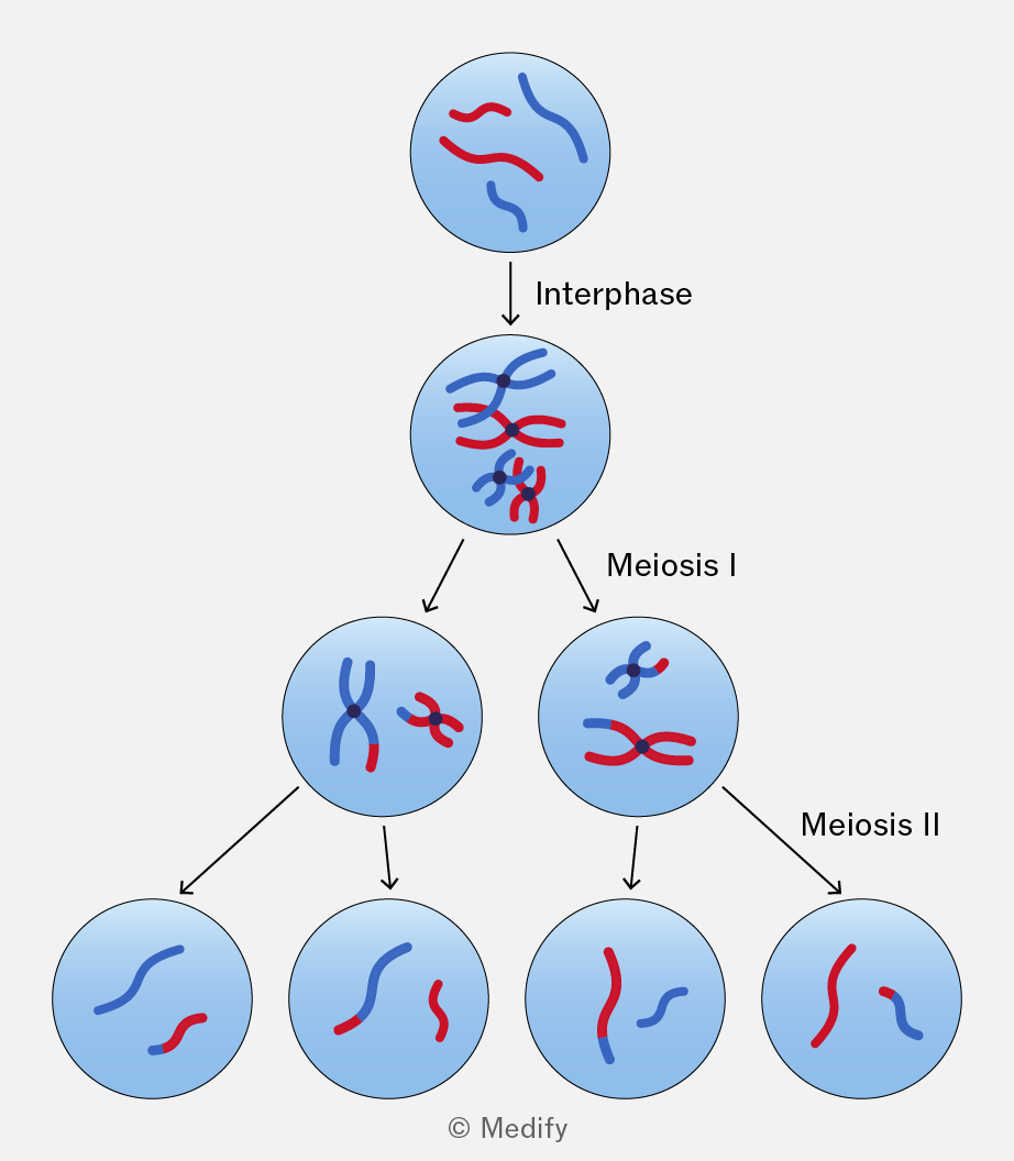
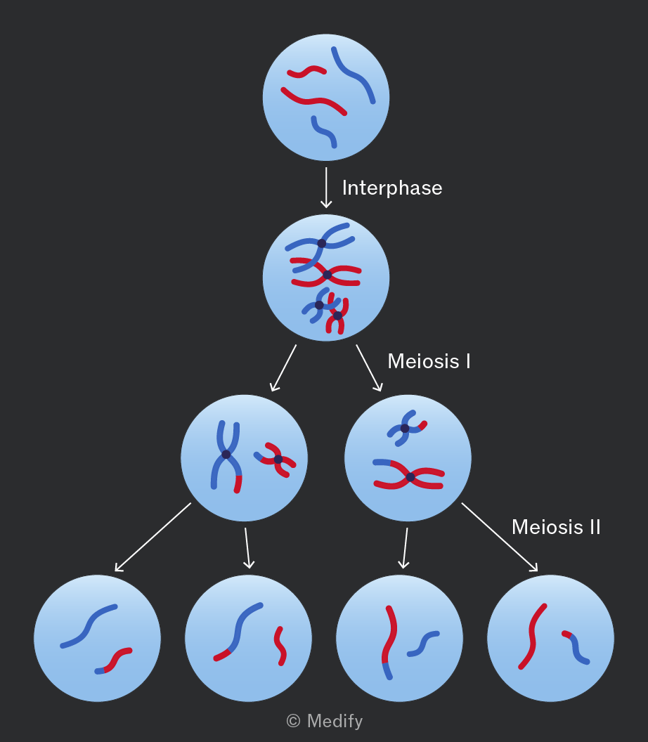
The diagram illustrates the stages of meiosis with labeled phases. At the top, a circle labeled 'Interphase' contains two pairs of loosely aligned red and blue squiggly lines. Below, an arrow points to a circle labeled 'Meiosis I' with more condensed and crossed red and blue lines. This circle splits into two circles; the left one has a pair of crossed red and blue lines, and the right one has two sets of red and blue lines. Two arrows point downward from each circle. The left-side arrow leads to a circle with separated red and blue lines. The right-side arrow, labeled 'Meiosis II,' leads to two circles, each containing separate red and blue lines. The entire diagram is labeled with the copyright '© Medify.'
Add to favourites

Genetic variation increases with random fertilisation of haploid gametes:

  • Independent assortment (Meiosis I).
  • Crossing over (Prophase I).
  • Random fertilisation (after meiosis). The randomness of which particular gametes combine during fertilisation.
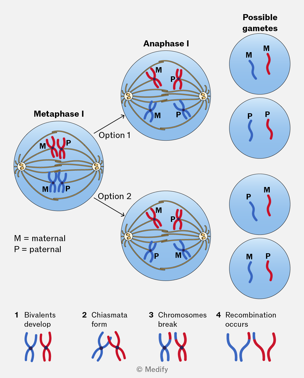
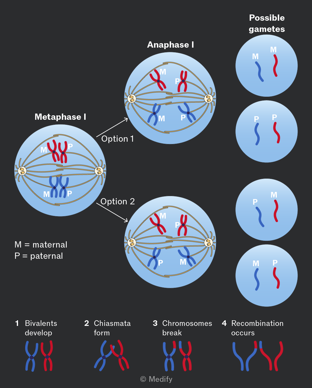
The image illustrates stages of meiosis focusing on metaphase I and anaphase I, along with possible gametes. The top shows two circles labeled 'Anaphase I', each with spindle fibers pulling apart chromosomes. The left circle is labeled 'Option 1' and shows maternal (M) and paternal (P) chromosomes separating. The right circle is labeled 'Option 2' and shows a different arrangement of M and P chromosomes. Below, 'Metaphase I' is depicted with paired chromosomes at the cell's equator, labeled M and P. To the right, 'Possible gametes' shows four circles with variations of M and P chromosomes, indicating different genetic outcomes. At the bottom, four stages are shown: 1) Bivalents develop, 2) Chiasmata form, 3) Chromosomes break, 4) Recombination occurs, with corresponding diagrams of red and blue chromosomes. A key clarifies M as maternal and P as paternal.
Add to favourites

Gene mutations are changes in the base sequence of DNA within a gene. They can occur spontaneously during DNA replication.

  • Base deletion occurs when a nucleotide is removed. This causes a frameshift and often results in an altered amino acid sequence.
  • Base substitution occurs when one nucleotide is swapped for another. This mutation type does not cause a frameshift. A substitution may or may not change the amino acid specified by the codon. If not, this is called a silent mutation.

Mutagenic agents (e.g., radiation or chemicals) can increase the rate of mutation by damaging DNA or interfering with DNA replication.

Add to favourites

Mutations in chromosome number can arise spontaneously through chromosome non-disjunction during meiosis.

Non-disjunction occurs when homologous chromosomes or sister chromatids fail to separate properly, producing gametes with an abnormal number of chromosomes (aneuploidy).

Add to favourites