Topic 4: Biodiversity and natural resources›Mass transport in plants (4.11, 4.12)

Mass transport in plants (4.11, 4.12)

An overview of mass transport in plants (4.11, 4.12) from Edexcel A level Biology A including: plant tissues and transpiration
12 min

Dicotyledonous plants (dicots) produce seeds with two cotyledons (‘seed leaves’), which store food for the developing embryo and form the first leaves.

Dicots have a vascular system of xylem and phloem running through their stems, roots, and leaves. These transport tissues are arranged in vascular bundles.
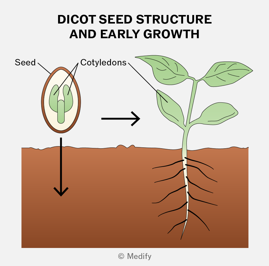
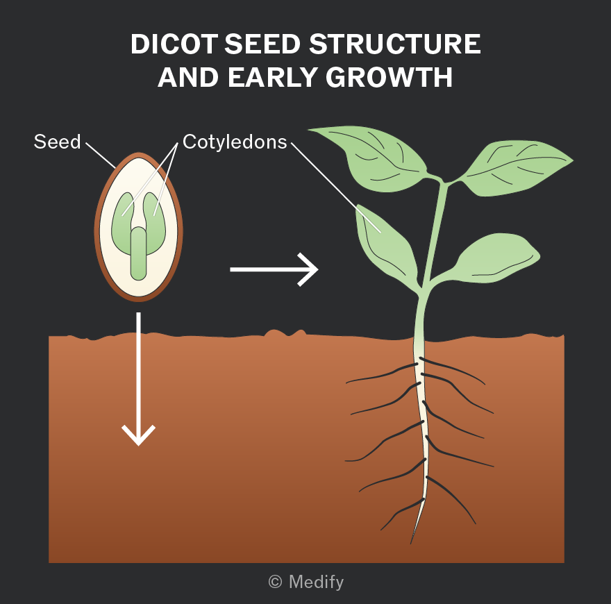
Illustration titled 'Dicot Seed Structure and Early Growth'. On the left, an oval-shaped seed with a brown outer layer is labeled 'Seed'. Inside, two green structures labeled 'Cotyledons'. An arrow points downwards from the seed to the soil. To the right, an arrow points from the seed to a small plant emerging from the soil. The plant has a stem with two pairs of green leaves. Roots extend downward into the soil.
Add to favourites

In a transverse cross-section of dicot stems, the vascular bundles are located near the outside of the stem in a ring arrangement.

The xylem is on the inner side of each vascular bundle, while the phloem is on the outer side, often with sclerenchyma fibres surrounding the phloem for extra support.

This arrangement provides a scaffolding effect, reducing bending while still allowing some flexibility, so the stem does not snap.
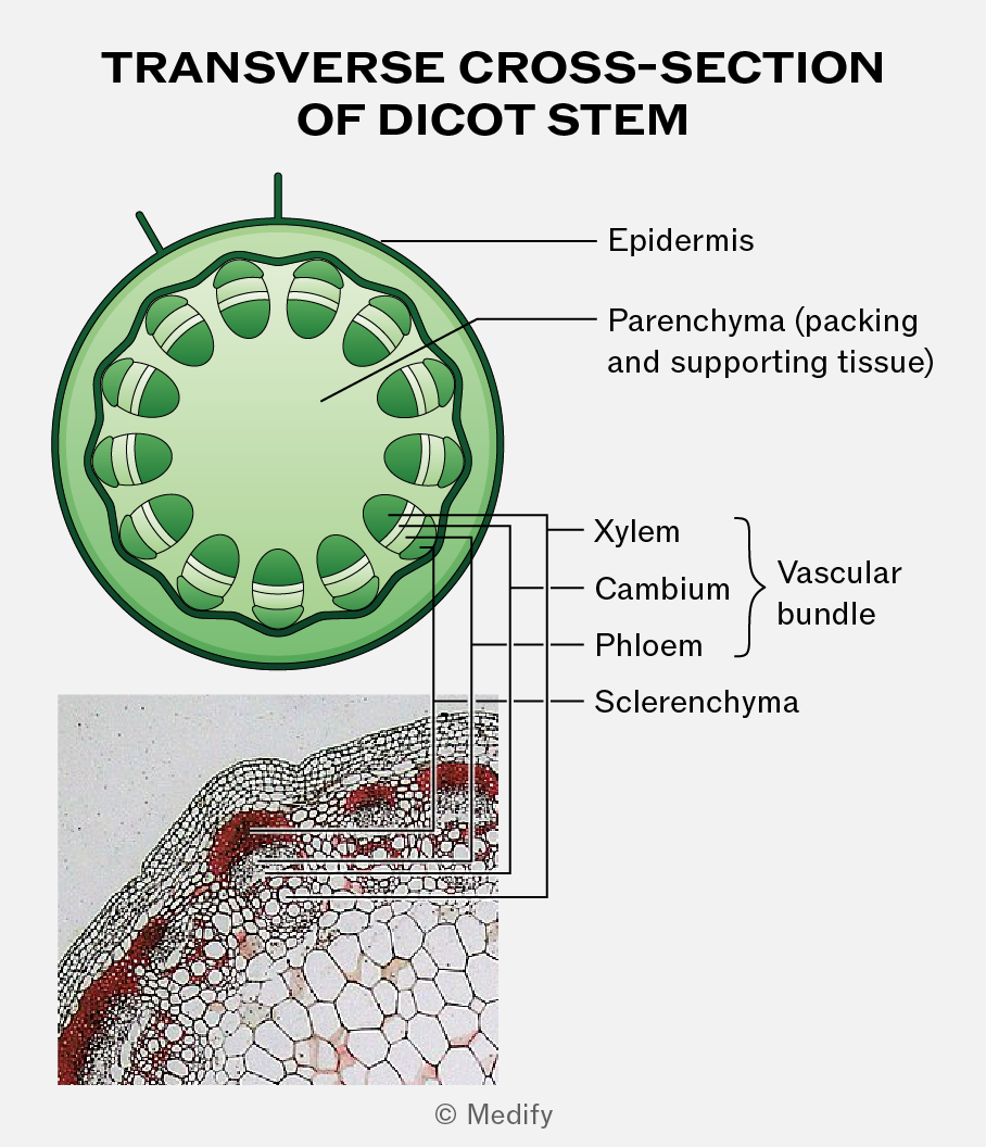
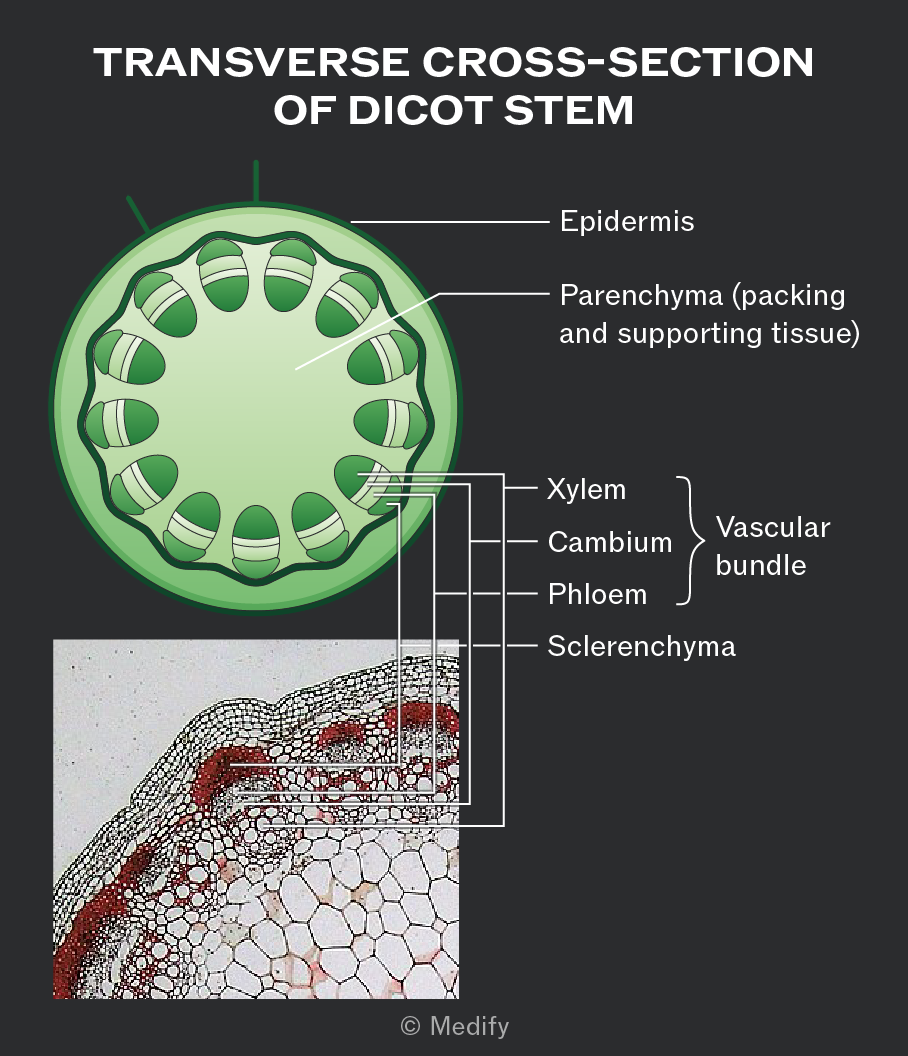
The image shows a labeled diagram of the transverse cross-section of a dicot stem. The diagram highlights various parts with corresponding labels: 'Epidermis' on the outer layer, 'Parenchyma (packing and supporting tissue)' in the inner section, and 'Vascular bundle' containing 'Xylem', 'Cambium', 'Phloem', and 'Sclerenchyma'. A detailed close-up image of the same cross-section is shown in the bottom left, with lines connecting to the labeled parts in the diagram. The title at the top reads 'TRANSVERSE CROSS-SECTION OF DICOT STEM'.
Add to favourites

In a longitudinal section of a dicot stem, the xylem and phloem run as continuous strands along the length of the stem.

The xylem is on the inner side of each vascular bundle, while the phloem is on the outer side, often with sclerenchyma fibres surrounding the phloem on the outside of the bundle.
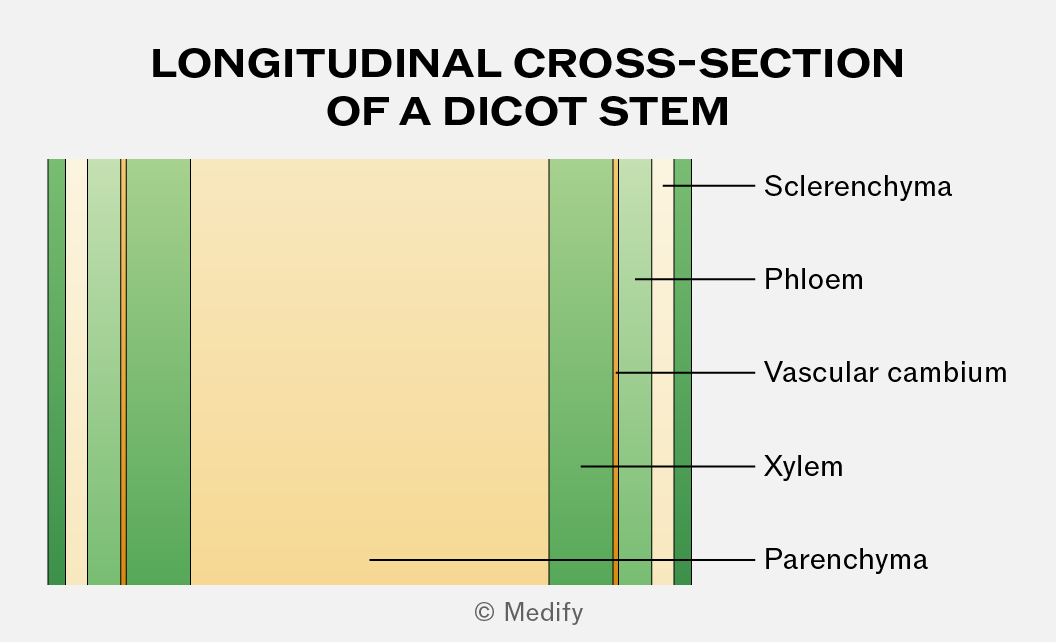
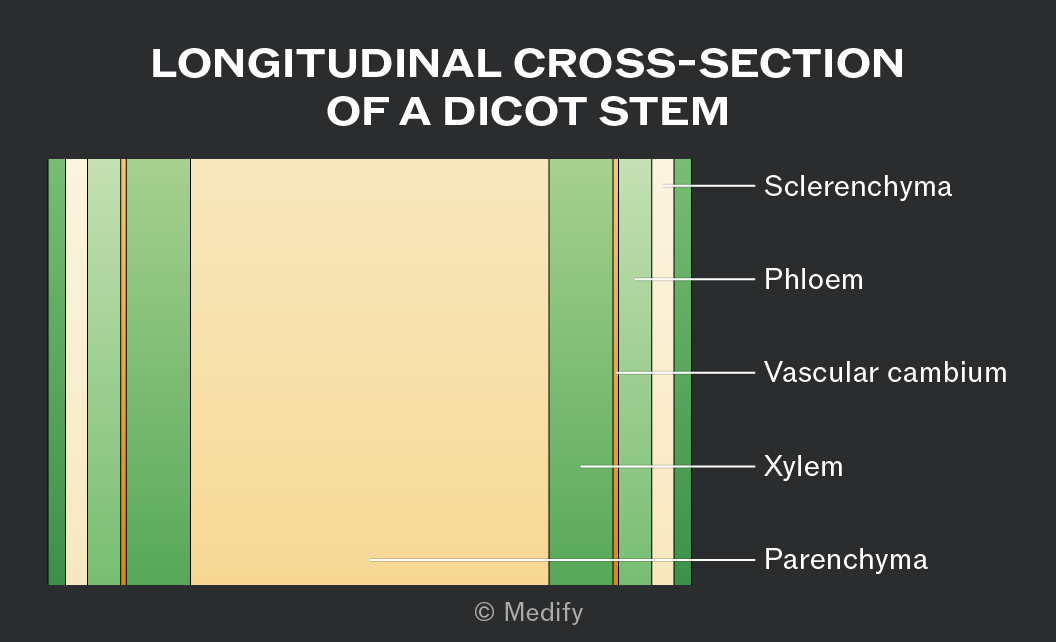
Diagram titled 'Longitudinal Cross-Section of a Dicot Stem'. It shows various layers from left to right: a dark green outer layer, followed by a thin light yellow layer, a thin dark green layer, a thin light yellow layer, a thin orange layer, and a wide gradient layer transitioning from light to dark green. On the right, there are labels pointing to specific layers: 'Sclerenchyma' points to the outermost dark green layer, 'Phloem' to the thin dark green layer, 'Vascular cambium' to the thin orange layer, 'Xylem' to the wide gradient layer, and 'Parenchyma' to the innermost light yellow area.
Add to favourites

Under the microscope, xylem vessels can be identified as hollow, thick-walled, and lacking cytoplasm. They have a wider lumen than other vessels.

Add to favourites

Xylem vessels:

  • transport water and mineral ions from roots to leaves through the stem (one-way flow) via the transpiration stream
  • provide mechanical support to the plant.
Add to favourites

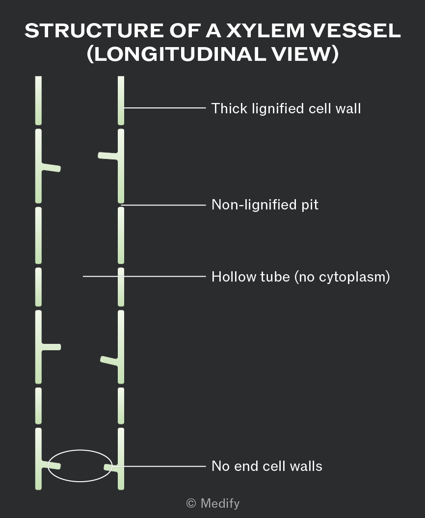
Xylem is a tissue made of cells joined end to end with no end walls between them. This creates a long, hollow tube structure that allows continuous, uninterrupted water transport.

The xylem cells that make up the vessels are dead, meaning they contain no cytoplasm or organelles. This gives the xylem an empty lumen, which reduces resistance to the flow of water.

The vessels are narrow, helping water rise by capillary action.
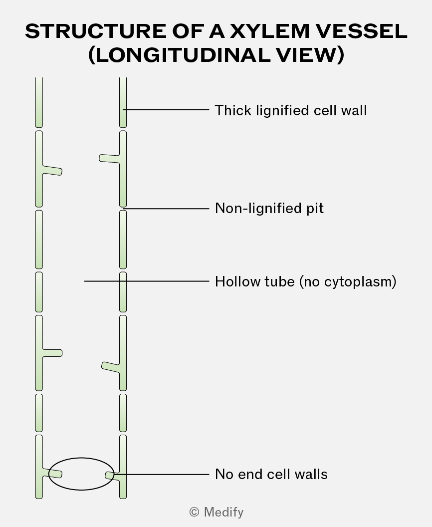
The image shows a diagram of the structure of a xylem vessel in longitudinal view. It consists of a vertical arrangement of elongated tube-like structures. Labels are pointing to specific parts: 'Thick lignified cell wall' points to the outer wall of the tube, 'Non-lignified pit' points to a small gap in the tube wall, 'Hollow tube (no cytoplasm)' indicates the empty interior of the tube, and 'No end cell walls' points to the open ends of the tube segments. The overall structure is depicted with overlapping segments resembling a stack of cylindrical cells without horizontal partitions.
Add to favourites

Xylem vessel walls are impregnated with spirals of lignin around the lumen of the vessel. The spiral pattern provides flexibility, while lignin provides support and additional mechanical strength, preventing the vessels from collapsing inwards under the tension generated during water transport.

Lignin is also waterproof, which minimises water loss from the vessel.

Pits within the walls of the xylem are non-lignified, allowing lateral movement of water between other cells and xylem vessels.
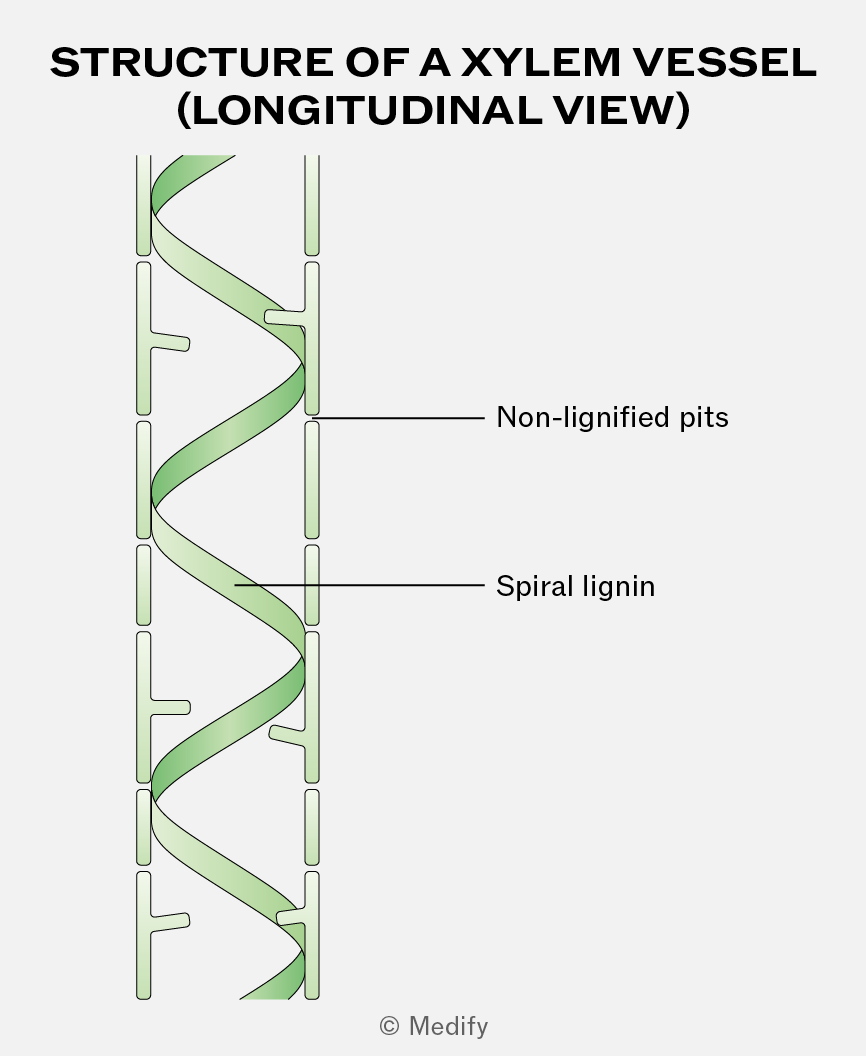
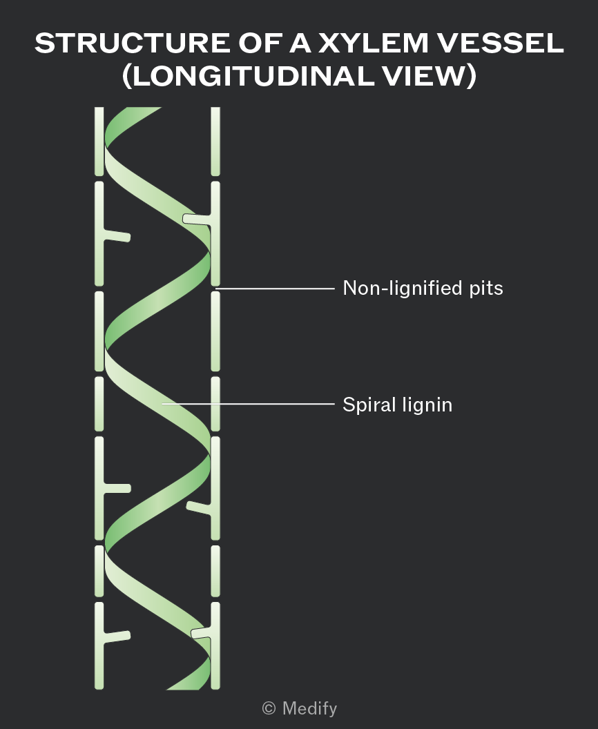
Illustration titled 'Structure of a Xylem Vessel (Longitudinal View)'. The diagram shows a tubular structure with a spiral green band labeled 'Spiral lignin'. The tube has horizontal lines at intervals, labeled 'Non-lignified pits'.
Add to favourites

Columns of dead sclerenchyma cells with lignified, thickened cell walls form hollow sclerenchyma fibres.

These fibres are located on the outer side of the vascular bundle and provide mechanical support.
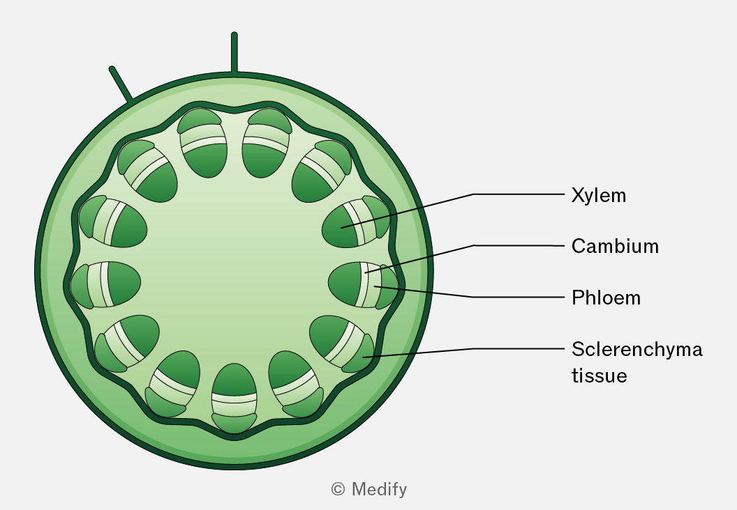
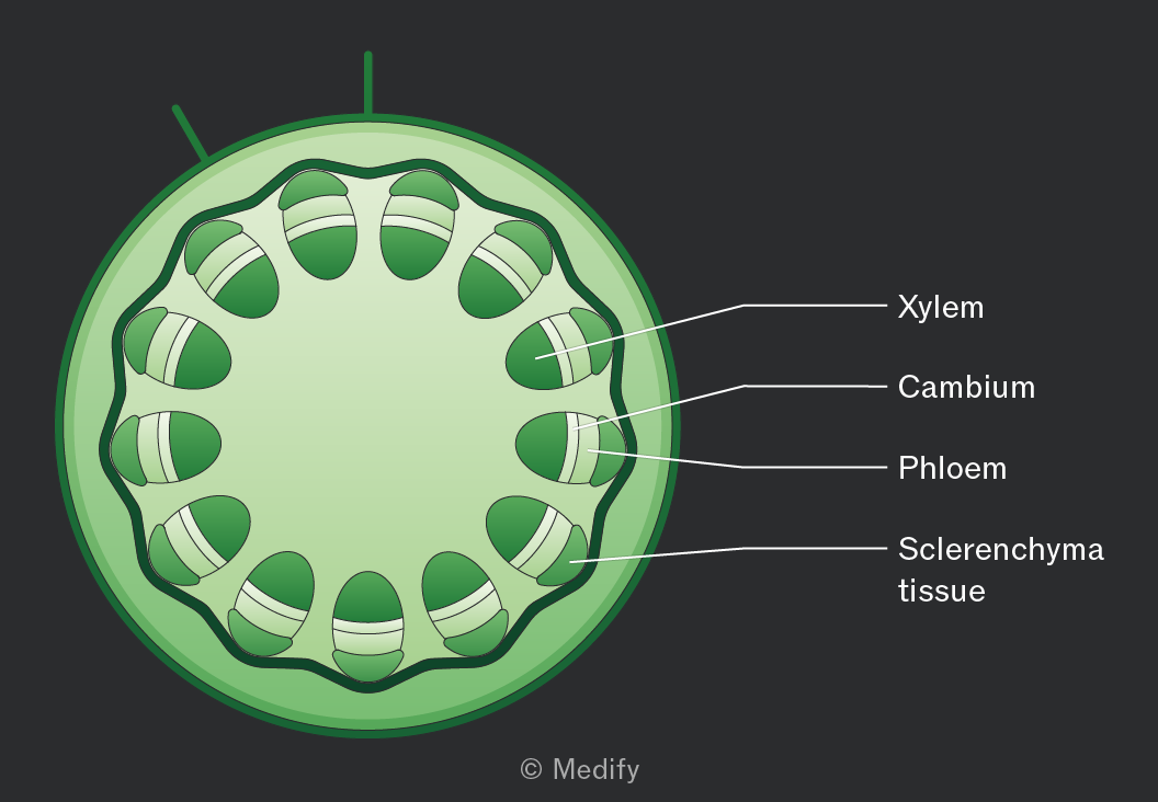
Cross-sectional diagram of a plant stem illustrating four labeled layers. The outermost layer is labeled 'Sclerenchyma tissue,' followed by 'Phloem,' then 'Cambium,' and the innermost layer is labeled 'Xylem.' Each label points to its respective section within the circular arrangement of tissues.
Add to favourites

Water is essential in plants for both structural and metabolic reasons:

  • It provides support and turgidity, helping cells maintain shape, and is used in photosynthesis to produce glucose and oxygen.
  • Water acts as a solvent, dissolving mineral ions for transport in the xylem and assimilates (sugars) for transport in the phloem.
  • Evaporation of water helps to keep the plant cool.
  • It is a metabolite used in many hydrolysis reactions.
Add to favourites

Mineral ions are essential for plant growth and metabolism:

  • Nitrate ions () – used to make amino acids, proteins, enzymes, and nucleic acids for growth.
  • Magnesium ions () – component of chlorophyll, required for photosynthesis.
  • Calcium ions () – important for cell wall stability and cell membrane function.
  • Phosphate ions () – used to make ATP, DNA, RNA, and phospholipids.
Add to favourites

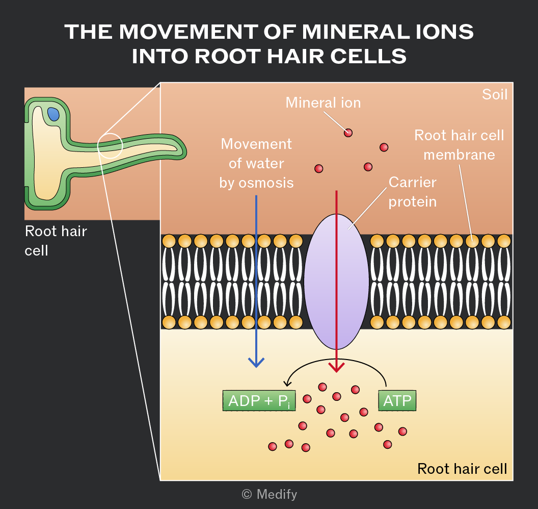
Mineral ions are actively transported from the soil into root hair cells, against their concentration gradient. This uses carrier proteins / ion pumps, which require ATP from respiration.

Root hair cells are adapted for this by having many mitochondria to supply energy.
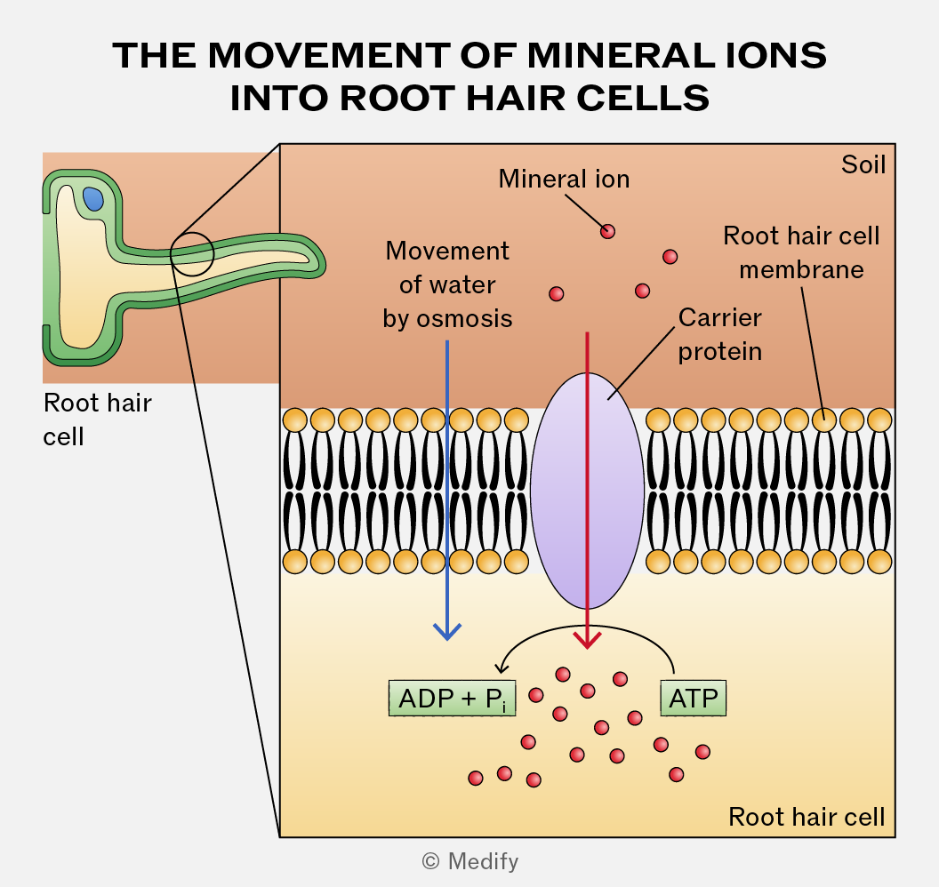
Diagram titled 'The Movement of Mineral Ions into Root Hair Cells.' The image shows a root hair cell with an enlarged section of its membrane. To the left, a root hair cell is depicted with an arrow pointing to an expanded view of the root hair cell membrane. The membrane consists of a double layer with black lines and yellow circles representing lipid bilayers. Above the membrane, labeled 'Soil,' small red circles labeled 'Mineral ion' are shown. A red arrow pointing downward indicates the movement of mineral ions through a 'Carrier protein,' which is an oval shape embedded in the membrane. The text 'Root hair cell membrane' is labeled above the membrane. Below the red arrow, red circles accumulate inside the root hair cell, with an arrow pointing from 'ADP + P_i' to 'ATP,' indicating energy conversion. A blue arrow points downward labeled 'Movement of water by osmosis,' showing water movement across the membrane. The bottom right corner is labeled 'Root hair cell.'
Add to favourites

Method for investigating the effect of a mineral on plant growth:

  1. Select plants that are clones / the same variety / the same age to reduce genetic variation.
  2. Prepare a range of at least five different mineral concentrations in the growth medium.
  3. Control abiotic variables such as light intensity, temperature, and water availability to ensure they remain constant.
  4. Plant the seeds or seedlings in the prepared mineral solutions and grow them for a set period of time under controlled conditions.
  5. Measure a relevant growth parameter, such as increase in height, leaf number, or dry biomass.
  6. Include repeats at each mineral concentration to calculate mean growth values.
  7. Record and compare growth across the different mineral concentrations to determine the effect of mineral deficiency.
Add to favourites

Stomata are pores mainly on the underside of leaves that allow carbon dioxide to enter and oxygen to leave the plant.

Each stomatal pore is surrounded by kidney-shaped guard cells with a thicker inner wall and thinner outer wall.

When guard cells take in water and become turgid, they swell and bend, opening the stomata. This happens during the day when photosynthesis occurs in the presence of sunlight.

When guard cells lose water and become flaccid, the stomata close. This happens at night when photosynthesis cannot occur.
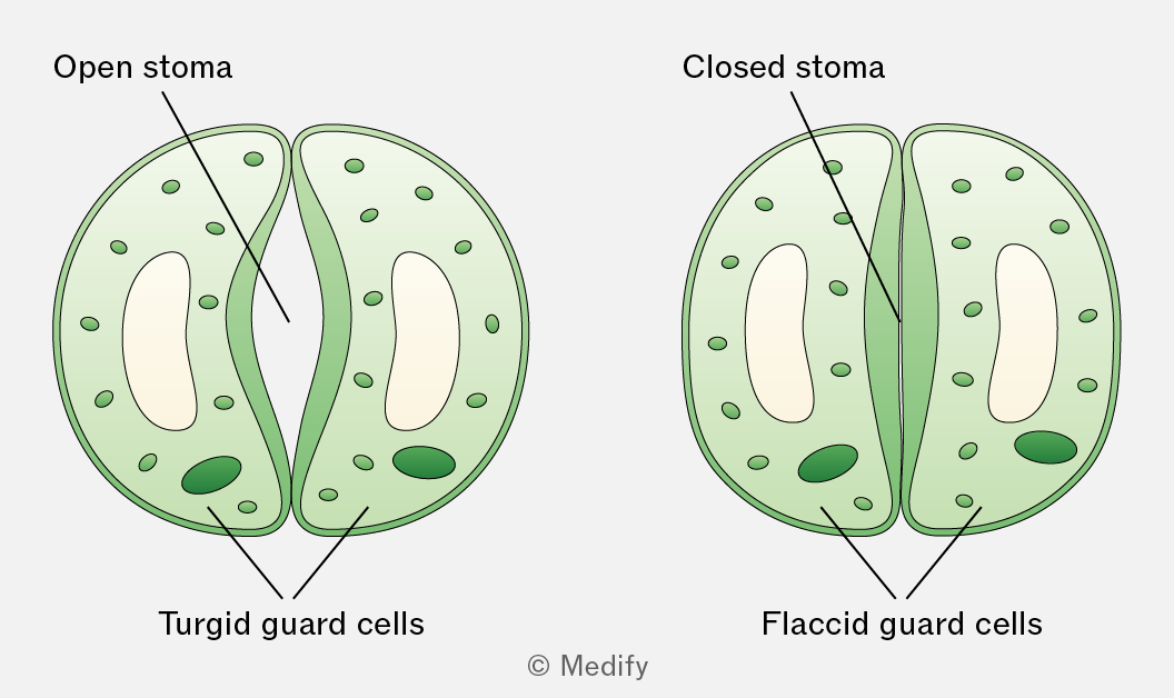
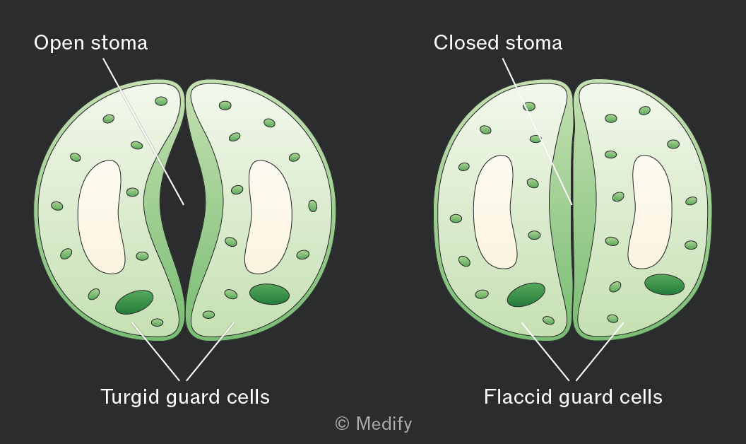
The image shows two diagrams of plant stomata. On the left, labeled 'Open stoma,' there are two turgid guard cells with a visible gap between them. On the right, labeled 'Closed stoma,' the guard cells are flaccid and touching, resulting in no gap. Both diagrams have labels pointing to the guard cells: 'Turgid guard cells' for the open stoma and 'Flaccid guard cells' for the closed stoma.
Add to favourites

Transpiration is the loss of water vapour from a plant by evaporation from the leaf and diffusion into the atmosphere through the stomata.

It occurs as a consequence of gas exchange through the stomata.

Water evaporates from the surfaces of the spongy mesophyll cells into the air spaces within the leaf and then diffuses out of the leaf through the open stomata down a water vapour potential gradient.
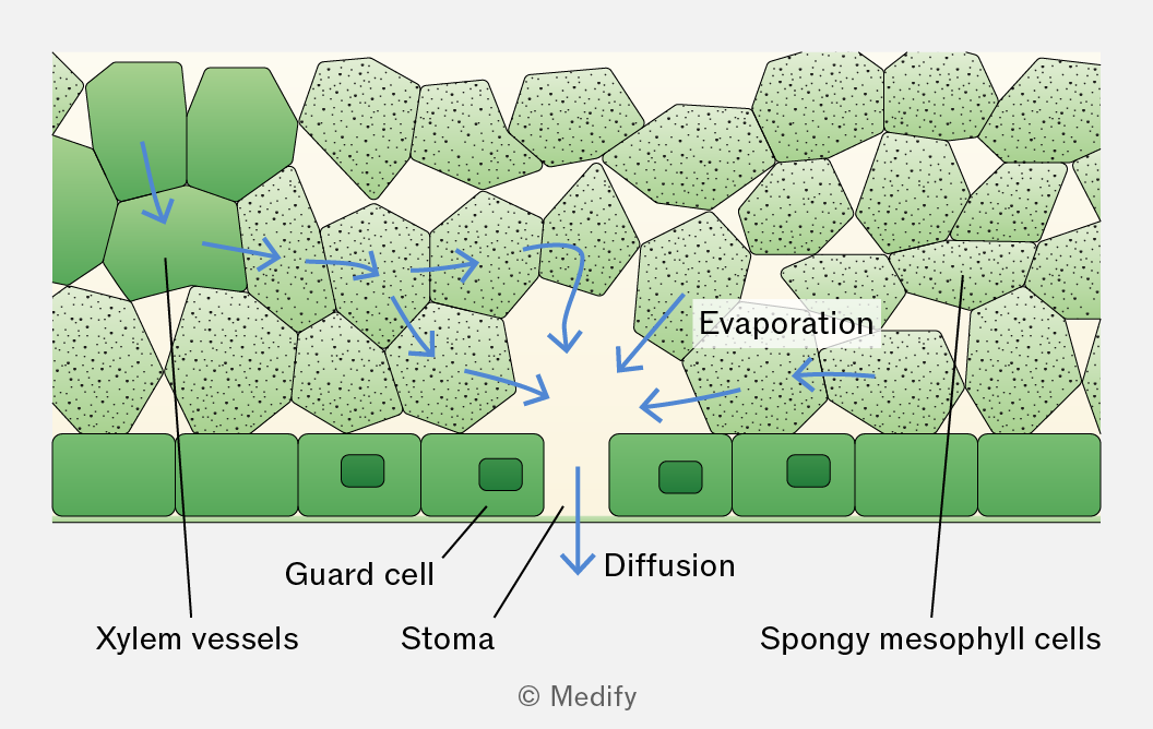
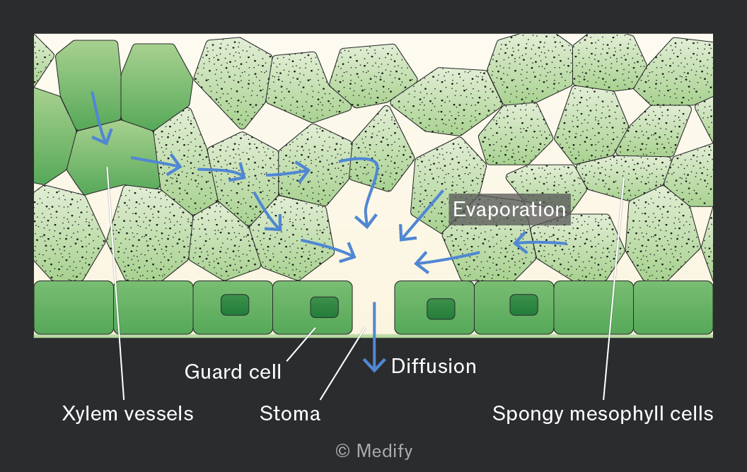
The image depicts a cross-section of a leaf showing the process of transpiration. At the top are polygonal spongy mesophyll cells, labeled 'Spongy mesophyll cells', with dotted textures. Blue arrows indicate the movement of water vapor, labeled 'Evaporation', moving through these cells and downwards. Below the mesophyll cells are rectangular guard cells, labeled 'Guard cell', with small openings called 'Stoma'. Below the stoma is a line labeled 'Diffusion' indicating the movement of water vapor out of the leaf. To the left are vertically oriented structures labeled 'Xylem vessels'.
Add to favourites

The transpiration stream is the movement of water from the roots to the leaves through the xylem in the stem. This process is explained by the cohesion–tension theory.

Add to favourites

In the cohesion–tension theory, transpiration reduces the water potential in the mesophyll cells, causing water to move by osmosis from the xylem into these cells. This creates tension (negative pressure) in the xylem, causing water to move down a pressure gradient by mass flow from roots to leaves.
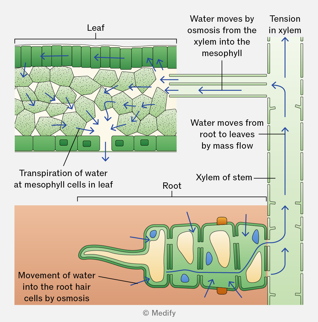
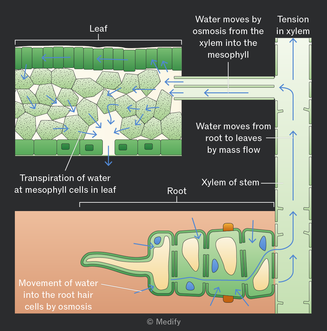
The diagram illustrates the movement of water in plants. At the top left, labeled 'Leaf,' water moves by osmosis from the xylem into the mesophyll. Blue arrows indicate the direction of water movement, with labels such as 'Transpiration of water at mesophyll cells in leaf.' On the right, a vertical section labeled 'Tension in xylem' shows arrows moving upwards. In the middle, 'Water moves from root to leaves by mass flow' is displayed with horizontal arrows. At the bottom, labeled 'Root,' a section illustrates 'Movement of water into the root hair cells by osmosis' with arrows entering the root cells. The central section is labeled 'Xylem of stem,' showing the pathway of water through the plant.
Add to favourites

In the cohesion–tension theory, the cohesion between water molecules, forms a continuous column of water in the xylem, which is pulled upwards as a result of the tension created by transpiration.

Adhesion occurs between water molecules and the walls of the xylem vessels, helping the water column move up the vessel by capillary action and resist the effects of gravity.
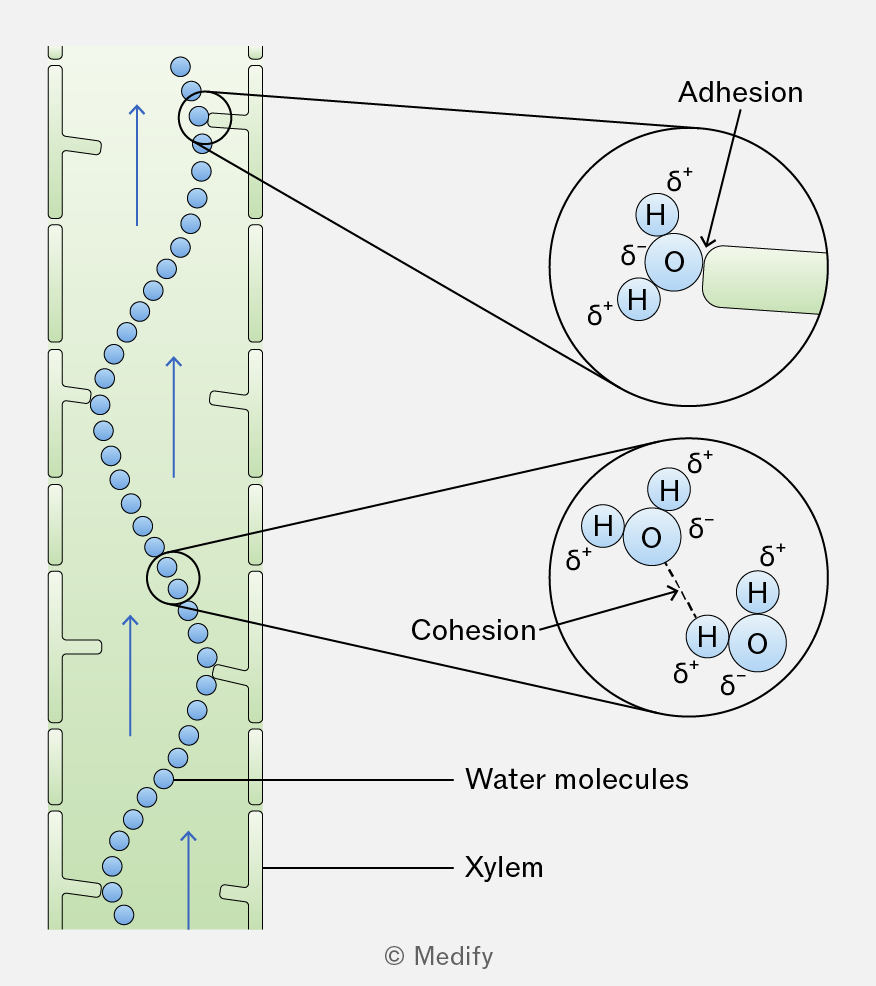
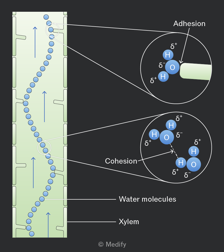
The image illustrates the movement of water molecules in the xylem, represented by a green column with blue circles and upward arrows, indicating upward movement. Two magnified sections show molecular interactions. The top magnified circle labeled 'Adhesion' depicts a water molecule (H2O) with partial charges, δ+ on hydrogen and δ- on oxygen, interacting with the xylem wall. The bottom magnified circle labeled 'Cohesion' shows two water molecules, with hydrogen bonds indicated by dashed lines, illustrating the attraction between molecules. Annotations include 'Water molecules,' 'Xylem,' and the company name 'Medify' at the bottom.
Add to favourites

The function of the phloem is the movement of assimilates (such as sucrose, amino acids, hormones and salts), which are dissolved in water from source (site of production) to sink (site of usage). This process is called translocation and it requires energy.

Translocation is a two-way flow, moving assimilates both up and down the plant by the process of mass flow, down a pressure gradient.

Phloem is a tissue made of two types of cells – sieve tube elements and companion cells.

Add to favourites

Sieve tube elements are cells stacked end to end to form long, hollow, continuous tubes, which allow mass flow of assimilates with minimal resistance. Although sieve tube elements are living cells, they contain very little cytoplasm, no nucleus, and few organelles, further reducing resistance and facilitating the flow of substances.

The end walls of sieve tube elements, called sieve plates, contain pores that allow assimilates to pass through and connect the cytoplasm of adjacent cells. The sieve plates also limit the size of molecules that can pass and slightly reduce the rate of flow.

The cell walls of sieve tube elements are thin, not lignified (cellulose only), and do not contain pits.
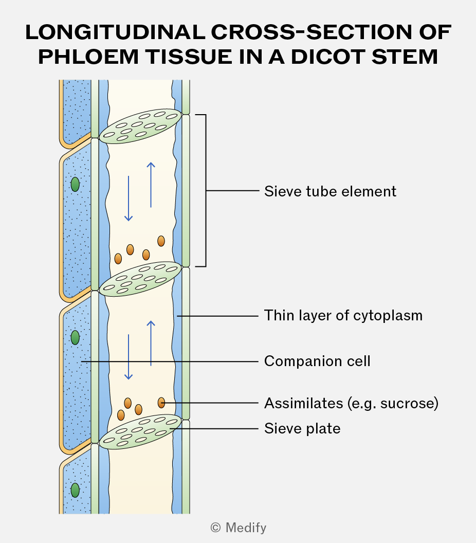
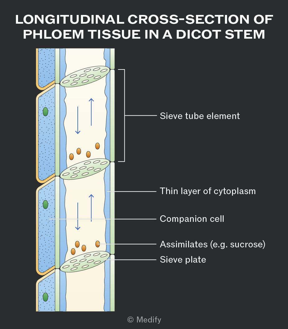
Diagram titled 'Longitudinal Cross-Section of Phloem Tissue in a Dicot Stem'. The image shows a vertical section of phloem tissue with several labeled components. On the left, there are labeled compartments: 'Companion cell' with a green circular structure and 'Thin layer of cytoplasm' appearing as a thin blue layer. The central column labeled 'Sieve tube element' contains arrows indicating flow direction and several small brown oval shapes labeled 'Assimilates (e.g. sucrose)'. At the top and bottom of this column are structures labeled 'Sieve plate', depicted as horizontal ovals with multiple perforations. The background features a pale yellow area indicating the phloem tissue structure.
Add to favourites

Companion cells act as the “life support” for sieve tube elements, carrying out the living functions. They contain dense cytoplasm, a large nucleus, and many mitochondria, and are connected to the sieve tube elements via plasmodesmata.

The mitochondria and other organelles provide energy for active transport, allowing the companion cells to load sucrose from the source (where assimilates originate) into the sieve tube elements through active loading.
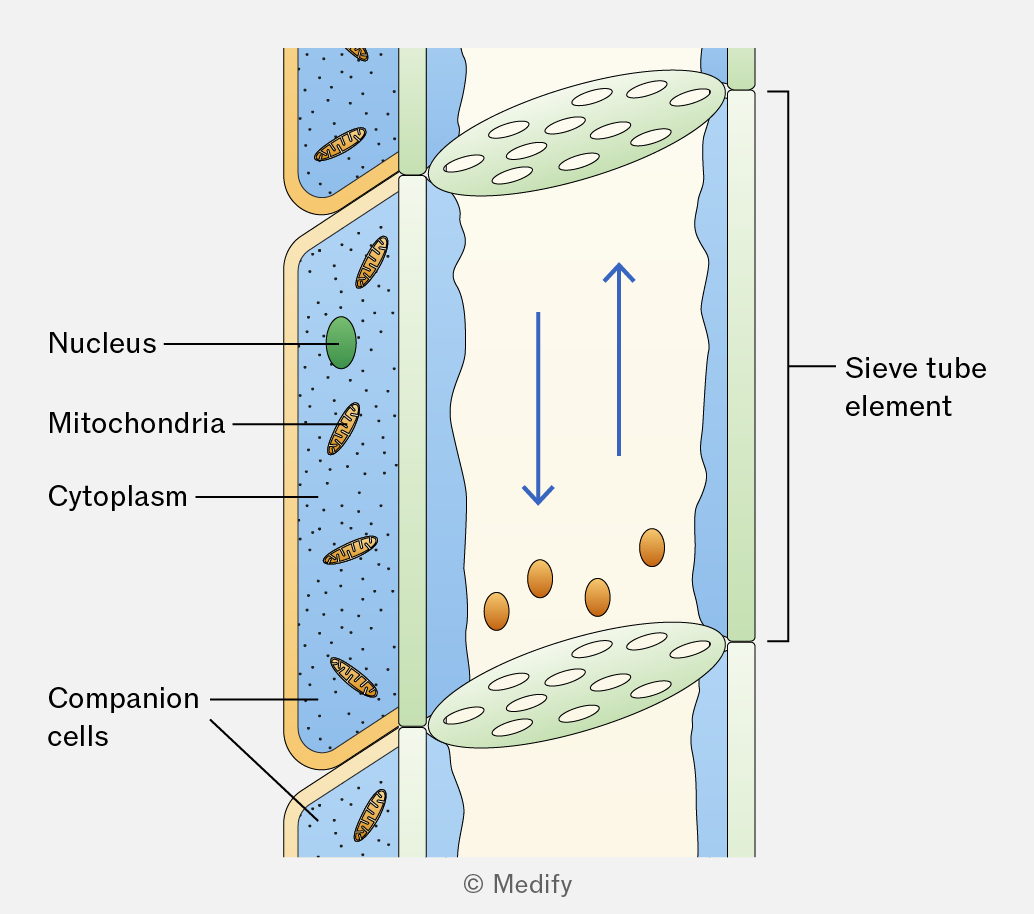
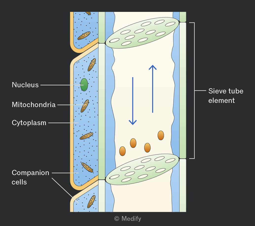
The image shows a cross-section of a plant cell with labeled parts. On the left, a cell labeled 'Companion cells' contains a 'Nucleus' depicted as a green oval, 'Mitochondria' shown as brown, oval-shaped structures, and 'Cytoplasm' filled with small dots. Adjacent to this is another section labeled 'Sieve tube element', shown as a larger, elongated area with a light background. Within this section, there are arrows indicating flow in both upward and downward directions, and several oval shapes representing cellular components. Both sections are enclosed within a cell wall. The image is © Medify.
Add to favourites

A source is a part of the plant where assimilates are produced.

Examples include green leaves, storage organs such as tubers that are unloading their stores, and food reserves in germinating seeds.

Add to favourites

Glucose produced in photosynthesising cells is converted into an assimilate, such as sucrose, for translocation in the phloem.

Sucrose is soluble, allowing it to move in sap, and relatively metabolically inactive, so it is not used in respiration. Transportation as sucrose also reduces diffusion out of the sieve tubes, as entry and exit are controlled by transport proteins.

Add to favourites

For the active loading of sucrose into the companion cells from the surrounding tissue, protons
() are actively pumped out using ATP and a proton pump. This creates a proton gradient.

Protons then re-enter the cell via co-transport proteins, carrying sucrose molecules with them. This process actively loads sucrose into companion cells, from where it diffuses into sieve tube elements for translocation.
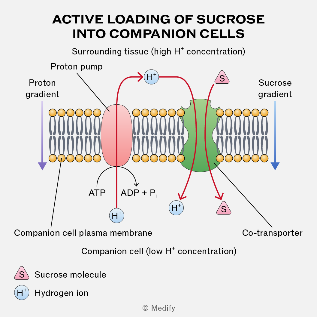
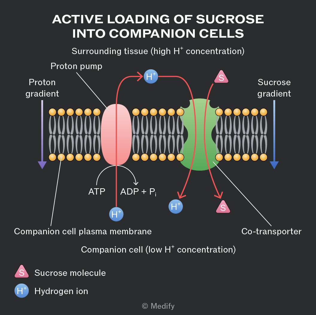
Diagram titled 'Active Loading of Sucrose into Companion Cells'. It shows a membrane with a proton pump and co-transporter. The membrane is labeled 'Companion cell plasma membrane'. The top region is labeled 'Surrounding tissue (high H⁺ concentration)' and the bottom 'Companion cell (low H⁺ concentration)'. The proton pump is depicted in red, moving H⁺ ions from the companion cell to the surrounding tissue, using ATP which converts to ADP + Pᵢ. A purple arrow labeled 'Proton gradient' points downward. The co-transporter is shown in green, moving H⁺ ions and sucrose (S) into the companion cell, with a downward blue arrow labeled 'Sucrose gradient'. The legend indicates S as 'Sucrose molecule' and H⁺ as 'Hydrogen ion'.
Add to favourites

The entry of sucrose into sieve tube elements at the source decreases the water potential in the sieve tube elements. This causes water to enter the phloem by osmosis from the surrounding cells and xylem vessels. This results in higher hydrostatic pressure within the sieve tube elements.
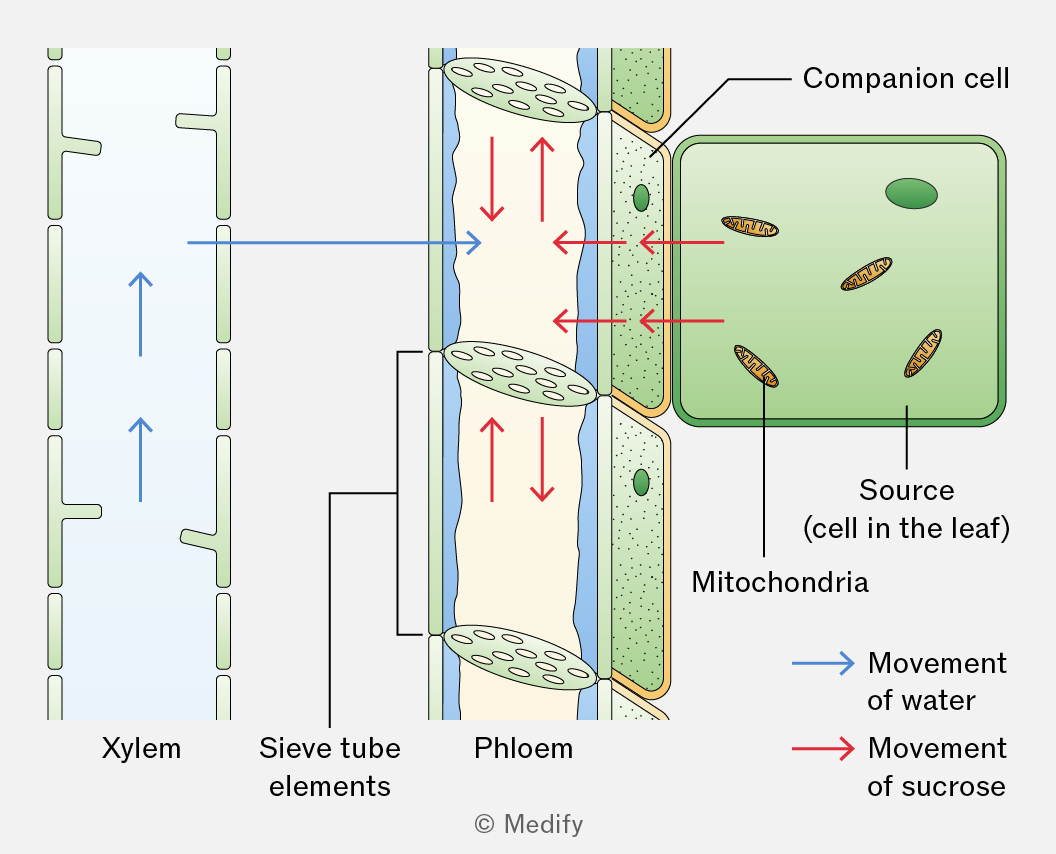
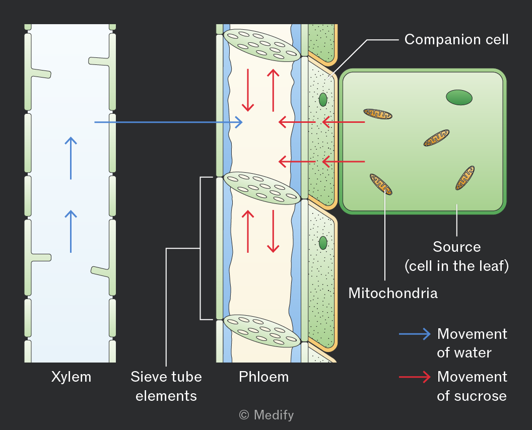
The diagram illustrates the movement of substances in plant vascular tissue. On the left, a vertical section labeled 'Xylem' shows upward blue arrows indicating the 'Movement of water.' Adjacent to it, a section labeled 'Sieve tube elements' is part of a larger area labeled 'Phloem.' Red arrows indicate the 'Movement of sucrose' downwards and laterally. On the right, a green labeled area shows a 'Companion cell' and a 'Source (cell in the leaf)' containing mitochondria. Blue arrows point from the xylem towards the phloem, and red arrows show movement from the source cell into the phloem. The diagram also includes the label 'Mitochondria' inside the source cell.
Add to favourites

When the sucrose arrives at the sink, it diffuses out of the sieve tube elements into the surrounding cells. This causes the water potential inside the sieve tube elements to increase so water leaves by osmosis down a water potential gradient. This lowers the hydrostatic pressure.
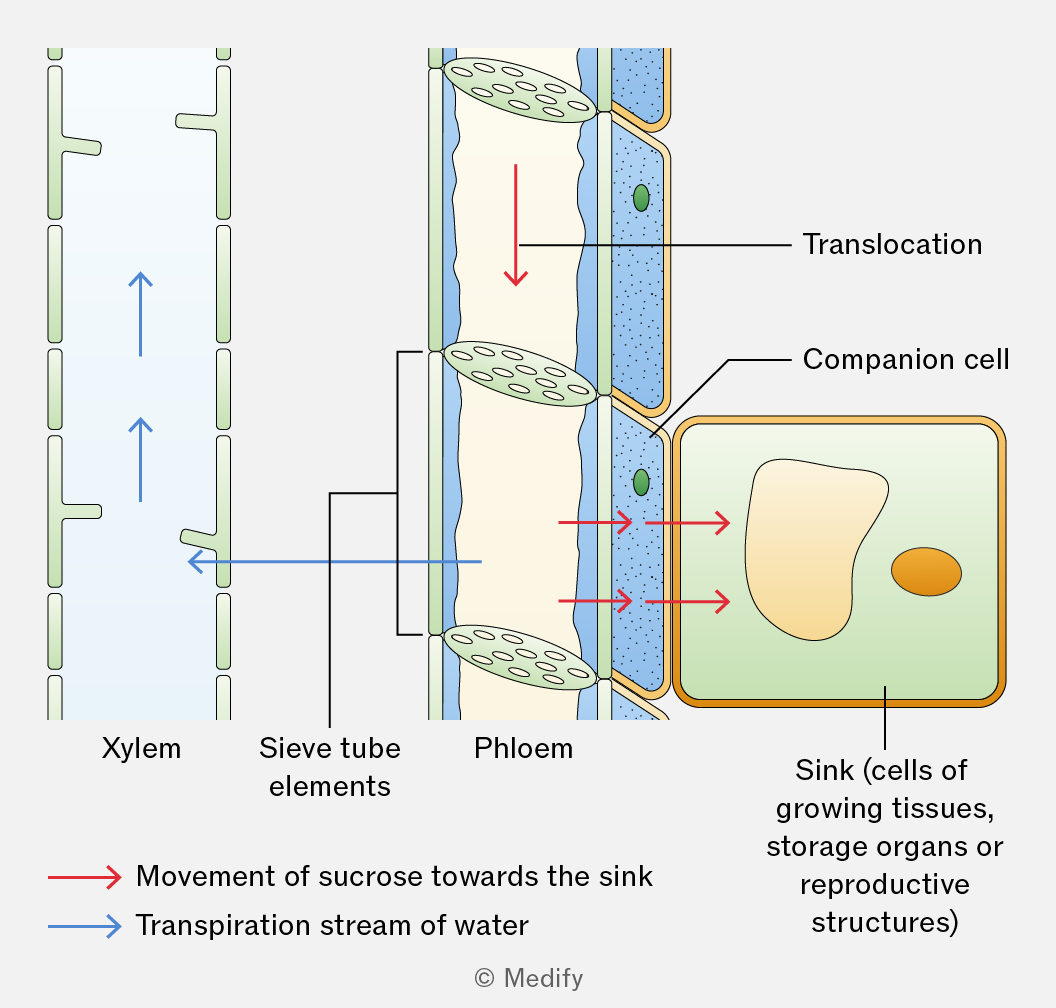
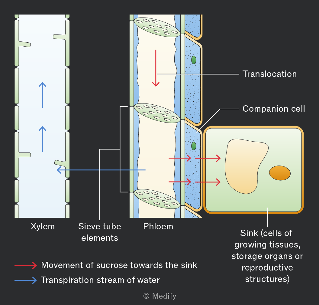
The image illustrates the process of translocation in plants with a focus on the xylem and phloem tissues. On the left, the xylem is depicted with blue arrows indicating the upward transpiration stream of water. In the center, the phloem is shown with sieve tube elements and red arrows representing the movement of sucrose towards the sink. The right side of the image includes a detailed view of a companion cell and a box labeled 'Sink (cells of growing tissues, storage organs or reproductive structures)'. Labels include 'Xylem', 'Sieve tube elements', 'Phloem', 'Translocation', and 'Companion cell'. Annotations at the bottom denote 'Movement of sucrose towards the sink' with red arrows and 'Transpiration stream of water' with blue arrows.
Add to favourites

A hydrostatic pressure gradient drives the mass flow of sucrose dissolved in water from source to sink. The high hydrostatic pressure in the phloem at the source enables rapid transport over long distances.

Add to favourites

A sink is a part of a plant where assimilates are used or stored. Assimilates are translocated from source to sink down a concentration gradient in the phloem. At the sink, assimilates are converted into glucose for respiration or starch for storage, maintaining a concentration gradient between the phloem and cells.

Examples include growing roots, dividing meristems, developing seeds, fruits, or storage organs.

Add to favourites