
Gas exchange (2.1)
On this page
Small and single–celled organisms have:
- High surface area to volume (SA:V) ratio – can rely on diffusion to obtain oxygen and nutrients and remove wastes.
- Low metabolic activity – requirements for oxygen and nutrients are lower and production of waste is slower.
Large organisms have:
- Low SA:V ratios – diffusion cannot provide the organism with enough oxygen and nutrients.
- Higher metabolic activity – higher requirements for oxygen and nutrients and faster production of toxic waste.
Larger organisms require specialised exchange surfaces to meet their oxygen, nutrient and waste removal needs.
The larger the SA:V ratio, the faster the rate of diffusion can occur.
Organisms can often be shapes that make it difficult to calculate their SA:V ratio. Their shapes are often modelled as simple geometric shapes such as spheres, cylinders or cubes to enable their SA:V ratio to be estimated more easily.
Efficient exchange surfaces have the following features to increase the rate of diffusion:
- high surface area
- thin layers
- maintaining a diffusion gradient
- selectively permeable.
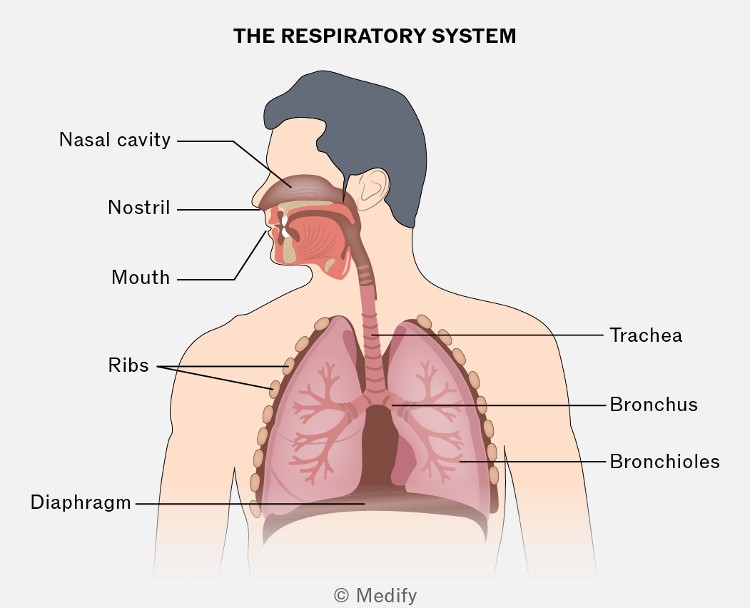
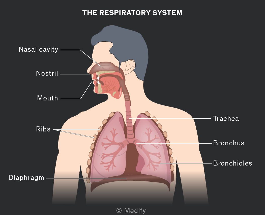
The trachea (windpipe) connects the mouth to the bronchi. The trachea is lined with incomplete rings of strong, flexible cartilage which maintains its shape and prevents collapse. The trachea has elastic fibres, which allow it to stretch and recoil during ventilation.
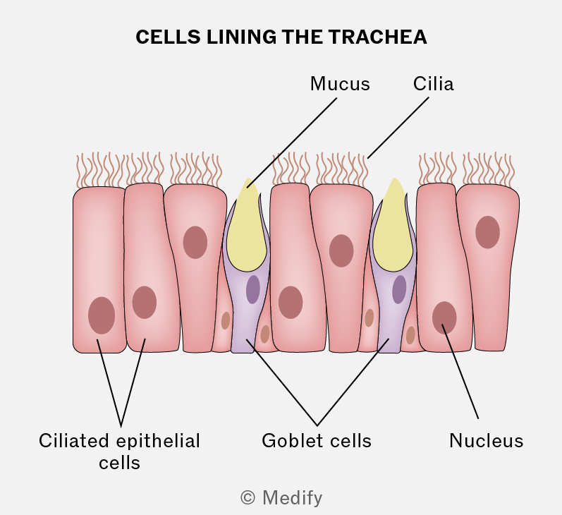
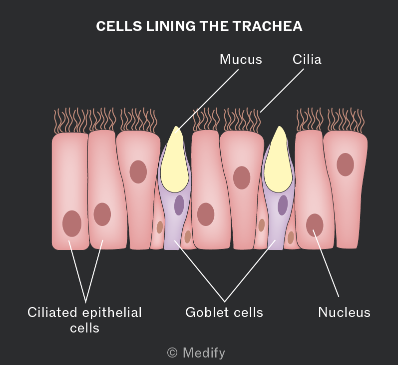
The cells lining the trachea are mostly ciliated epithelial cells, with goblet cells in between. Goblet cells produce mucus, which trap dirt and microorganisms. The ciliated epithelial cells waft and move mucus away from the lungs and towards the throat, where it is swallowed.
The trachea branches into two smaller bronchi, which lead to each lung. The bronchi have a similar function to the trachea and also contain supporting rings of cartilage. The bronchi then branch into bronchioles.
The smaller bronchioles do not have cartilage rings and contain smooth muscle. The smooth muscle can contract and relax, controlling the air reaching the lungs. Bronchioles are lined with a thin layer of flattened epithelial cells. Due to the thin layer of cells, a small amount of gas exchange can happen in the bronchioles.
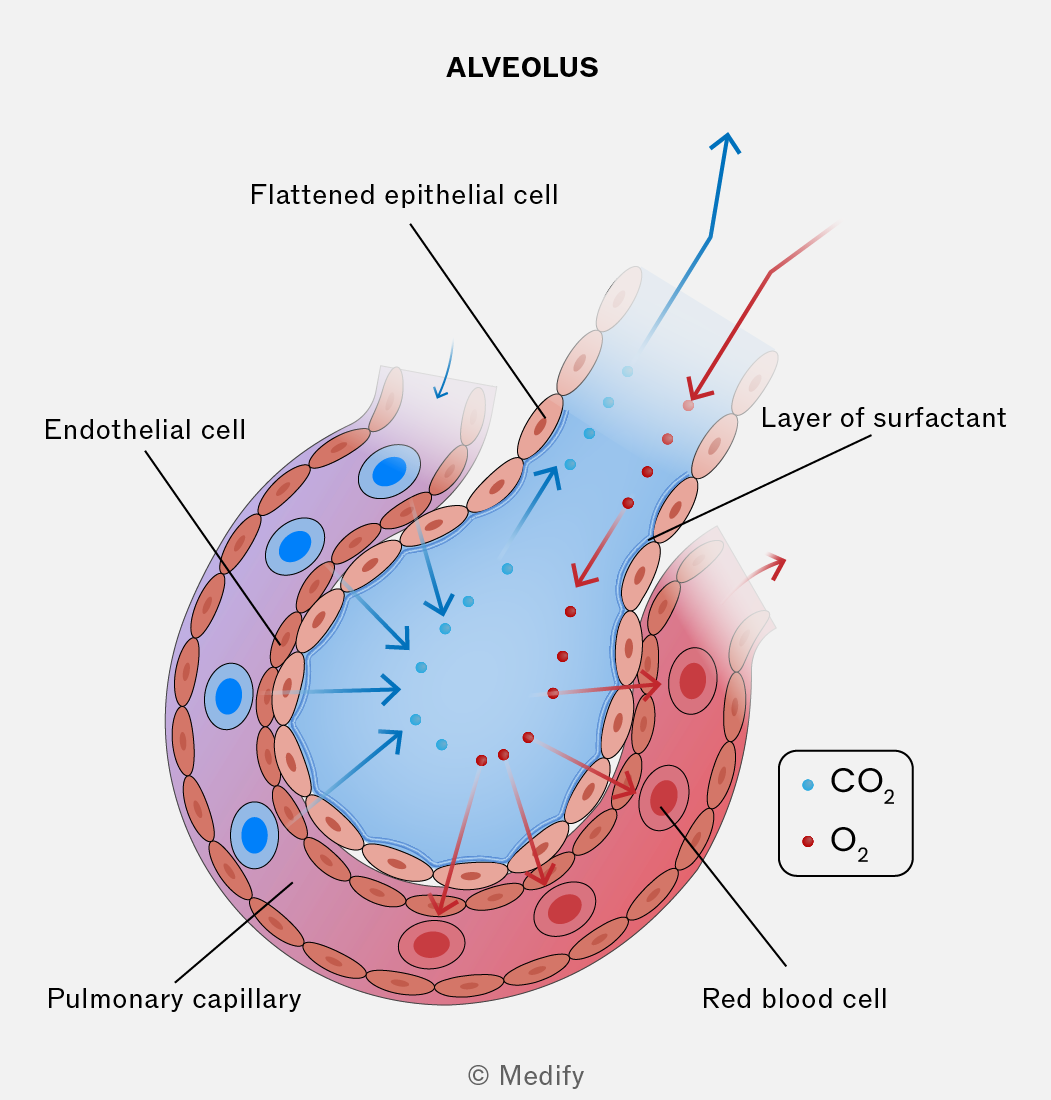
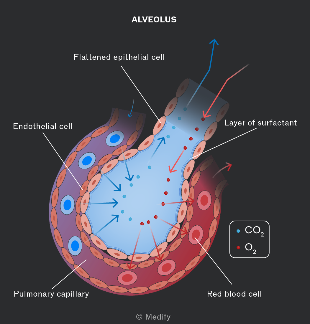
The branches of the small bronchioles end at the alveoli. The alveoli are tiny air sacs in the lungs where the majority of gas exchange takes place.
The alveoli have many adaptations:
- Lined with elastic tissue, allowing them to stretch and recoil during ventilation.
- Large surface area for increased rate of diffusion.
- Good ventilation and blood supply maintain a steep concentration gradient, which increases the rate of diffusion.
- Surfactant makes the surface of the alveoli moist, which aids diffusion.
- Each alveoli is lined with flattened epithelial cells, and the capillaries are lined with a thin layer of endothelial cells. This makes the distance required for diffusion short, increasing the rate of gas exchange.
The factors that affect the rate of diffusion can be combined into an equation called Fick’s law to determine: