
DNA replication and mutation (2.11, 2.12, 2.14)
On this page
DNA replication is the process by which DNA is copied.
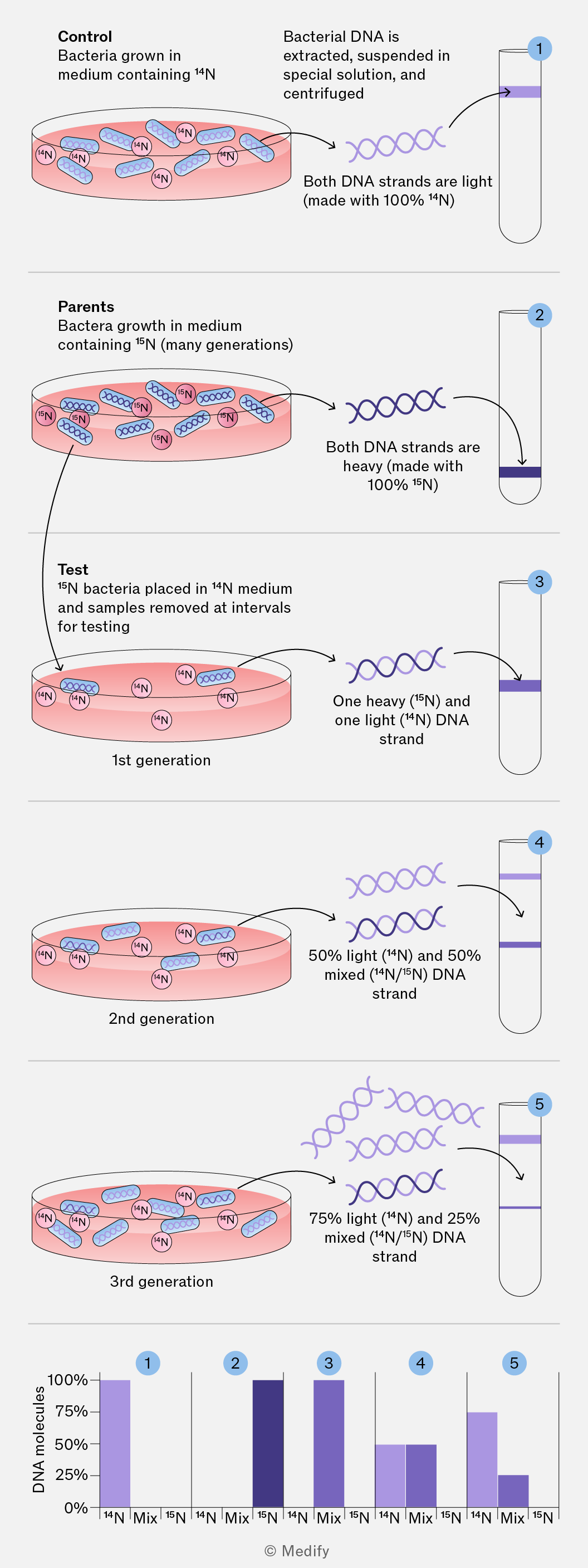
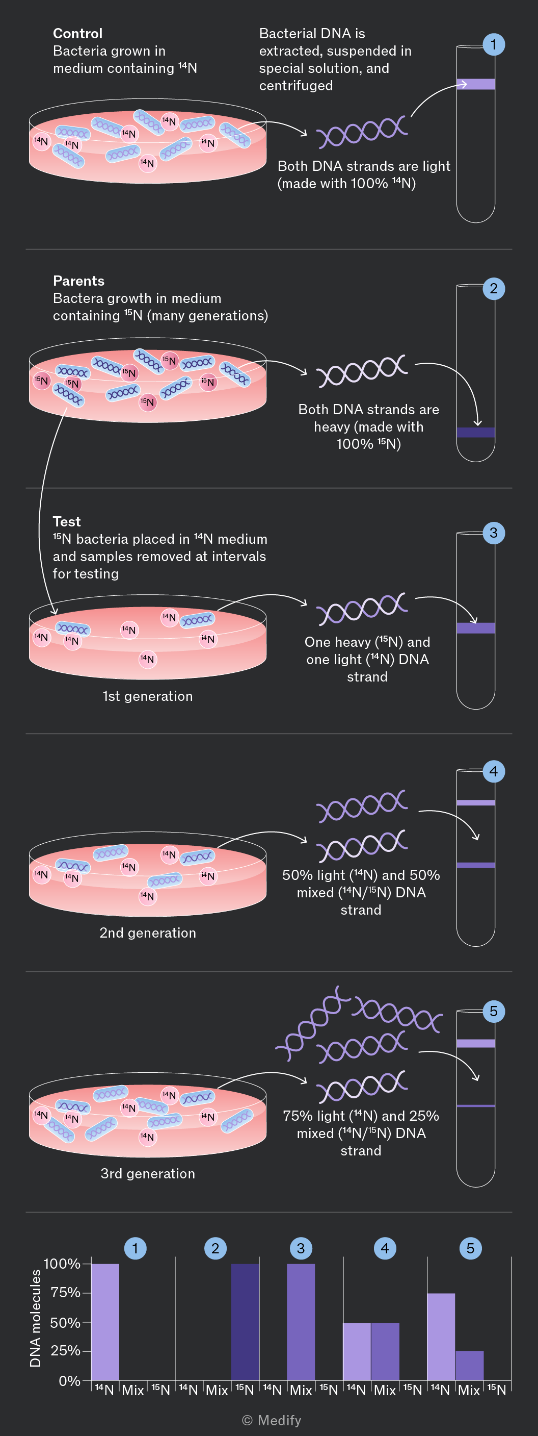
It follows a semi–conservative mechanism; each new DNA molecule consists of one original (parental) strand and one newly synthesised strand.
This ensures that each daughter cell receives an exact copy of the DNA, maintaining genetic continuity between generations of cells.
Watson and Crick proposed the semi–conservative model of DNA replication. This replication mechanism was validated by the Meselson–Stahl experiment (1958) using two different isotopes of nitrogen: and
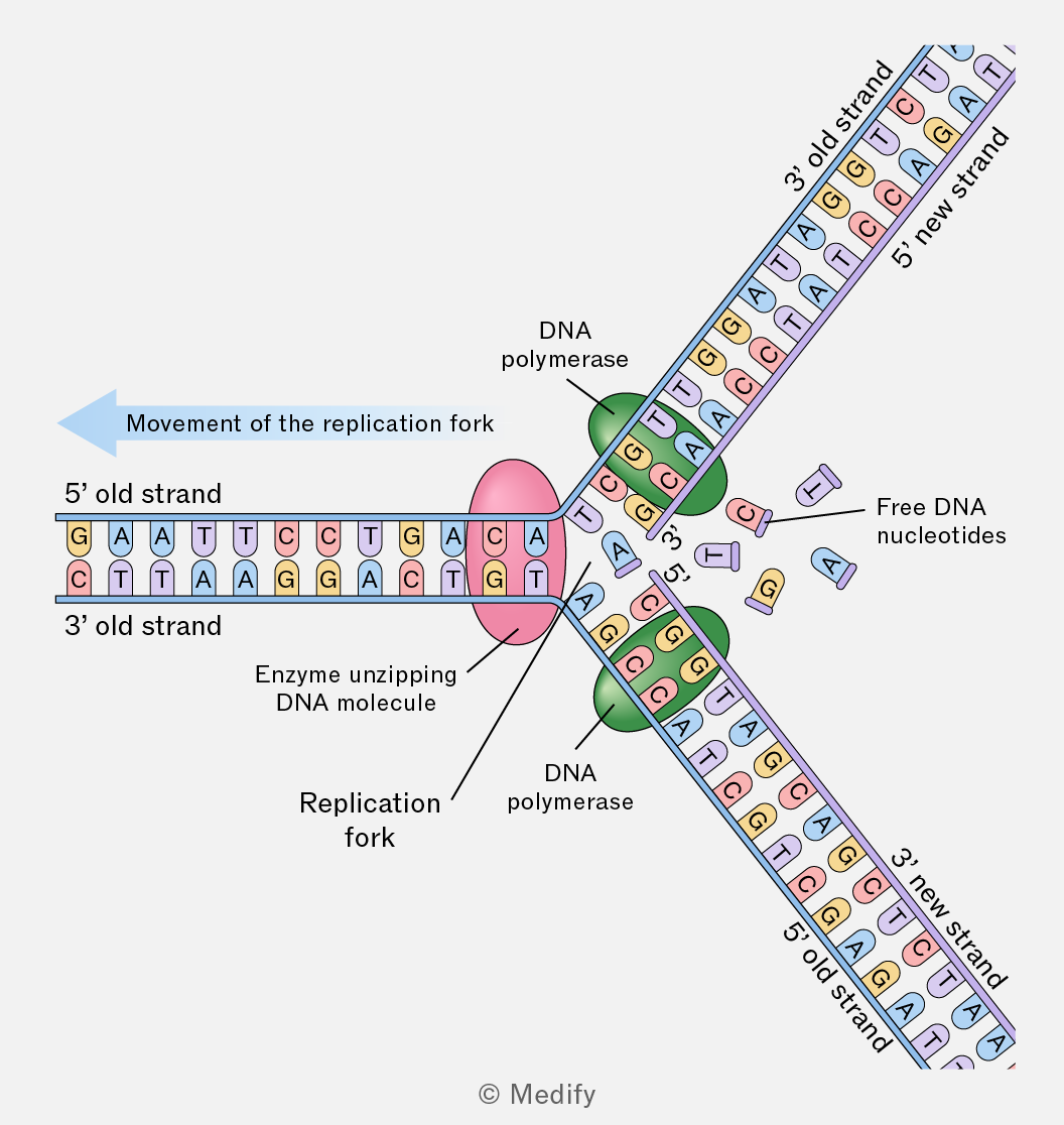
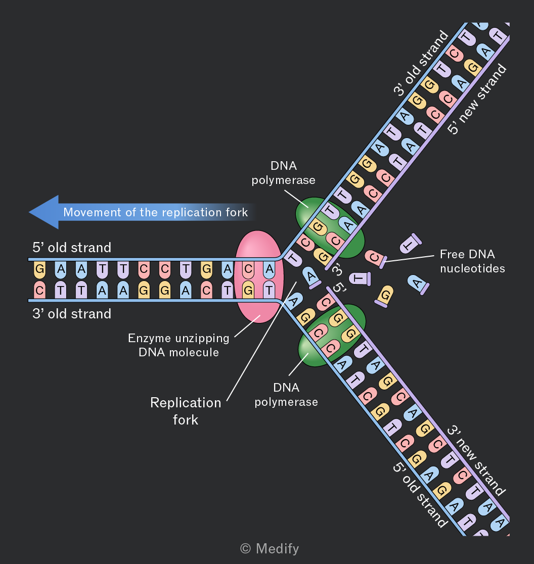
DNA replication begins with the unwinding of the DNA double helix and the breakage of the hydrogen bonds between complementary base pairs.
This separation creates two single–stranded templates that can both be used for synthesising new DNA strands.
Free DNA nucleotides bond to their complementary base pairs on the parent strands of DNA. DNA polymerase joins these new adjacent nucleotides via condensation reactions, which form phosphodiester bonds.
Two identical daughter DNA molecules are created, each containing one parent strand and one newly synthesised strand.
Errors in DNA replication can cause spontaneous genetic mutations, which are changes to the base sequence of DNA.
Where mutations occur in cells that form gametes, they can be inherited.
A large proportion of DNA does not code for proteins; therefore, mutations in these regions may have no effect.
There are many types of genetic mutation, including:
- Base deletion, which occurs when a nucleotide is removed. This causes a frameshift and often results in an altered amino acid sequence. For example, this is the most common mutation associated with cystic fibrosis.
- Base substitution, which occurs when one nucleotide is swapped for another. This mutation type does not cause a frameshift. A substitution may or may not change the amino acid specified by the codon. For example, this is the mutation associated with sickle cell anaemia.
Cystic fibrosis (CF) is a genetic disorder caused by mutations in the CFTR gene, located on chromosome 7. This gene codes for the CFTR protein that regulates chloride ion transport across cell membranes.
There are hundreds of possible mutations in the CFTR gene, each impacting the CFTR protein in a different way, but all causing CF.
Faulty CFTR proteins cause thick, sticky mucus which:
- In the lungs blocks the airways and traps bacteria, leading to infections and reduced gas exchange.
- In the digestive system blocks pancreatic ducts, stopping enzyme release and reducing nutrient absorption.
- In the reproductive system, blocks the sperm duct in males and the cervix in females, causing infertility.