Topic 2: Genes and health›Proteins (2.9)

Proteins (2.9)

An overview of proteins (2.9) from Edexcel A level Biology A including: amino acids, levels of protein structure and types of protein
5 min

Amino acids are the monomers from which polypeptides and proteins are made.

Polypeptides are linear chains of more than two amino acids, whereas proteins consist of one or more polypeptides folded into a specific 3D functional structure.

Add to favourites

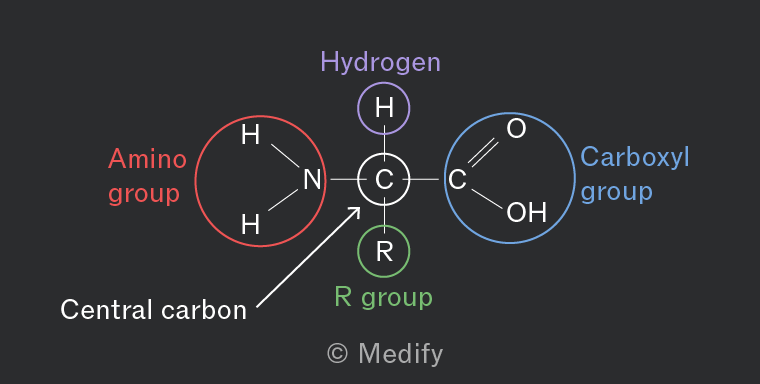
Amino acids contain a central carbon atom, attached to a hydrogen atom, an amino group (), a carboxylic acid group () and a variable side chain group (R group).

There are 20 amino acids common to all organisms and they differ only by their R group.

Essential amino acids come from the diet and non-essential ones can be synthesized in the body.
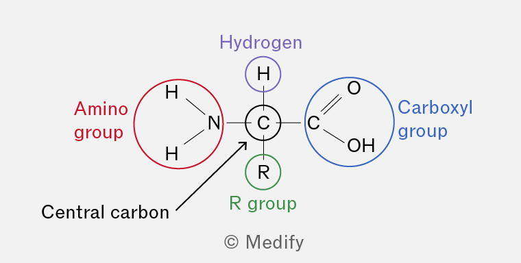
The image shows a structural diagram of an amino acid with a central carbon atom labeled 'Central carbon'. The central carbon is connected to four groups: an amino group, a hydrogen atom, a carboxyl group, and an R group. The amino group, labeled in red as 'Amino group', consists of a nitrogen atom bonded to two hydrogen atoms. The hydrogen atom is labeled in purple as 'Hydrogen'. The carboxyl group, labeled in blue as 'Carboxyl group', consists of a carbon atom double-bonded to an oxygen atom and single-bonded to a hydroxyl group (OH). The R group is labeled in green as 'R group'.
Add to favourites

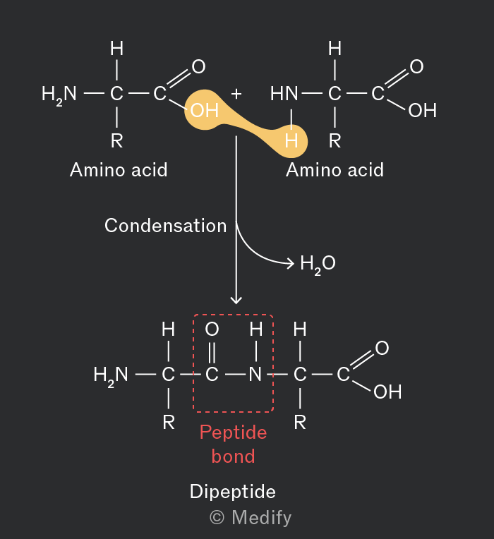
Polypeptides are formed when amino acid monomers are linked to each other during condensation reactions by peptide bonds (strong covalent bonds).

The peptide bond is formed between the carboxyl group of one amino acid and the amino group of another amino acid.

The reaction is catalysed by peptidyl transferase.
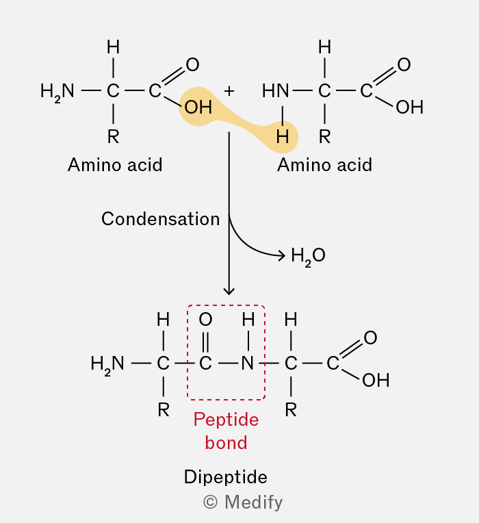
The image shows a chemical reaction diagram of two amino acids undergoing condensation to form a dipeptide. At the top, two amino acids are depicted. The left amino acid has the structure: H2N-C(R)-C(=O)-OH, and the right amino acid has the structure: HN-C(R)-C(=O)-OH. The 'R' represents a side chain. A highlighted bond between the OH group of the left amino acid and the H of the NH group of the right amino acid, indicating water (H2O) is removed. The label ‘Condensation’ with an arrow points downward to a dipeptide structure. The dipeptide has the structure: H2N-C(R)-C(=O)-NH-C(R)-C(=O)-OH. A red dashed box highlights the peptide bond between C(=O) and NH, labeled ‘Peptide bond’ in red. The term 'Dipeptide' is labeled below the structure. An arrow points to 'H2O', indicating the removal of water. The diagram includes the text labels 'Amino acid', 'Condensation', 'Peptide bond', and 'Dipeptide'.
Add to favourites

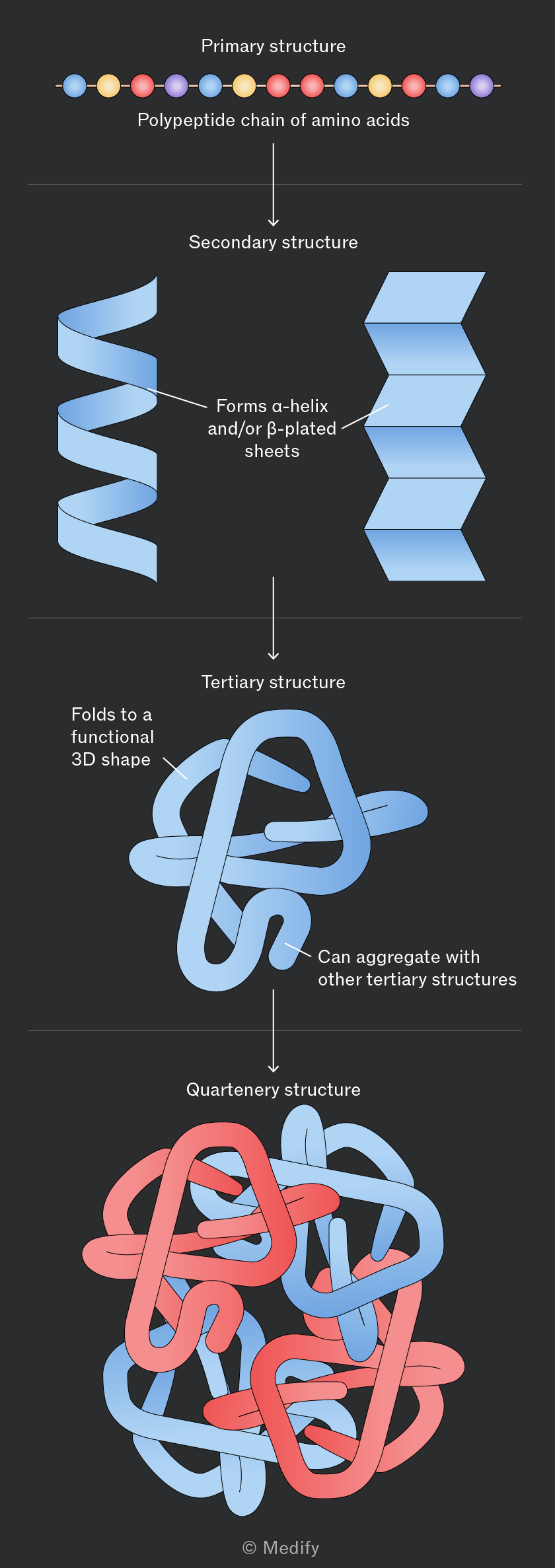
There are four levels of protein structure:
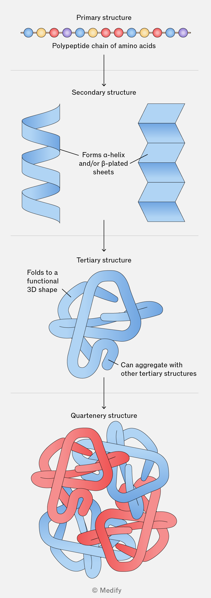
Diagram illustrating the four levels of protein structure. At the top is the 'Primary structure', shown as a linear 'Polypeptide chain of amino acids' with colored circles. An arrow points down to the 'Secondary structure', featuring a blue alpha-helix and beta-plated sheet with the label 'Forms α-helix and/or β-plated sheets'. Another arrow points down to the 'Tertiary structure', depicting a blue folded shape labeled 'Folds to a functional 3D shape'. A further arrow leads to the 'Quaternary structure', showing a complex of intertwined red and blue shapes labeled 'Can aggregate with other tertiary structures'. The diagram is credited to © Medify.
Add to favourites

Primary structure refers to the specific sequence and number of amino acids in a polypeptide chain, determined by the DNA base sequence.

This linear sequence determines how the protein folds into its three-dimensional structure and therefore its function. A change in the amino acid sequence can alter the protein’s structure and affect its function.
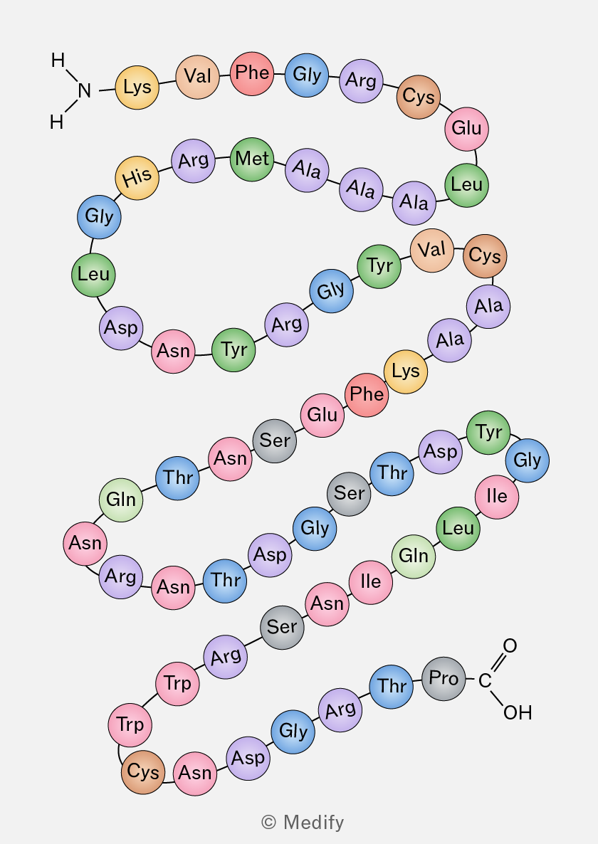
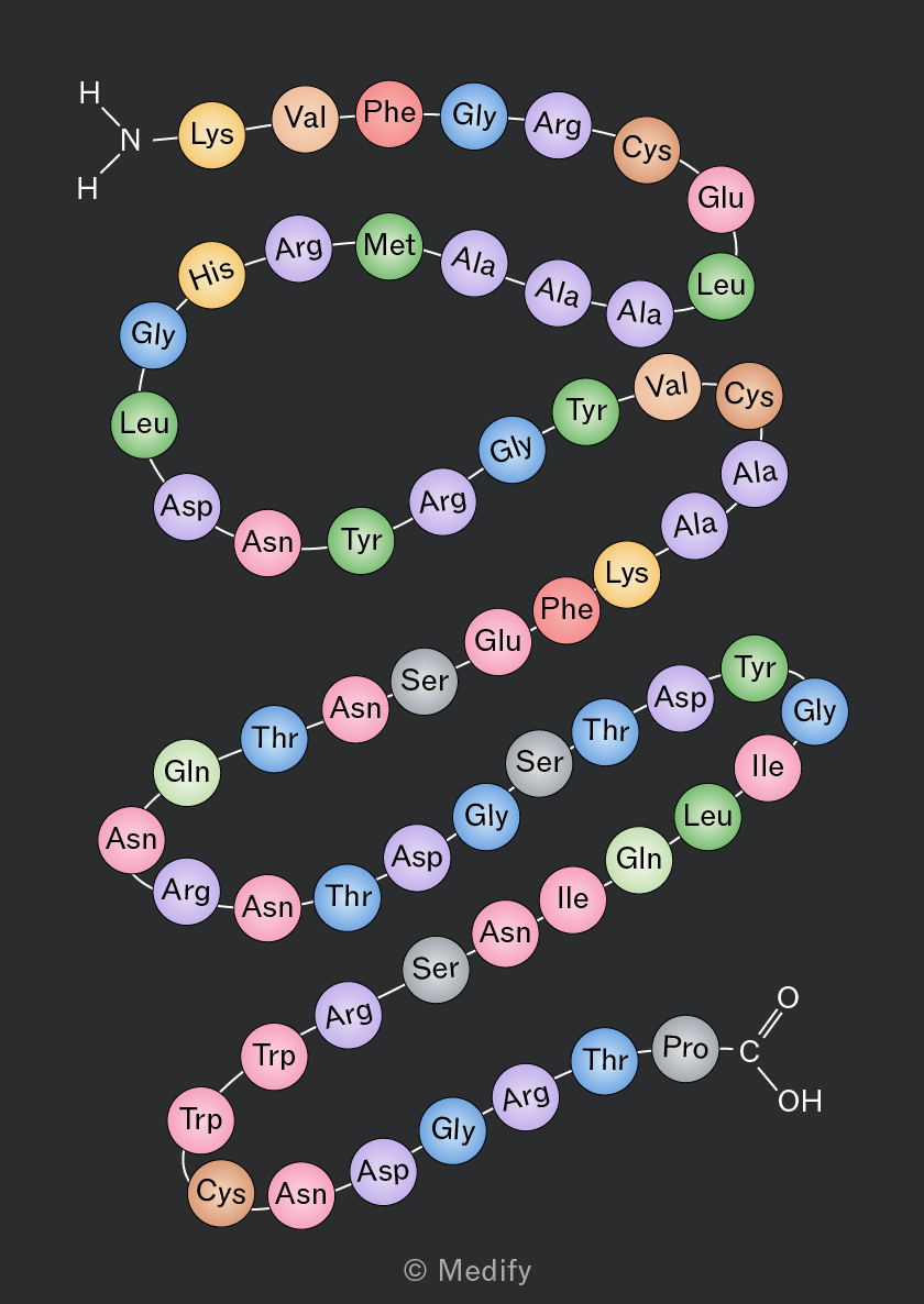
A diagram of a protein structure showing a sequence of amino acids represented as colored circles connected by lines. The sequence starts with an NH2 group and includes the following amino acids: Lys, Val, Phe, Gly, Arg, Cys, Glu, Leu, Ala, Ala, Ala, Met, Arg, His, Gly, Leu, Asp, Asn, Tyr, Arg, Gly, Tyr, Val, Cys, Ala, Ala, Lys, Phe, Glu, Ser, Asn, Thr, Asn, Gln, Leu, Ile, Gly, Asp, Tyr, Ser, Thr, Ser, Gly, Asp, Thr, Asn, Arg, Asn, Gln, Leu, Ile, Gly, Thr, Arg, Gly, Thr, Pro, Cys, Asn, Asp, Gly, Arg, Trp, Trp, and ends with a COOH group.
Add to favourites

Secondary structure refers to the localised folding of a polypeptide chain into -helices and -pleated sheets

In an -helix, hydrogen bonds form between atoms in the polypeptide backbone, causing the chain to coil into a spiral.

In a -pleated sheet, several polypeptide chains lie parallel to one another and are held together by hydrogen bonds. These hydrogen bonds stabilise the secondary structure of the protein.
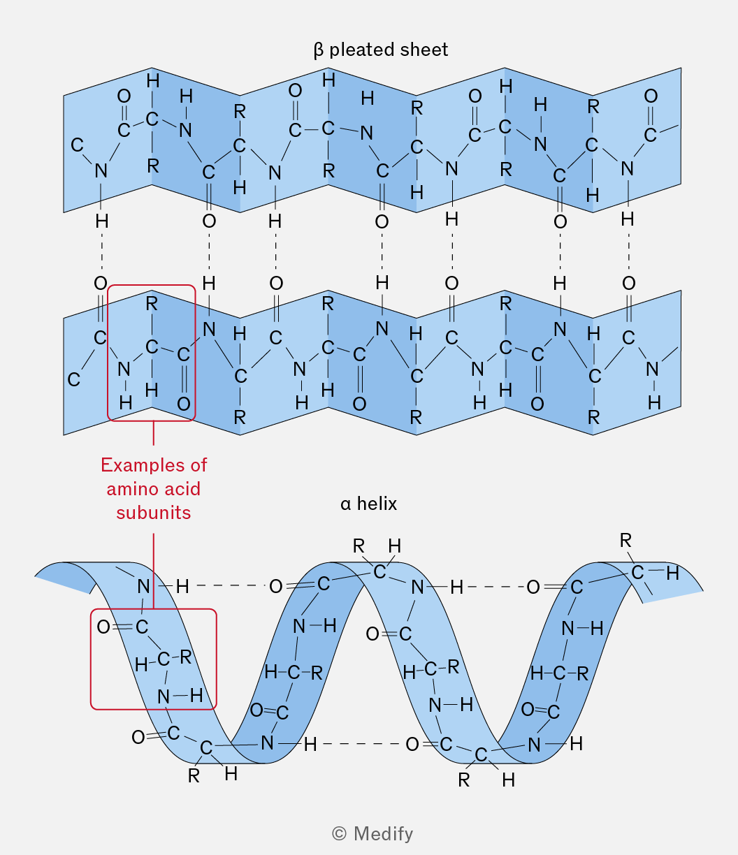
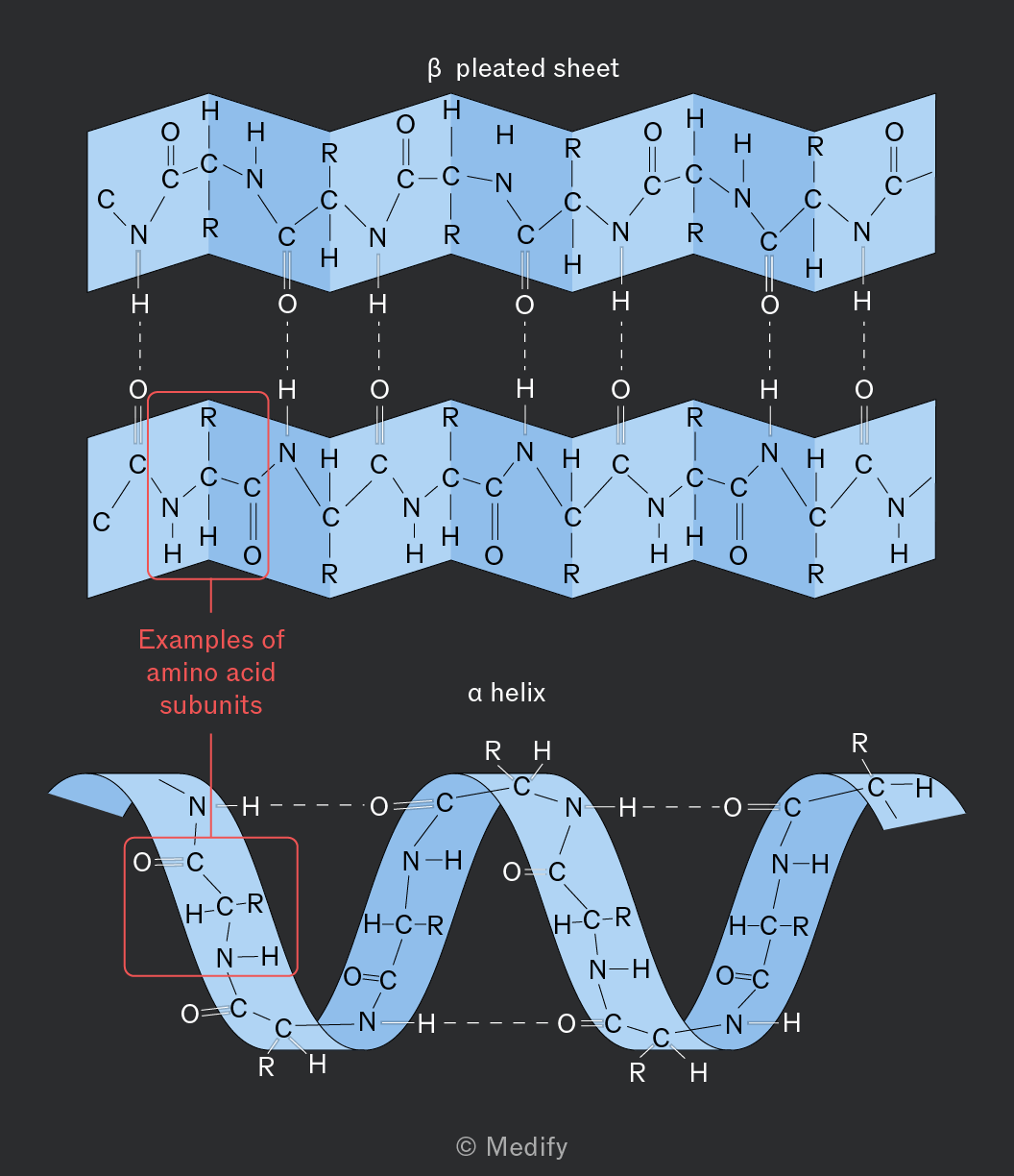
The image shows two diagrams representing protein structures. The top diagram is labeled 'β pleated sheet' and consists of two parallel zigzag patterns. Each zigzag pattern includes repeating units with nitrogen (N), carbon (C), hydrogen (H), and oxygen (O) atoms, connected by dashed lines representing hydrogen bonds. An annotation on the left highlights a section labeled 'Examples of amino acid subunits' with a red box around one of the repeating units. The bottom diagram is labeled 'α helix' and features a coiled ribbon structure. The coil is composed of similar repeating units with N, C, H, and O atoms, with dashed lines indicating bonds. Both diagrams contain labels for atomic groups, such as 'R' for side chains, and show the characteristic folding patterns of secondary protein structures.
Add to favourites

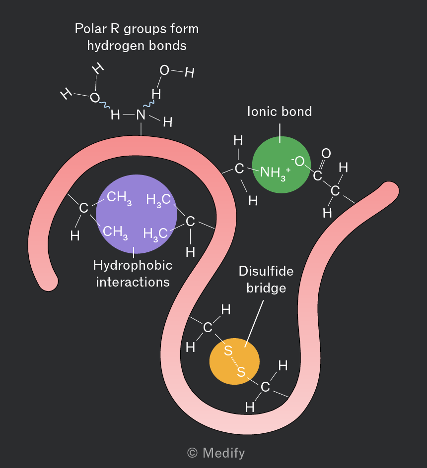
Tertiary structure is the overall three-dimensional shape of a protein, determined by interactions between the R groups of amino acids. The shape of a protein is closely related to its function.

Tertiary structure is stabilised by:

  • Hydrogen bonds between polar R groups.
  • Ionic bonds between oppositely charged R groups (impacted by ).
  • Disulfide bonds / bridges are covalent bonds between cysteine residues (impacted by ).
  • Hydrophobic interactions, where non-polar R groups cluster together away from water.
  • Hydrophilic interactions, where polar R groups are attracted to water and orientate towards the outside of the protein.
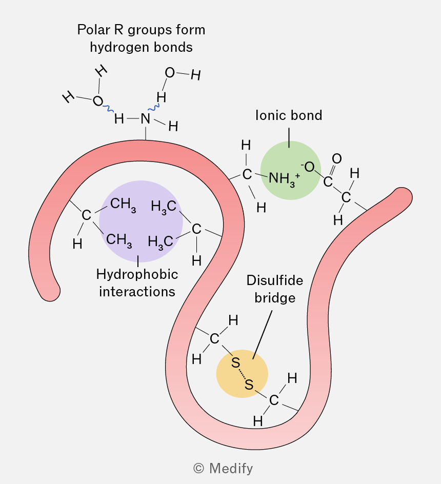
The image illustrates different types of interactions within a protein structure. At the top, there is a depiction of polar R groups forming hydrogen bonds, shown with a hydrogen bond between an OH group and an NH group. On the left, a purple circle labeled 'Hydrophobic interactions' encloses two CH3 groups bonded to a central carbon atom. In the center, a green circle labeled 'Ionic bond' shows an NH3+ group interacting with an O- group. On the right, a yellow circle labeled 'Disulfide bridge' shows two sulfur atoms (S) bonded together. The protein backbone is represented by a pink ribbon winding through these interactions.
Add to favourites

Quaternary structure occurs when two or more polypeptide chains (which may be identical or different) associate to form a functional protein.

The subunits are held together by the same interactions found in tertiary structure (hydrogen bonds, ionic bonds, disulfide bonds and hydrophobic / hydrophilic interactions), but these occur between polypeptides rather than within one polypeptide.

For example, haemoglobin consists of two -globin and two -globin subunits and is only functional when all four polypeptides are associated.

Add to favourites

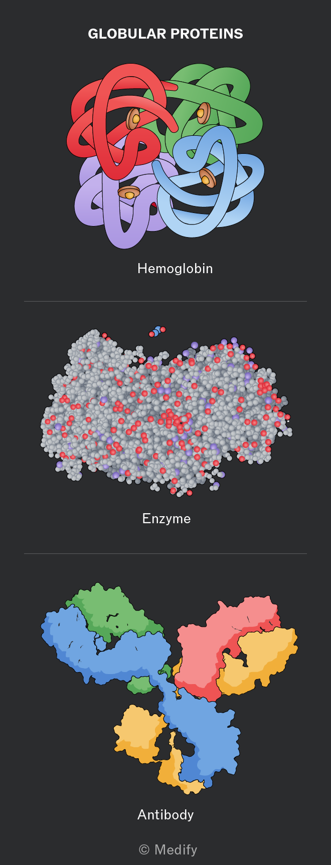
Globular proteins are compact, roughly spherical in shape, and soluble in water meaning that they can be transported in the circulatory system.

Examples include haemoglobin, enzymes and antibodies.
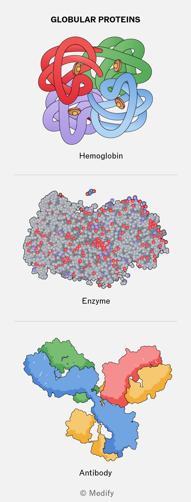
The image shows three illustrations of globular proteins. At the top, labeled 'Hemoglobin,' is a multicolored, knot-like structure with interwoven ribbons in red, green, blue, and purple, with small brown circular elements. In the middle, labeled 'Enzyme,' is a densely packed cluster of gray spheres with red and purple highlights. At the bottom, labeled 'Antibody,' is a structure with interconnected regions in blue, green, red, and yellow, forming a Y-shaped configuration. The title at the top reads 'GLOBULAR PROTEINS' and the bottom is marked © Medify.
Add to favourites

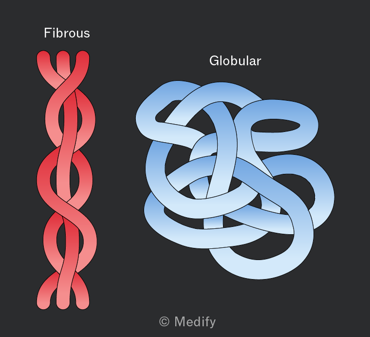
Fibrous proteins are long, insoluble polypeptide chains with structural roles, such as in keratin and collagen.

They are not folded into compact shapes and have repetitive amino acid sequences. Their strength comes from extensive hydrogen bonding and additional cross-links, such as disulfide bonds, between chains.

These proteins form strong fibres and are well suited to support and protection.
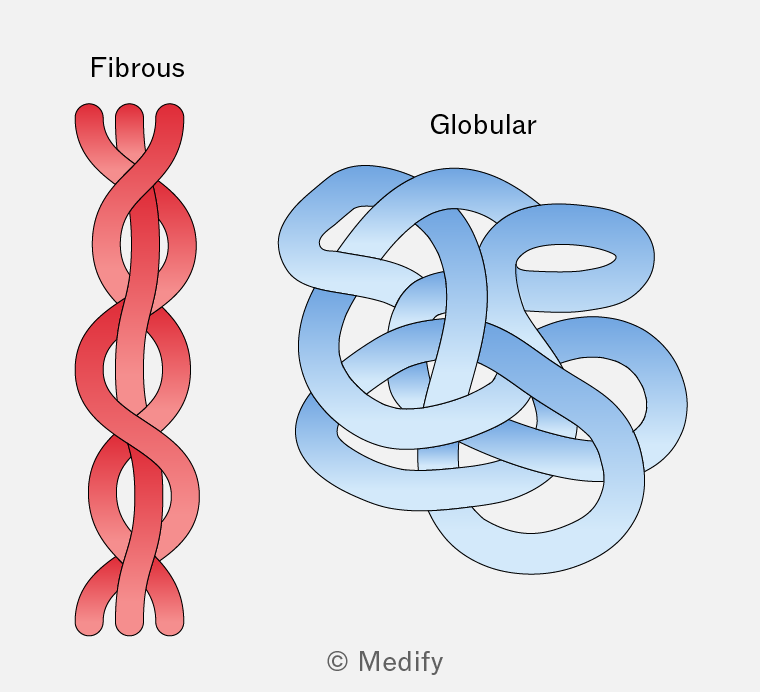
The image displays two structures labeled 'Fibrous' and 'Globular'. The fibrous structure on the left is shown as a red, twisted, rope-like form with long, intertwined strands. The globular structure on the right is depicted as a complex, blue, tangled mass with loops and curves, resembling a knotted ball. Both structures are visually distinct, with fibrous being linear and elongated, while globular is compact and spherical. The credit '© Medify' is at the bottom center of the image.
Add to favourites

Collagen is a fibrous protein found in connective tissues such as skin, tendons, ligaments, cartilage and the nervous system.

It consists of three polypeptide chains wound together in a triple helix, like a rope, making it strong yet flexible. The chains are held together by hydrogen bonds and covalent cross-links, and are staggered to avoid weak points.

Collagen molecules form fibrils and fibres and are insoluble in water.

Add to favourites