
Cell division, cell diversity and cellular organisation (2.1.6)
On this page
Not all eukaryotic cells retain the ability to divide, but those that can undergo the cell cycle, a series of stages involving growth and division. The outcome is two genetically identical daughter cells.
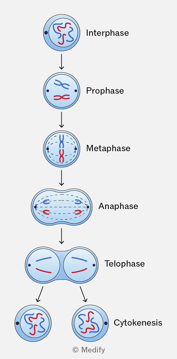
The cycle consists of interphase, followed by mitosis and cytokinesis (cytoplasmic division).
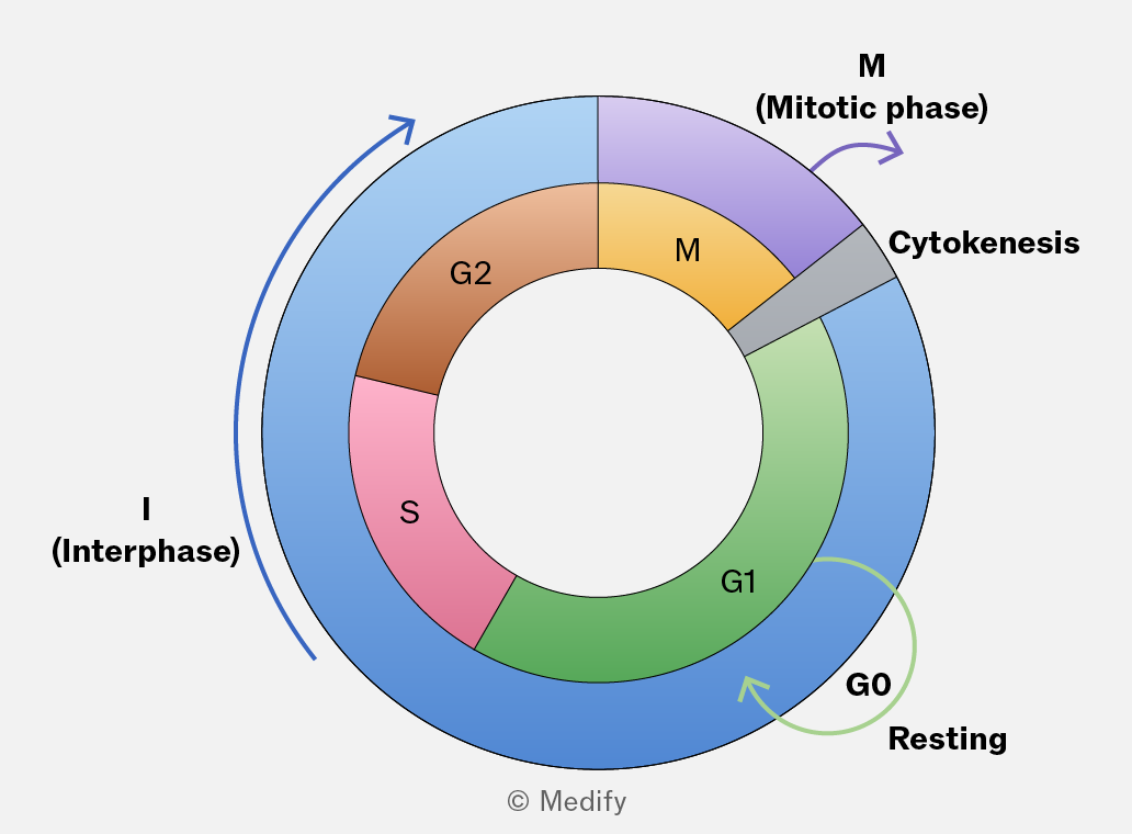
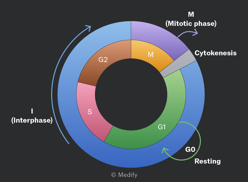
Interphase is the stage during which the cell grows and prepares for division. It occupies approximately 90 of the cell cycle and consists of three phases:
- G1 (first growth)
- S (DNA synthesis)
- G2 (second growth).
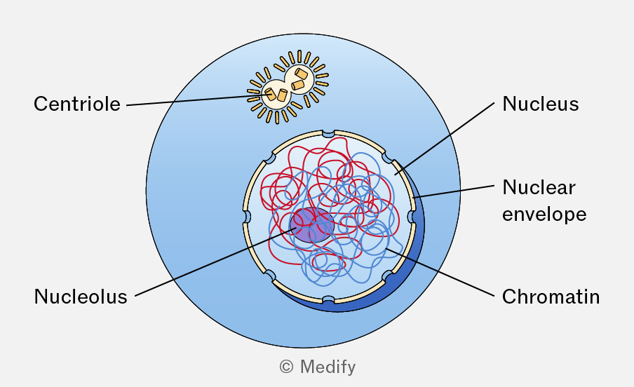
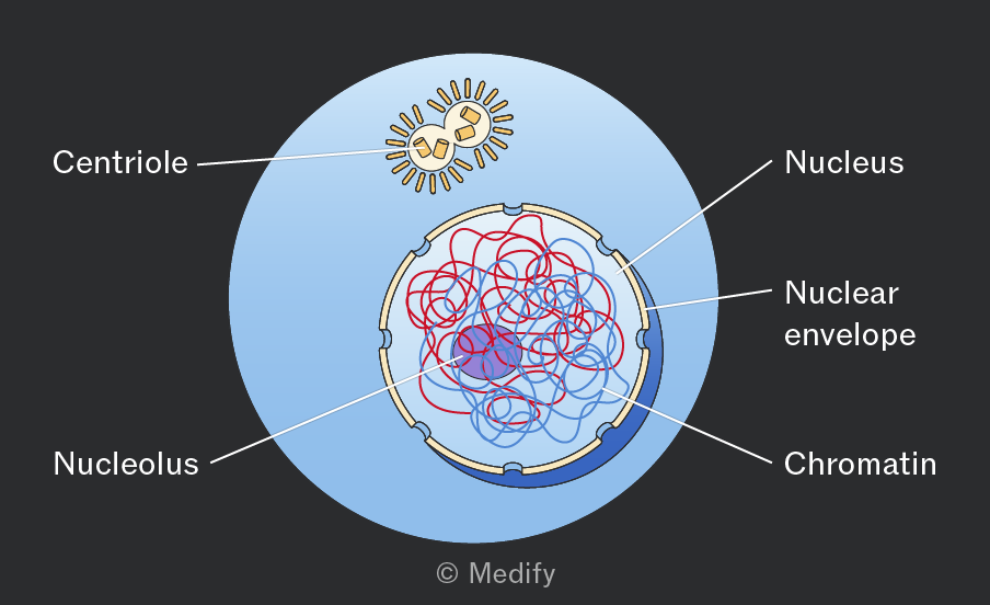
In the G1 Phase (first growth phase), the cell prepares for division, grows in size and synthesises proteins and organelles while carrying out its normal metabolic functions.
DNA replication occurs during the S Phase (synthesis), resulting in each chromosome consisting of two identical sister chromatids. This means that the DNA content of the cell doubles, though the number of chromosomes remains the same. The two daughter cells will each receive an equal amount of DNA, identical to the original cell.
Centrioles (centrosomes), which will organise the mitotic spindle, also duplicate during this phase.
During the G2 Phase (second growth phase), the cell continues to grow and builds energy stores to ensure it is ready to enter mitosis. The cell’s DNA is also checked for errors.
Synthesis of microtubules and other proteins necessary for spindle formation occurs.
Mitosis is the nuclear division process during which a eukaryotic cell separates its duplicated chromosomes into two identical nuclei. It consists of prophase, metaphase, anaphase and telophase and is followed by cytokinesis.
Cytokinesis is the division of the cytoplasm following nuclear division, resulting in the formation of two daughter cells.
Checkpoints control the cell cycle:
- G1 checkpoint: determines whether conditions such as cell size and nutrient levels are suitable for entry into the S phase or whether the cell exits the cycle into G0 (resting).
- G2 checkpoint: ensures that all DNA is replicated correctly and that the cell is ready to divide.
- Metaphase checkpoint: makes sure that all chromosomes have been attached to spindle fibres at the equator of the cell.
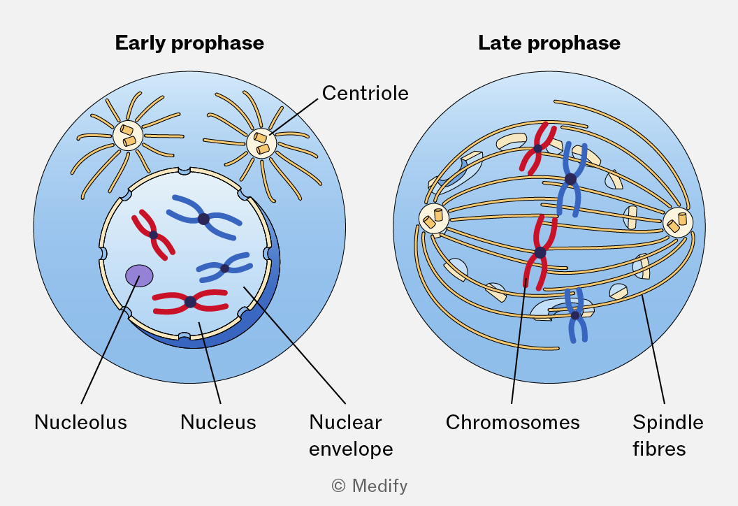
Prophase is the first stage of mitosis. Chromatin condenses into chromosomes, each consisting of two sister chromatids joined at the centromere.
The nuclear envelope breaks down and the nucleolus disappears.
Centrosomes (centrioles) move to opposite poles of the cell, forming spindle fibres that guide chromosome movement.
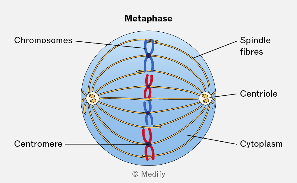
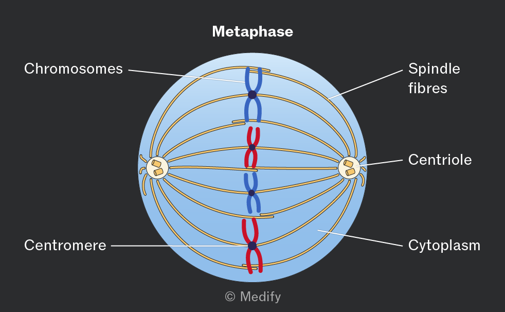
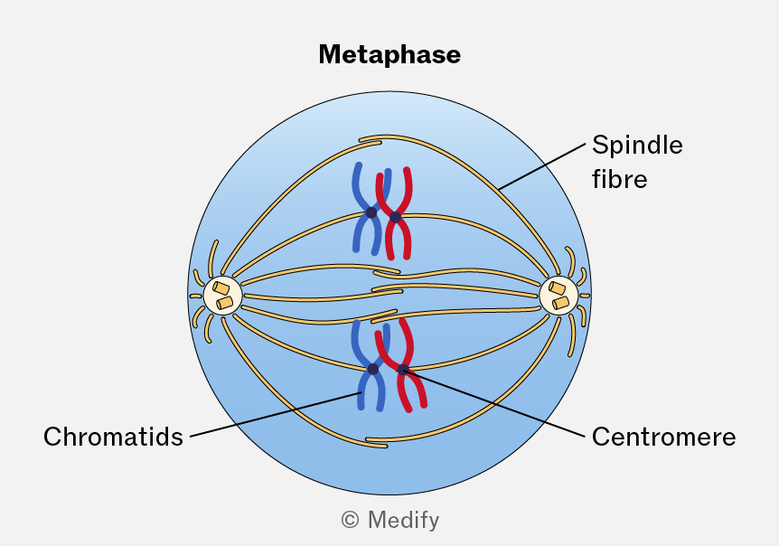
During metaphase, chromosomes align at the cell equator, also called the metaphase plate.
Spindle fibres from opposite poles attach to each centromere.
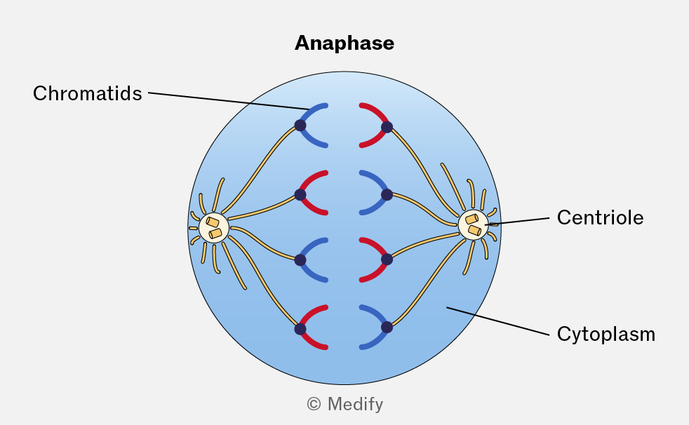
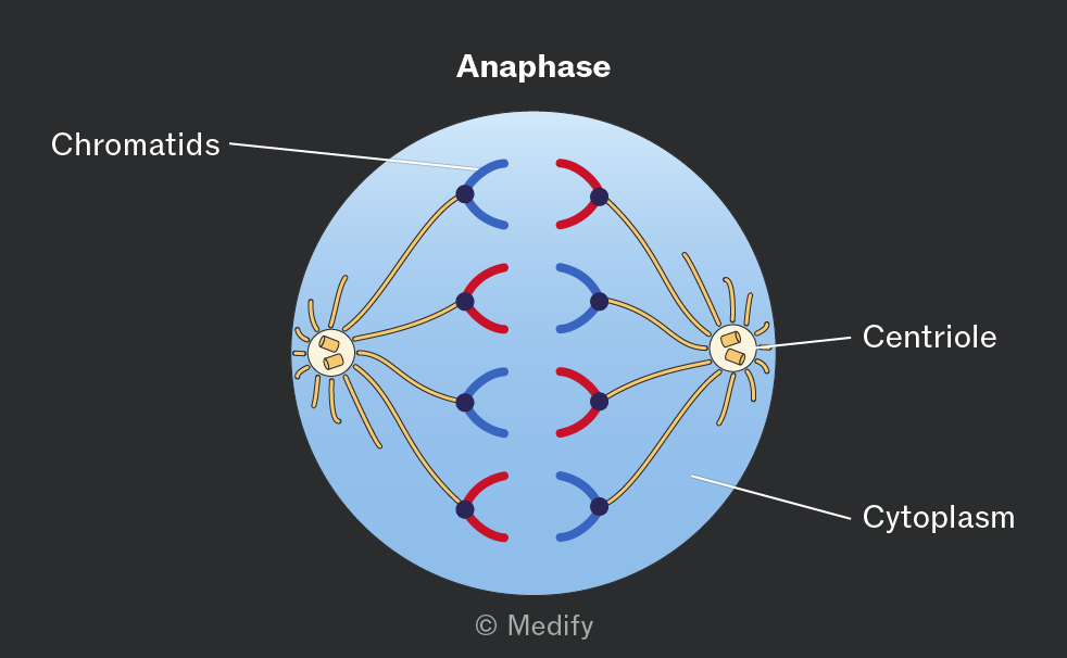
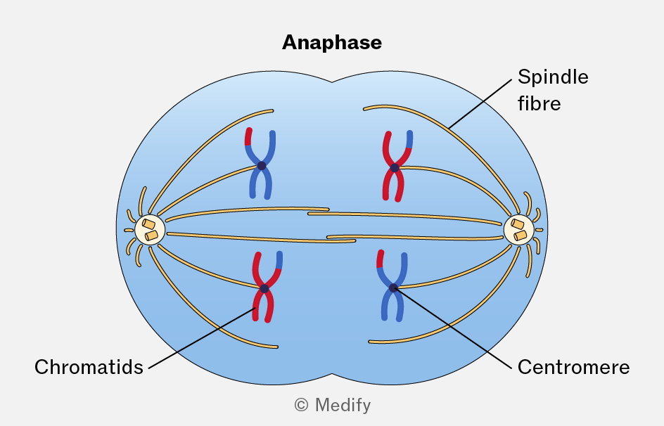
During the anaphase, the centromeres divide, allowing sister chromatids to separate.
The spindle fibres shorten, moving the chromatids towards opposite poles of the cell.
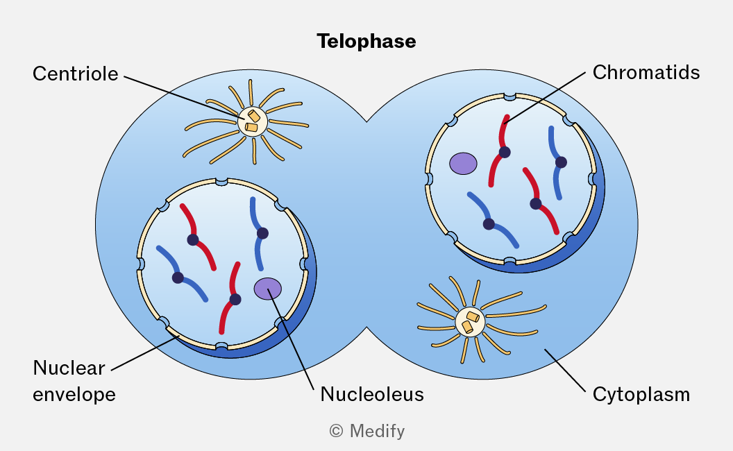
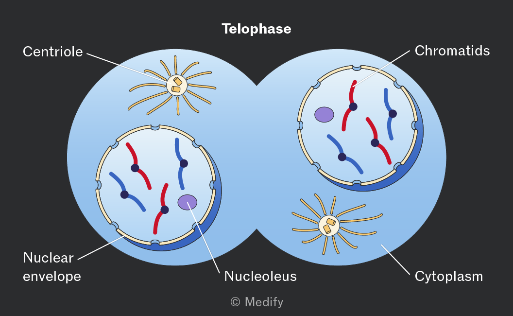
During telophase, the chromatids are at the cell’s poles and are now chromosomes. The nuclear envelope reforms around each set of chromosomes, creating two nuclei.
The chromosomes uncoil back into chromatin, and the nucleolus is formed.
The mitotic spindle breaks down as the cell prepares for cytokinesis.
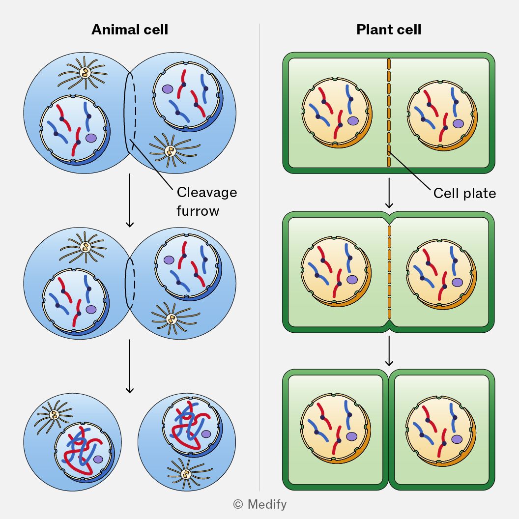
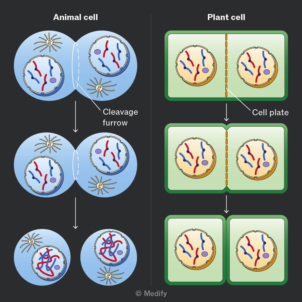
Cytokinesis is the final stage of the cell cycle in which the cytoplasm, cell membrane, and organelles divide, resulting in two daughter cells that are genetically identical.
- In animal cells, the plasma membrane folds, pinching the cell membrane inwards, forming a cleavage furrow, until the membrane meets in the middle and the cell is split into two.
- In plant cells, vesicles migrate to the equator of the cell, and fuse with each other and the cell membrane, dividing the cell in two. New sections of cell walls then form.
Mitosis is the process by which a cell divides to produce two genetically identical daughter cells.
This process is vital for asexual reproduction, growth and repair of tissue in plants, fungi and animals.
Meiosis is a type of cell division that produces four genetically distinct gametes (sperm and egg cells) and generates genetic variation through independent assortment of chromosomes and crossing over of alleles between chromatids.
It reduces the chromosome number from diploid to haploid.
Meiosis involves two sequential divisions:
- Meiosis I – separates homologous chromosomes.
- Meiosis II – separates sister chromatids.
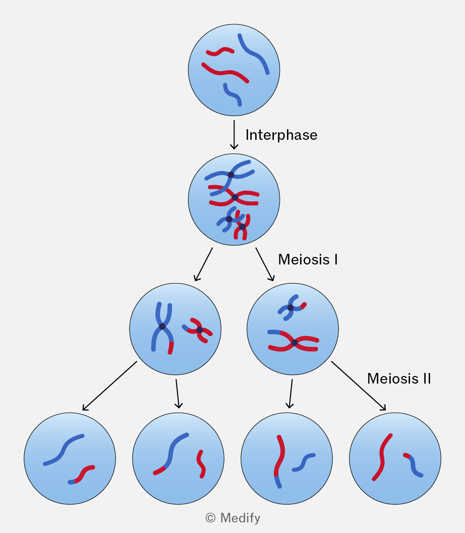
During interphase before meiosis, DNA is replicated during the S phase. Each chromosome is duplicated and consists of two genetically identical sister chromatids joined at the centromere.
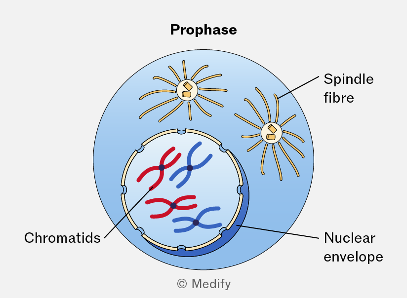
During prophase I of meiosis:
- Chromosomes condense and become visible.
- Homologous chromosomes pair to form bivalents.
- Crossing over occurs between non-sister chromatids of homologous chromosomes at chiasmata, exchanging sections of DNA and generating genetic variation.
- The nuclear envelope breaks down and spindle fibres form.
During metaphase I of meiosis:
- Bivalents line up along the cell equator.
- Each homologous pair aligns independently of other pairs, leading to independent assortment of maternal and paternal chromosomes, generating genetic variation.
During anaphase I of meiosis:
- Homologous chromosomes are pulled to opposite poles by spindle fibres.
- Sister chromatids remain joined at their centromeres.
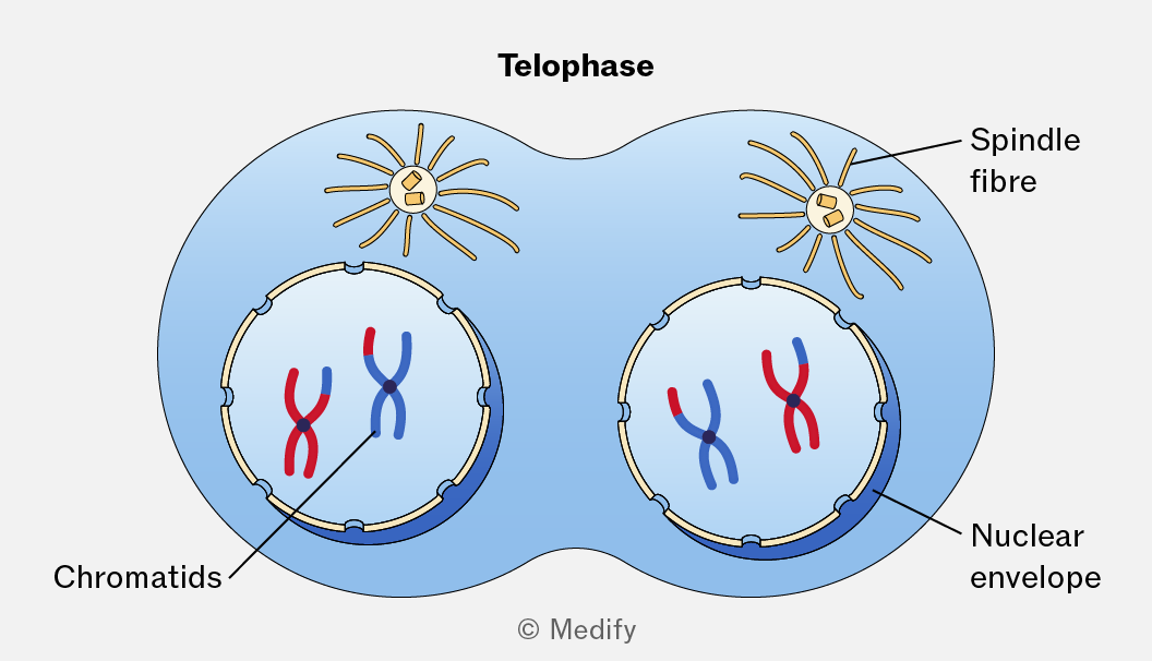
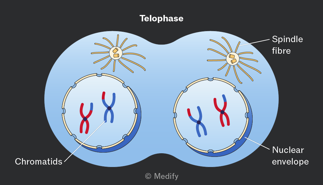
During telophase I of meiosis:
- Nuclear envelopes may reform around each set of chromosomes (depending on the species).
- The cytoplasm divides to form two haploid cells.
- Each chromosome still consists of two sister chromatids joined at the centromere.
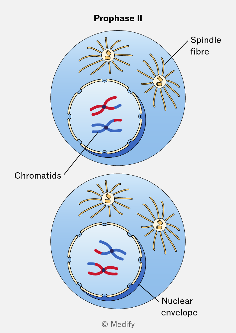
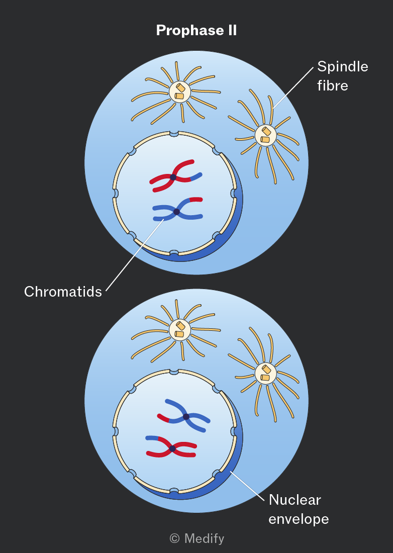
During prophase II of meiosis:
- Chromosomes condense (if they had decondensed after meiosis I).
- Spindle fibres form.
- The nuclear envelope breaks down.
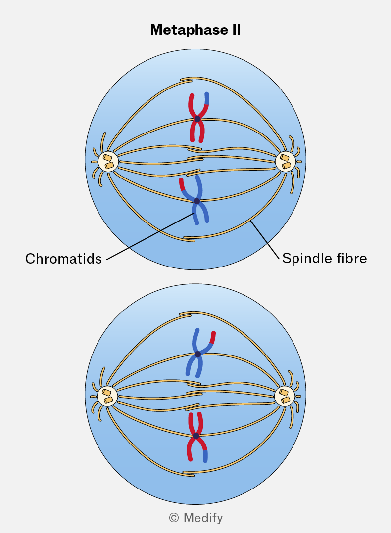
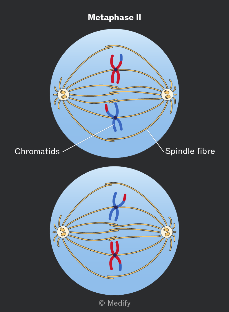
During metaphase II of meiosis:
- Chromosomes line up individually along the equator of the cell.
- Spindle fibres from opposite poles attach to the centromeres.
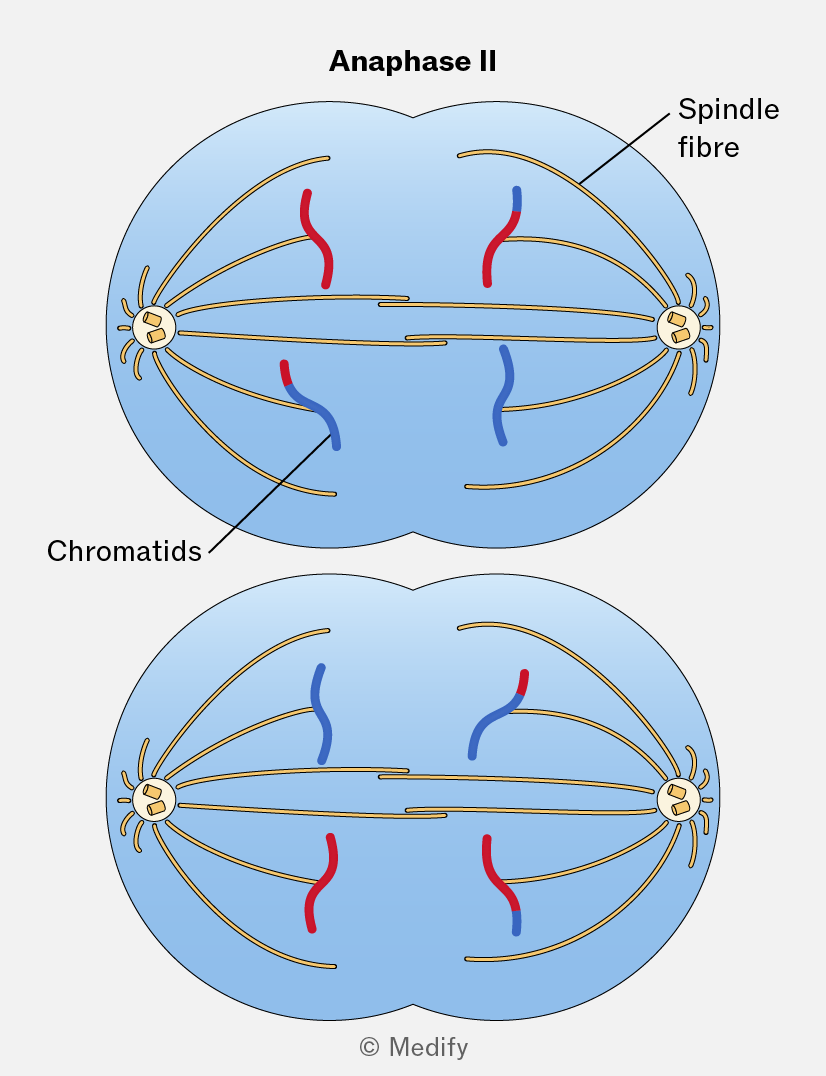
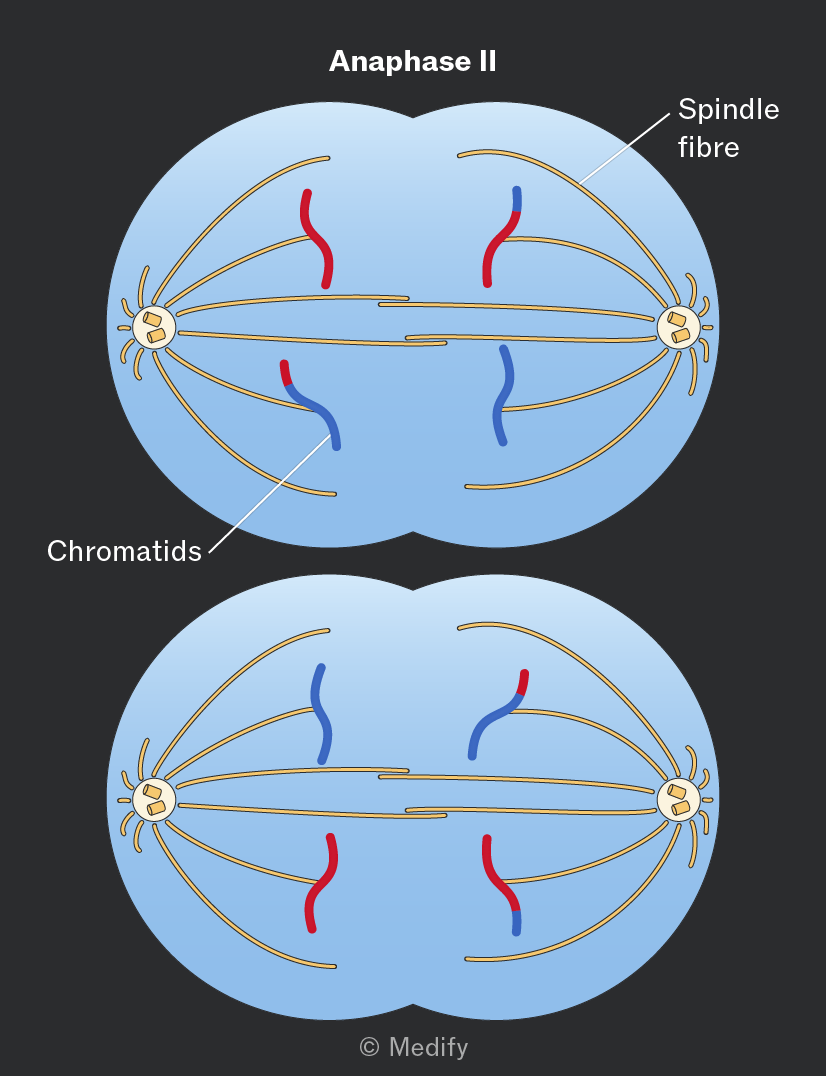
During anaphase II of meiosis:
- The centromeres divide.
- Sister chromatids separate and are pulled to opposite poles by the spindle fibres.
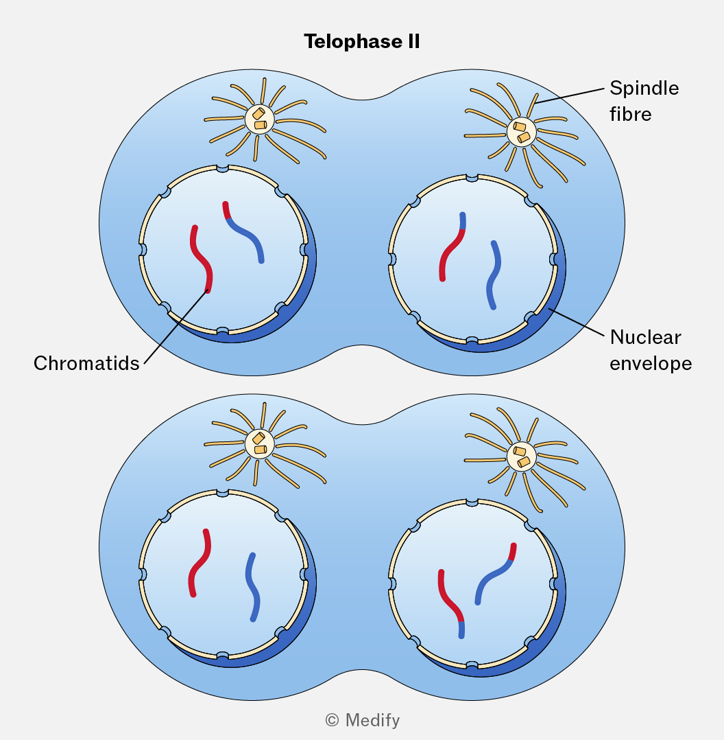
During telophase II of meiosis:
- Nuclear envelopes reform around each set of chromosomes.
- The cytoplasm divides (cytokinesis) resulting in four haploid daughter cells, each genetically unique.
When cells differentiate, they become specialised and adapted for specific functions.
Examples of specialised animal cells include:
- Spermatozoa / sperm cells: contain many mitochondria to provide ATP for movement. They are long and streamlined with a flagellum for swimming. The acrosome contains enzymes that digest the outer layers of the ovum. The head contains a haploid nucleus and very little cytoplasm.
- Squamous epithelial cells: are thin and flattened cells providing a short diffusion pathway and a smooth surface for fluids to move more easily across.
- Ciliated epithelial cells: possess cilia that move substances across a cell surface. They contain many mitochondria to supply ATP for ciliary movement.
Examples of specialised plant cells include:
- Palisade mesophyll cells: contain chloroplasts to maximise the rate of photosynthesis. Their elongated shape allows them to pack closely near the leaf surface, maximising light absorption.
- Root hair cells: have long, hair-like extensions that increase the surface area available for water and mineral ion absorption. They contain many mitochondria to provide ATP for the active transport of ions from the soil.
- Guard cells: are specialised cells that have unevenly thickened cell walls, with a thickened, less-elastic wall next to the stomata. Guard cells contain chloroplasts and use ATP to actively transport potassium ions into and out of the cells. This changes their water potential, causing water to enter or leave by osmosis. The resultant turgidity or flaccidity controls the opening and closing of the stomata.
Multicellular organisms are organised hierarchically, with increasing levels of complexity at each level.
Cells are the basic structural and functional units of life. Cells are specialised for particular functions. Examples include muscle cells and nerve cells in animals, and palisade mesophyll cells in plants.
A tissue is a group of similar cells working together to perform a specific function.
Tissue examples include:
- Squamous epithelium: thin, flat cells for diffusion (e.g. lining of lungs).
- Ciliated epithelium: cells with cilia to move substances (e.g. trachea).
- Muscle tissue: contracts for movement.
- Cartilage: provides support and flexibility.
- Xylem: transports water and minerals in plants.
- Phloem: transports sugars in plants.
An organ is made of different tissues working together to perform a specific function.
Organ examples include:
- Heart: composed of muscle, nerve, and connective tissue.
- Leaf: composed of palisade, spongy mesophyll, xylem and phloem tissue.
An organ system is a group of organs working together to perform major body functions.
Organ system examples include:
- digestive system
- circulatory system
- respiratory system.
All organ systems together form a complete organism, such as a human.
Stem cells are undifferentiated cells that can divide by mitosis to produce more stem cells.
Alternatively, they can differentiate into specialised cell types that make up tissues and organs.
There are several sources of stem cells:
- Embryonic stem cells, obtained from an early embryo.
- Umbilical cord blood.
- Adult stem cells- found in certain tissues such as bone marrow.
- Induced pluripotent stem cells (iPS cells) — cells developed in a lab to behave like embryonic stem cells.
Stem cells in bone marrow can differentiate into specialised cells. Examples include:
- Erythrocytes (red blood cells): specialised to transport oxygen. They have a large surface area to volume ratio for rapid gas diffusion, are flexible, so can travel easily through capillaries and contain fewer organelles (including lacking a nucleus), so there is more space for haemoglobin.
- Neutrophils: specialised to engulf and destroy pathogens. Larger than erythrocytes, they move towards sites of infection via chemotaxis.
Although both cells come from the same stem cell, different genes are expressed during differentiation, causing the cells to develop different structures and functions.
Plant growth occurs in specialised regions of active cell division called meristems. Cells produced by meristems differentiate to form specialised tissues such as:
- Xylem vessels: form when lignin is deposited in cell walls (killing the cell), and the end of the cells break down to form continuous tubes.
- Phloem sieve tube elements: lose their organelles and form sieve plates; the companion cells retain their organelles to be able to provide ATP to the sieve tube elements.
Stem cells can be used to repair or replace damaged tissues. For example, stem cells from bone marrow can be transplanted to treat blood disorders such as leukaemia.
Stem cells also offer potential treatments for neurological conditions and have uses in developmental biology research.