
Atomic structure (3.1.1)
On this page
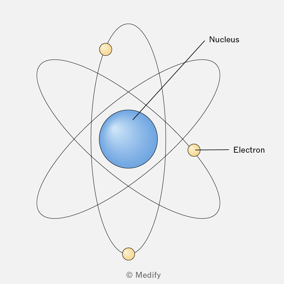
The structure of the atom:
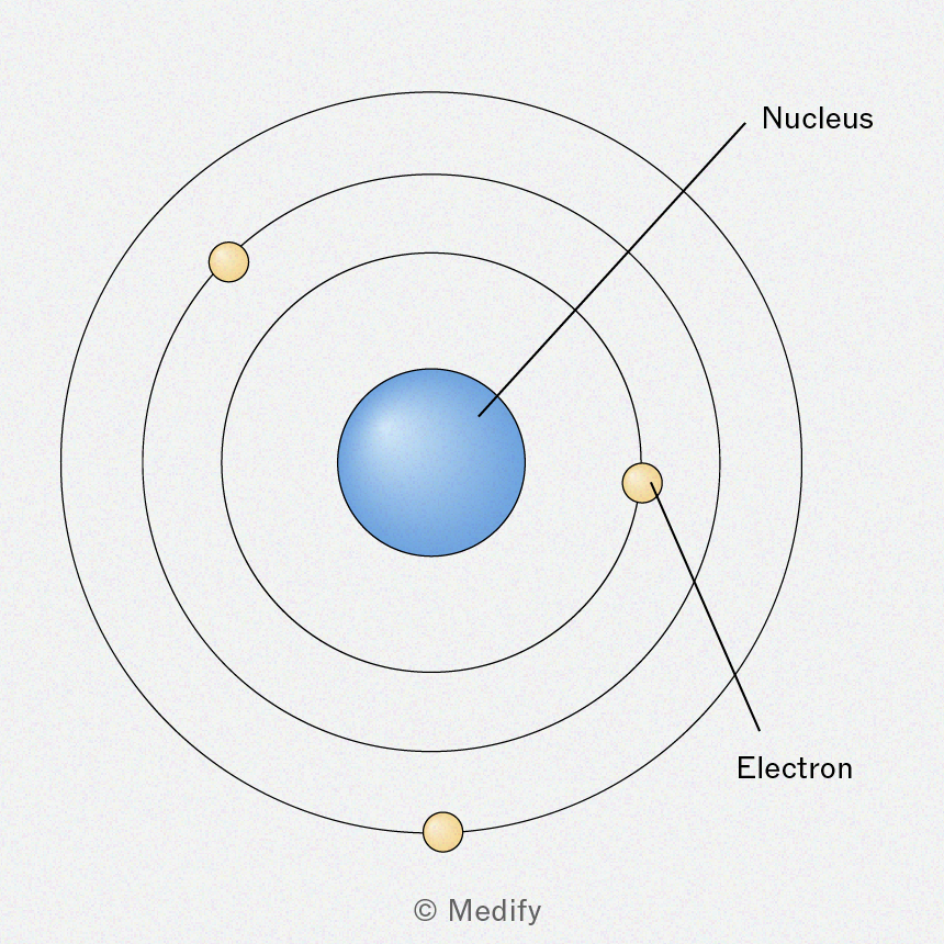
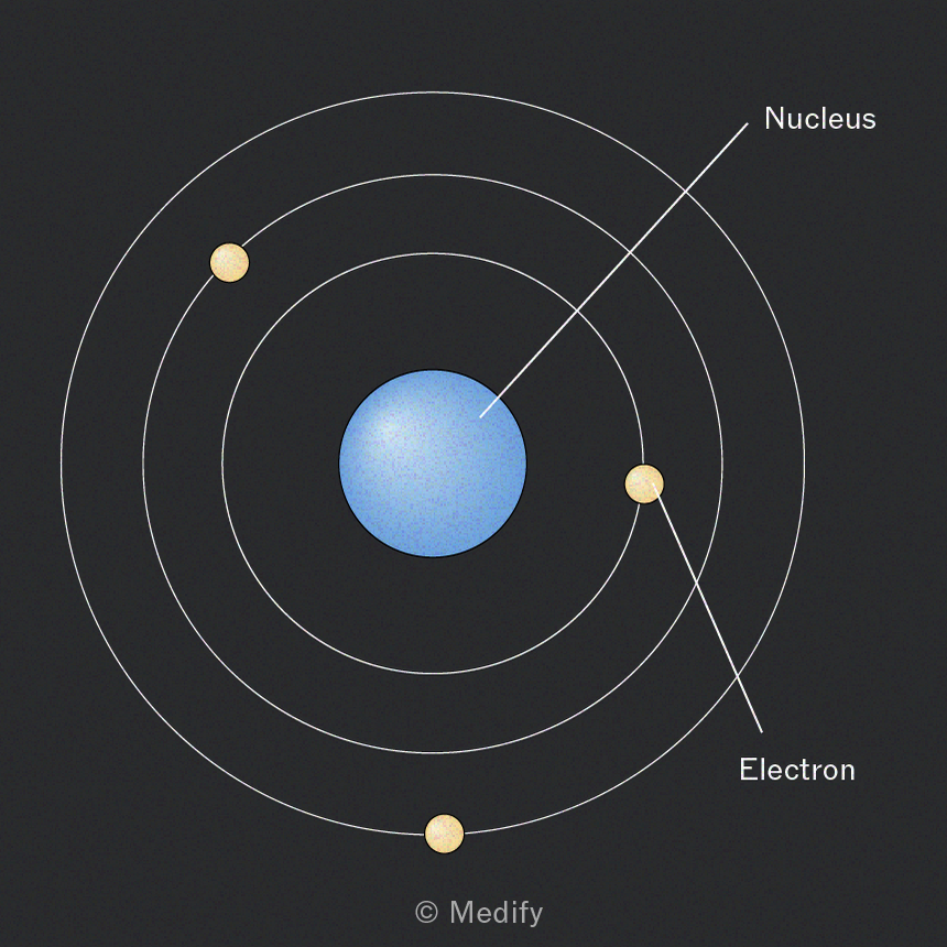
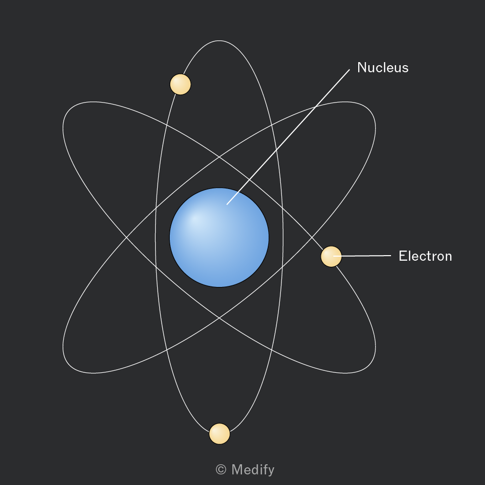
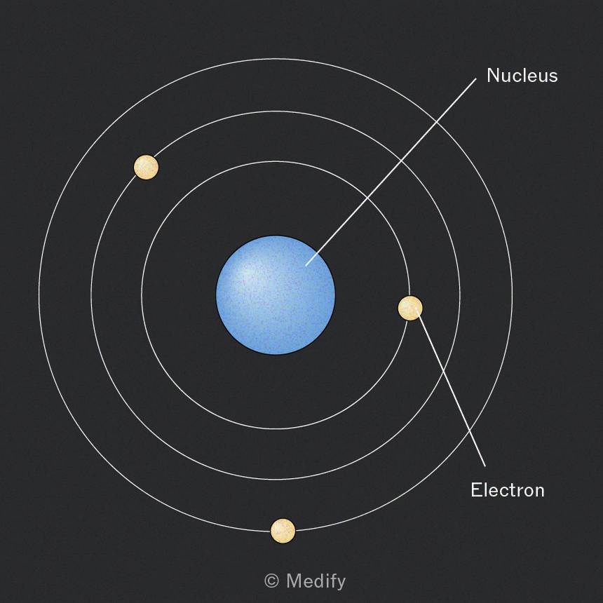
The nucleus contains the protons and neutrons.
The nucleus is surrounded by shells (orbitals) which contain the electrons.
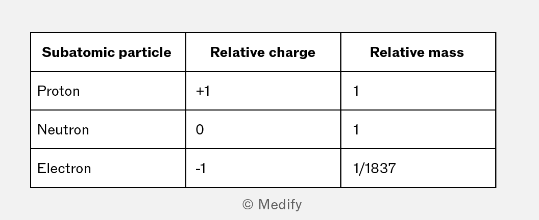
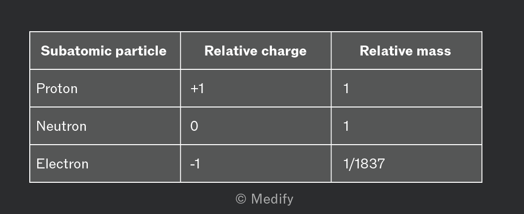
Subatomic particles can be described by their relative charge and relative mass.
Dalton (1800s):
- discovered that atoms can be combined to make compounds
- believed atoms cannot be broken into smaller parts
- disproved by J.J. Thomson’s cathode ray tube experiments.
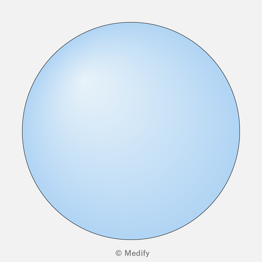

Thomson (1897):
- discovered the electron
- believed that atoms were a sphere of positive mass embedded with electrons: the plum pudding model
- was disproved by Rutherford’s gold foil experiment.
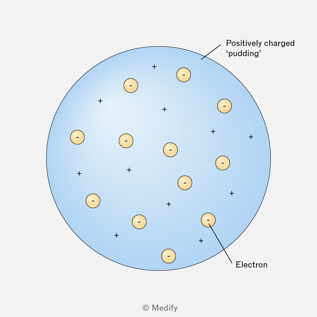
Rutherford (1905):
- discovered the nucleus with his gold foil experiment.
Rutherford’s gold foil experiment:
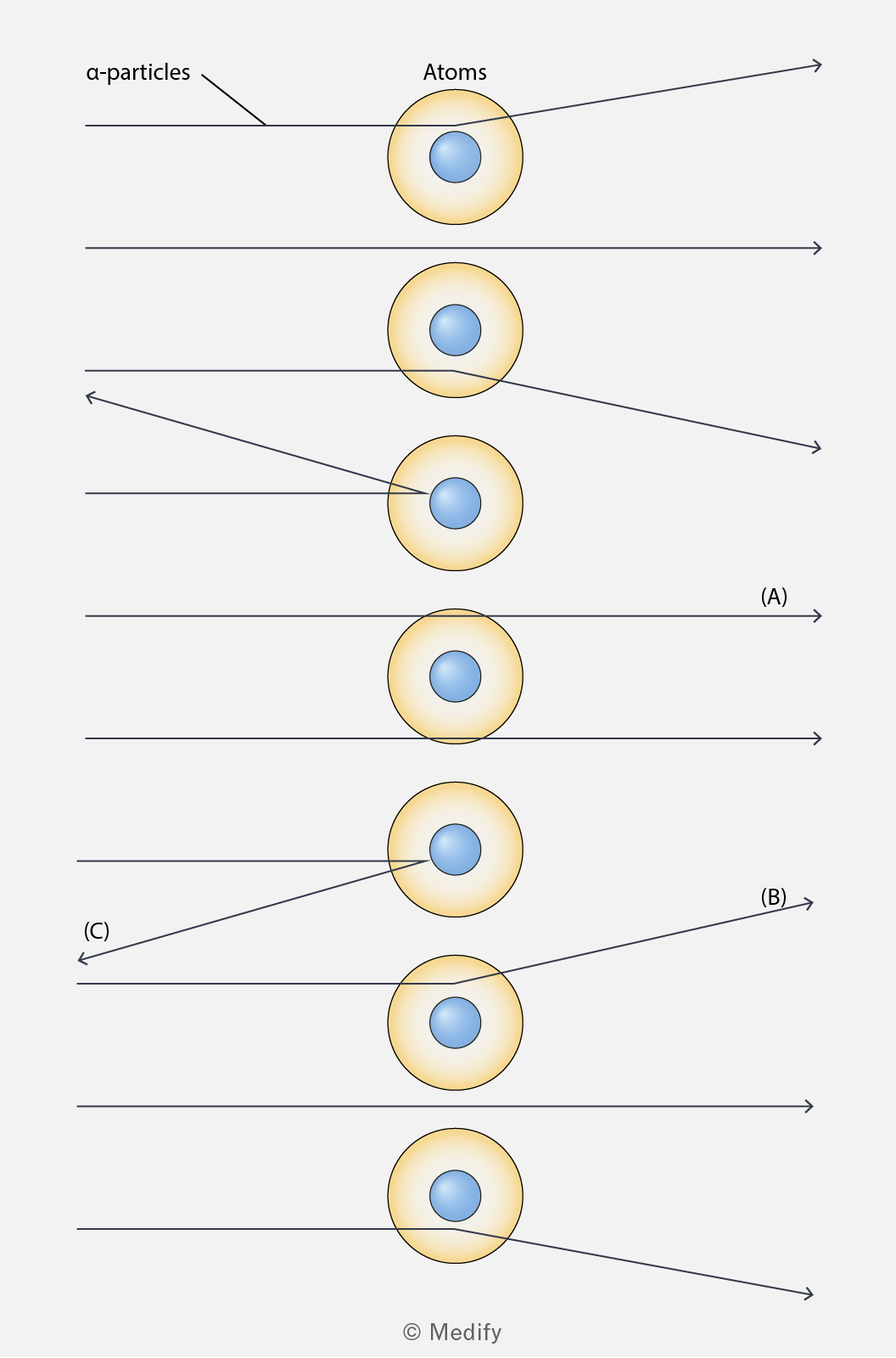
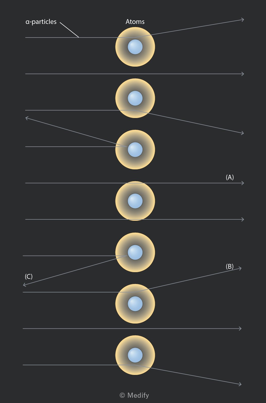
- A very thin piece of gold foil was bombarded with alpha particles.
- Most particles went straight through (A).
- Some were deflected (B) and a few were reflected (C).
- This proved that the vast majority of the mass of an atom is concentrated in a small central volume and that the central mass was positively charged.
Bohr (1913):
- discovered energy levels
- related atomic spectra to electronic energy levels.
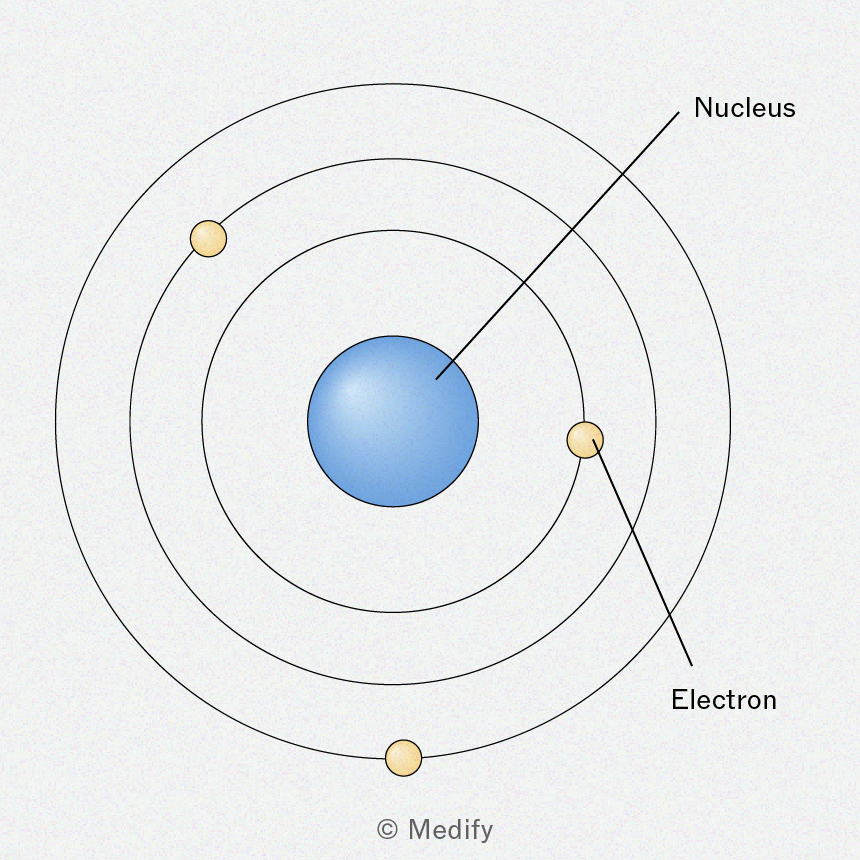
The atomic number represents the number of protons in the nucleus of an atom.
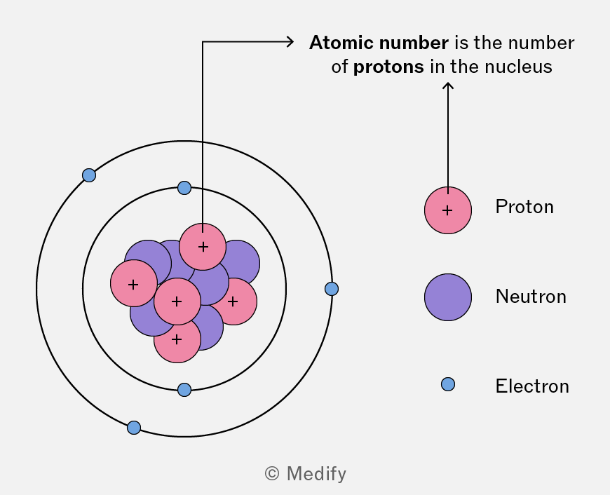
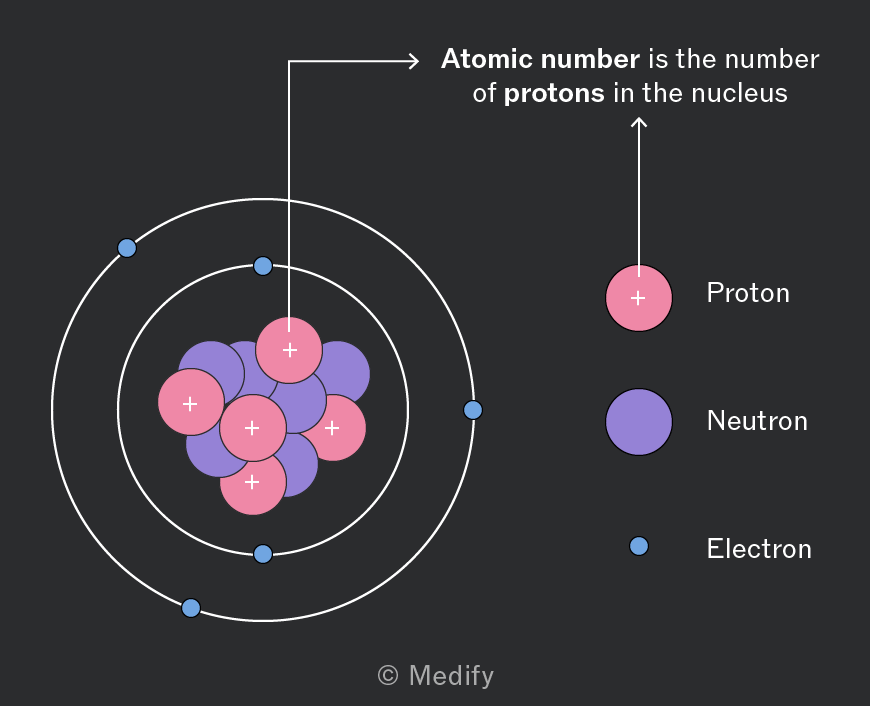
It is a unique identifier for each element and is a fundamental factor in the organisation of the periodic table.
Elements are arranged in increasing order of atomic number from left to right.
In a neutral atom, the number of protons is the same as the number of electrons.
Atomic number () = number of protons
Mass number () = number of neutrons + the atomic number
Ions are particles with a charge.
In an ion, the number of electrons = number of protons − the charge.
- The number of electrons in a
- The number of electrons in a
The number of neutrons = mass number − the atomic number.
Isotopes are atoms of the same element with different numbers of neutrons and different masses.
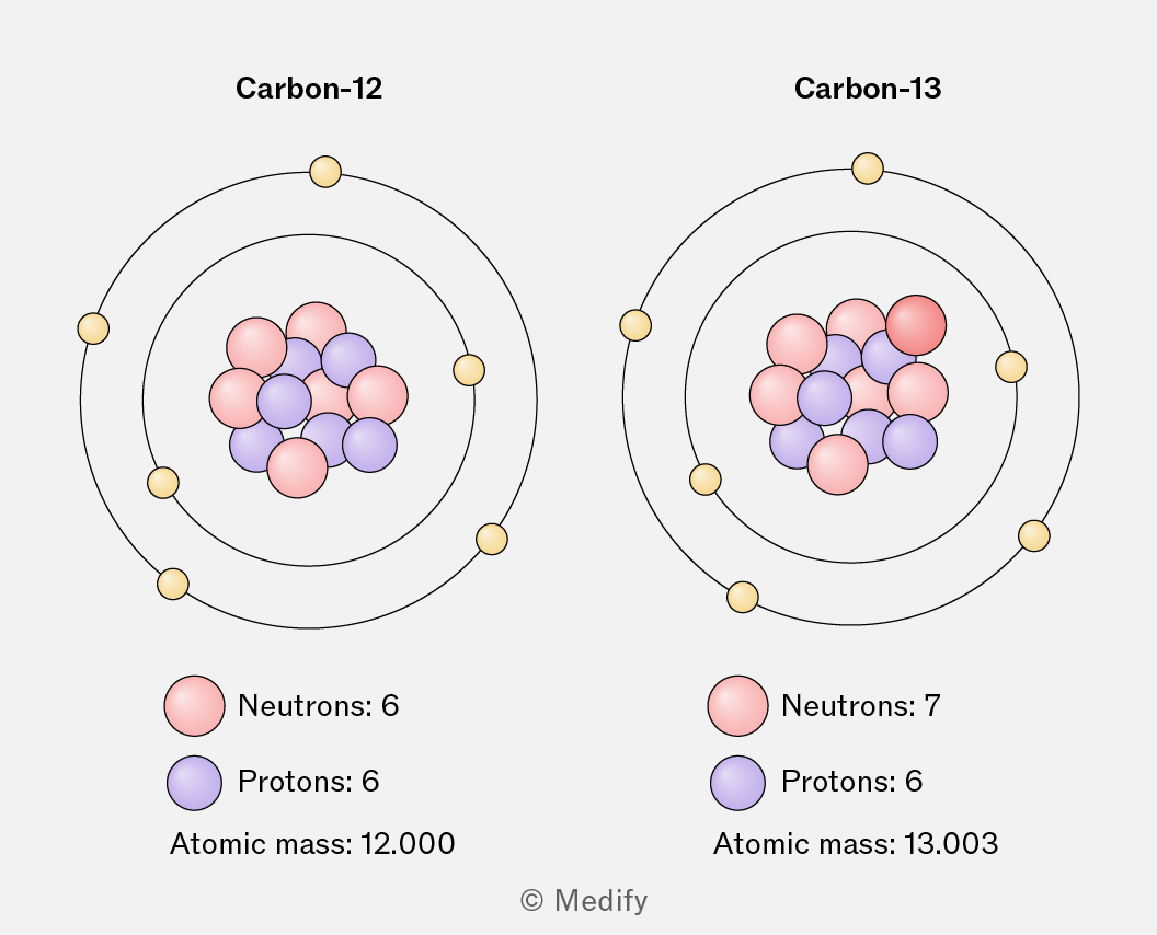
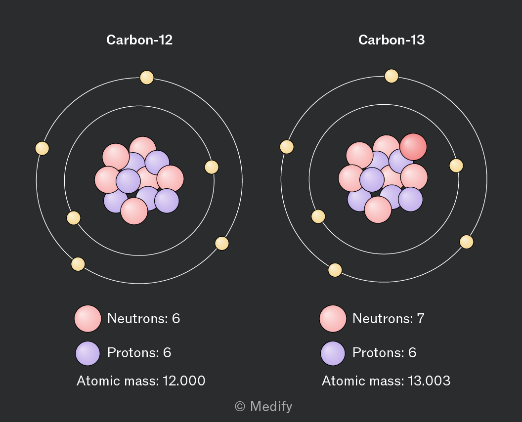
Carbon-12 and carbon-13 are isotopes of carbon.
- Both identify as carbon, with an atomic number of six.
- Carbon-12 has six neutrons and a relative atomic mass of 12 whereas carbon-13 has seven neutrons and a relative atomic mass of 13.
Relative atomic mass: weighted mean mass of all isotopes in an element, compared with the mass of carbon-12.
It is determined using the following equation:
The term relative formula mass (RFM) is used for compounds with giant structures like ionic compounds or giant covalent lattices.
The RFM and (relative molecular mass) can be found from the sum of the relative atomic masses, .
Taking as an example:
- Sodium has
- Sulfur has
- Oxygen has
RFM
A simple time of flight (TOF) mass spectrometer is used to identify the mass of ions.
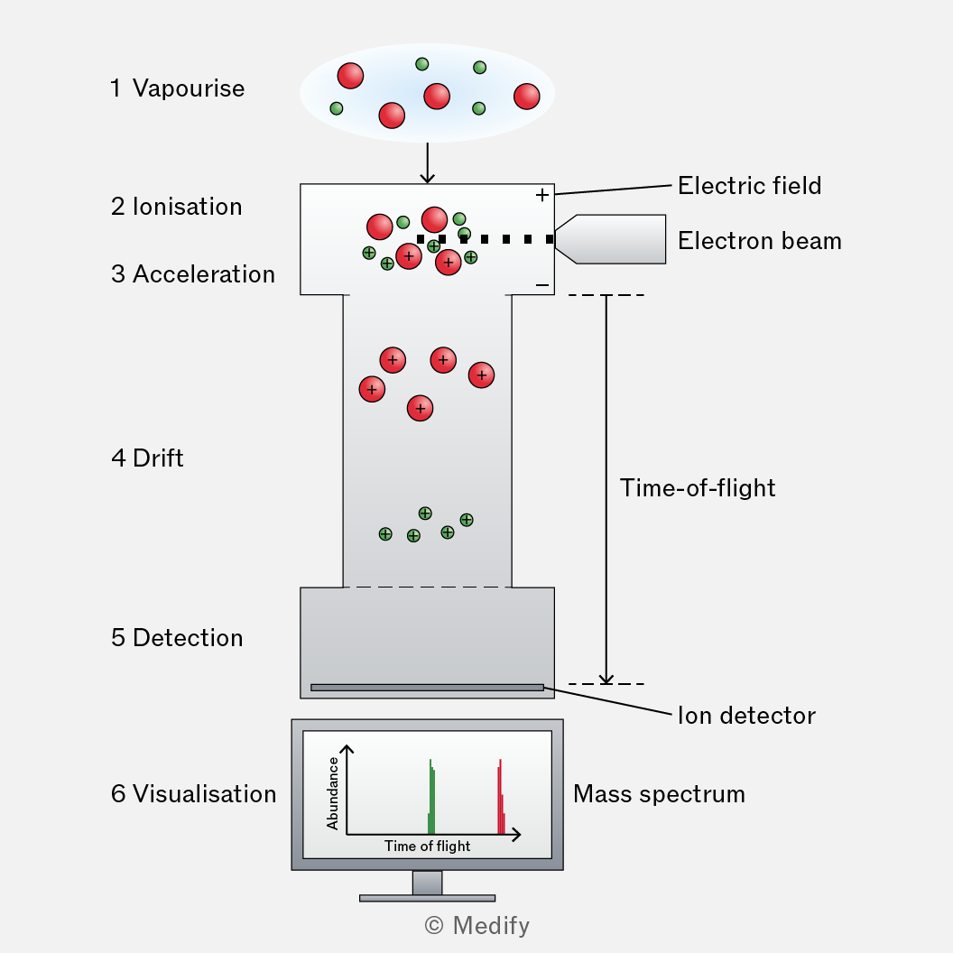
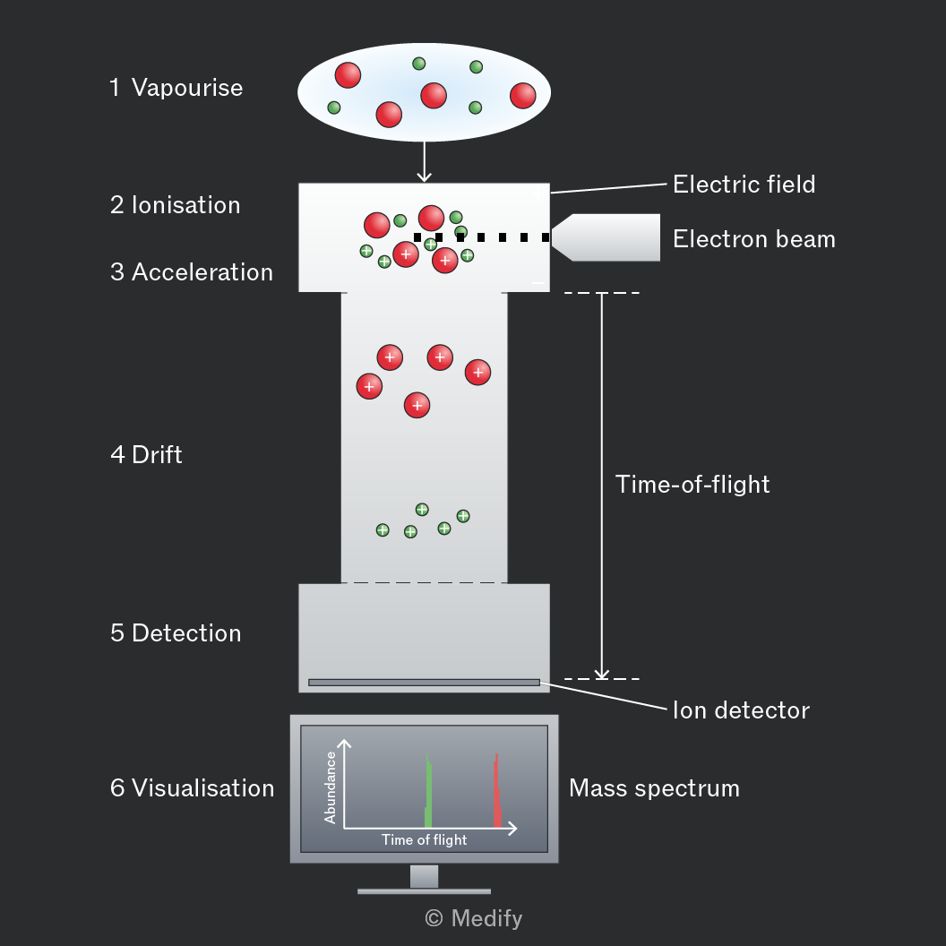
- A sample is vapourised.
- The vapourised sample is bombarded with high speed electrons that knock out an electron in the molecule, causing its ionisation.
- An electric field is applied, which accelerates each ion toward a drift tube.
- The ions all drift with the same kinetic energy, but their velocities differ depending on their mass-to-charge ratios (m/z). Ions with a smaller m/z travel faster and reach the detector first.
- The mass spectrometer detects the ions’ time of flight.
- An electric current proportional to the relative abundance of ions in the beam produces a peak on the mass spectrum at the time it was detected.
The raw data for a mass spectrum is the signal intensity paired with the time of flight.
The data is processed using information from calibration.
Signal intensity can be converted to relative abundance. This sits on the axis.
The conversion is not always normalised, (made so the peaks sum to ), so it is important to check.
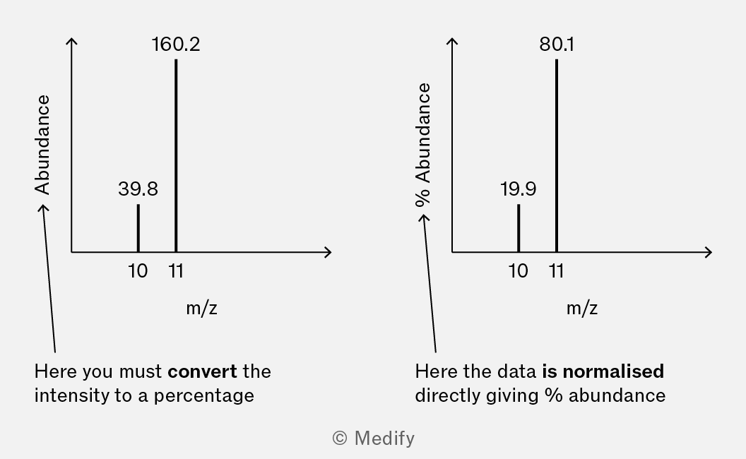
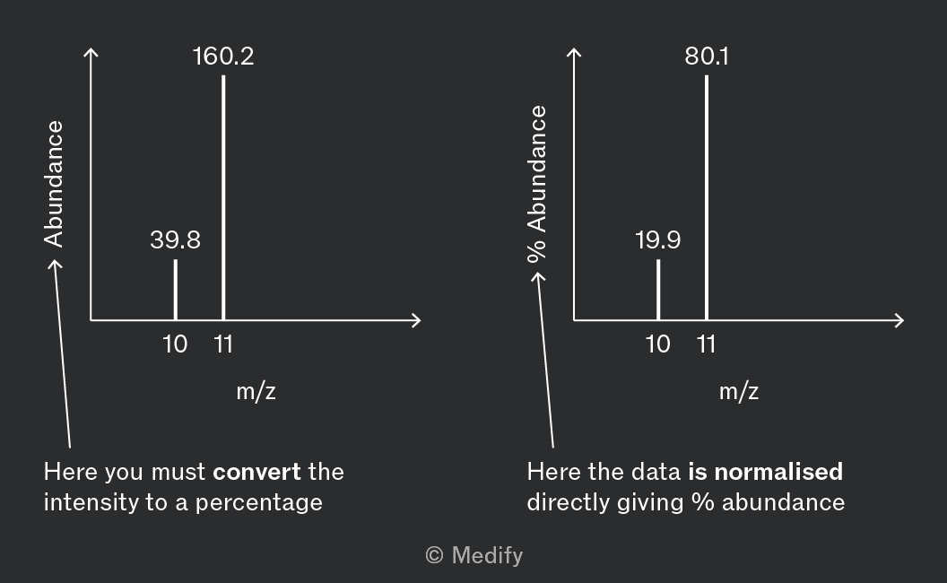
Time of flight is converted to mass to charge ratio (m/z). This sits on the axis.
The mass spectrum below shows the relative abundance of the two isotopes of boron.
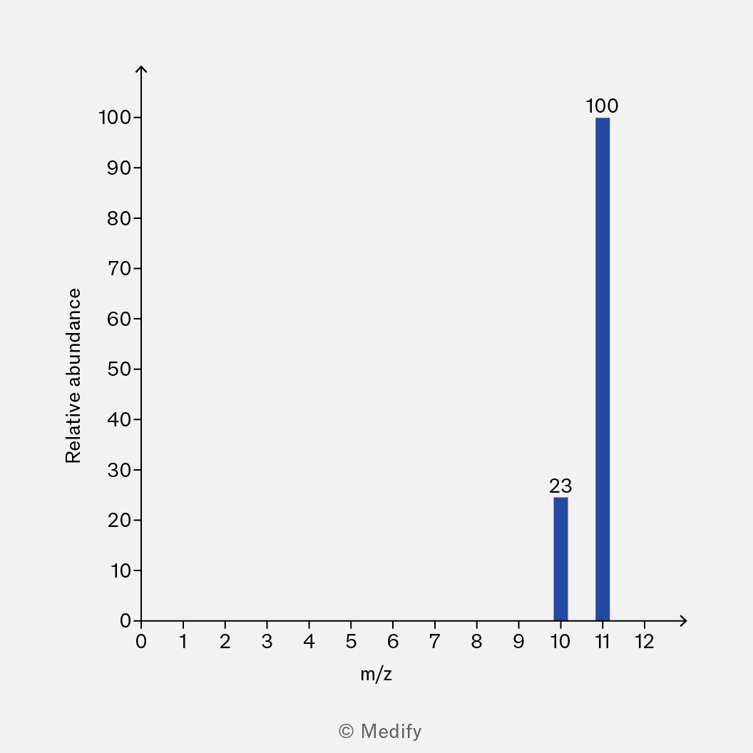
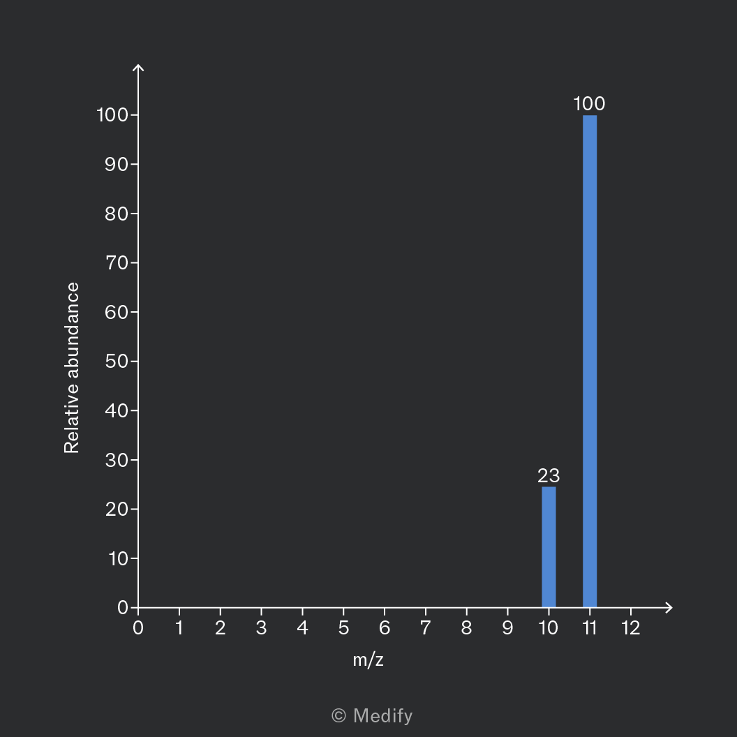
Isotope 1: m/z , relative abundance
Isotope 2: m/z , relative abundance
The relative atomic mass of boron can be calculated from this data:
Organic molecules in a mass spectrometer are vapourised then undergo ionisation, with the loss of an electron from one of the covalent bonds.
This can be represented as:
is the molecular ion, it represents the whole molecule minus one electron.
The singly charged molecular ion peak, , has an m/z equal to the molecule’s relative molecular mass ().
The molecular ion peak has the highest m/z value and is located on the far right of the mass spectrum.
Mass spectra of organic molecules will exhibit a tiny peak at 1 mass unit greater than the molecular ion.
This is called the M+1 peak and is caused by the presence of the carbon-13 isotope which comprises of all carbon atoms.
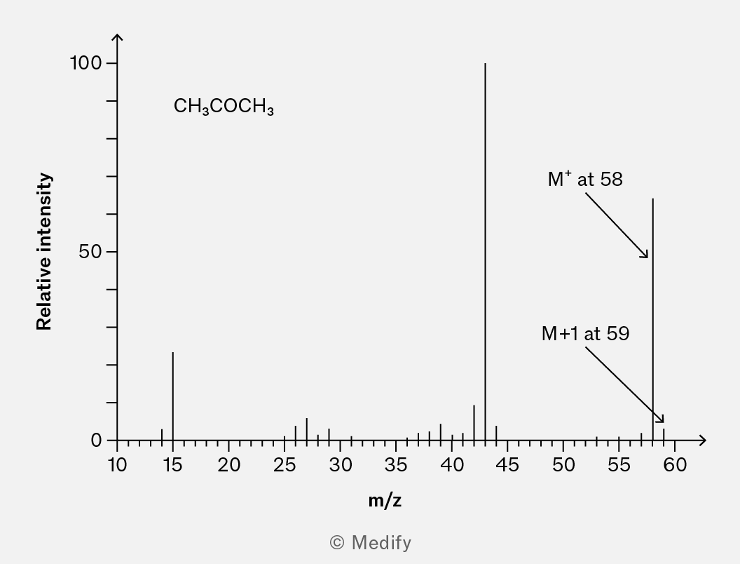
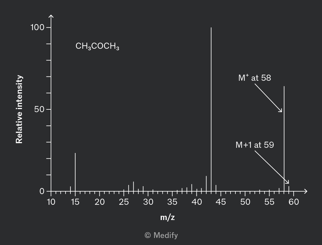
The of propanone, , is 58.
In the spectrum of propanone, the molecular ion peak, , is at 58 and a smaller M+1 peak at 59 shows the proportion of propanone featuring carbon-13.
Electrons occupy specific energy levels outside the nucleus of an atom.
The arrangement of electrons in these quantised energy levels in an atom is referred to as its electron structure or electronic configuration.
Principal energy levels or principal quantum shells (n) are numbered based on their distance from the nucleus.
The first principle quantum shell is closest to the nucleus and described as n = 1.
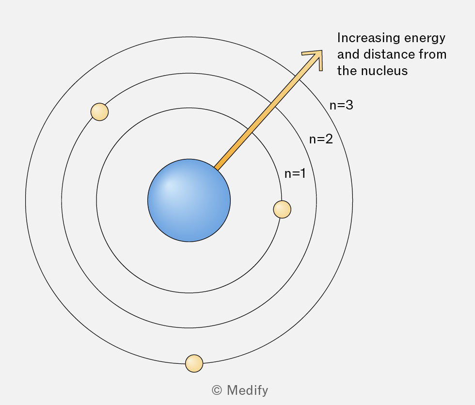
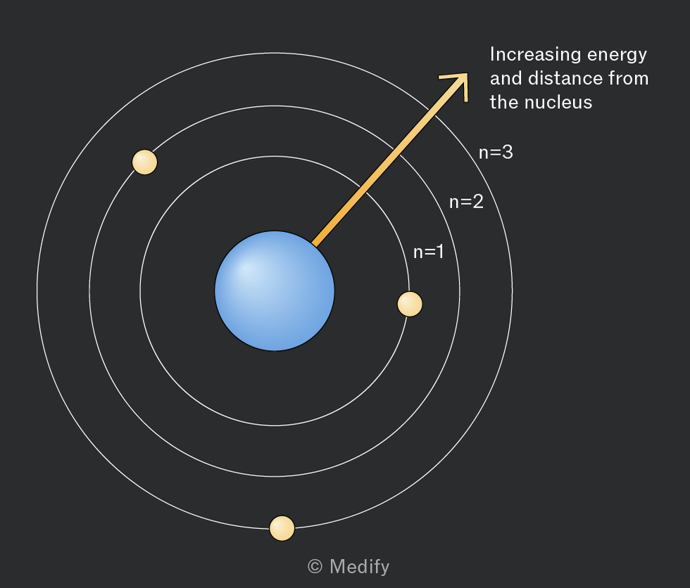
n is an integer value which increases as the shells move further from the nucleus.
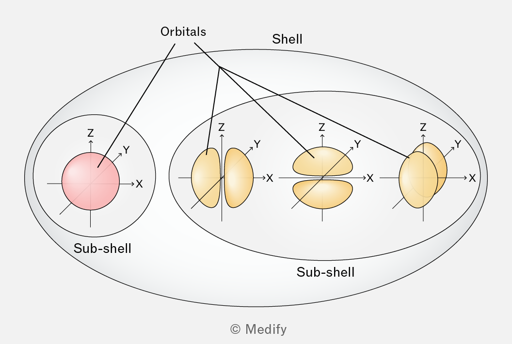
Shells are divided into subshells which can be further divided into atomic orbitals.
The atomic orbital is a three-dimensional region around the nucleus of an atom, where there is a maximum probability of finding an electron.
Each orbital can have a maximum of 2 electrons, with opposite spins.
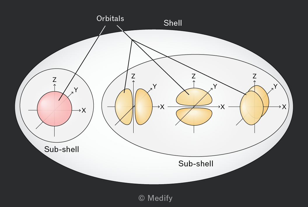
Electrons always fill up atomic orbitals in an order of increasing orbital energy. This leads to the most stable electronic configuration of the atom; the one that has the lowest overall energy.
Orbital energy increases with increasing principle quantum number (shell number). 4 > 3 > 2 > 1.
The subshells within a shell have slightly different energy levels with the trend f > d > p > s.
All orbitals in the same subshell are degenerate; they have an equal energy. Electrons-electron repulsion means all degenerate orbitals are filled singly before any electrons are paired.
The first electron is always placed in the 1s orbital, the lowest energy orbital.
Subshell notation describes the electrons contained in each subshell of an atom:
- X denotes the principal quantum number (shell number)
- y shows the subshell type (s, p, d, f)
- z represents the number of electrons occupying the subshell
Using the subshell notation the electronic configuration of carbon (Z = 6) is:
1s2 2s2 2p2
Make use of your periodic table to remember the order in which the subshells are filled.
List the subshells in order as you move in increasing atomic number from hydrogen to the element of interest.
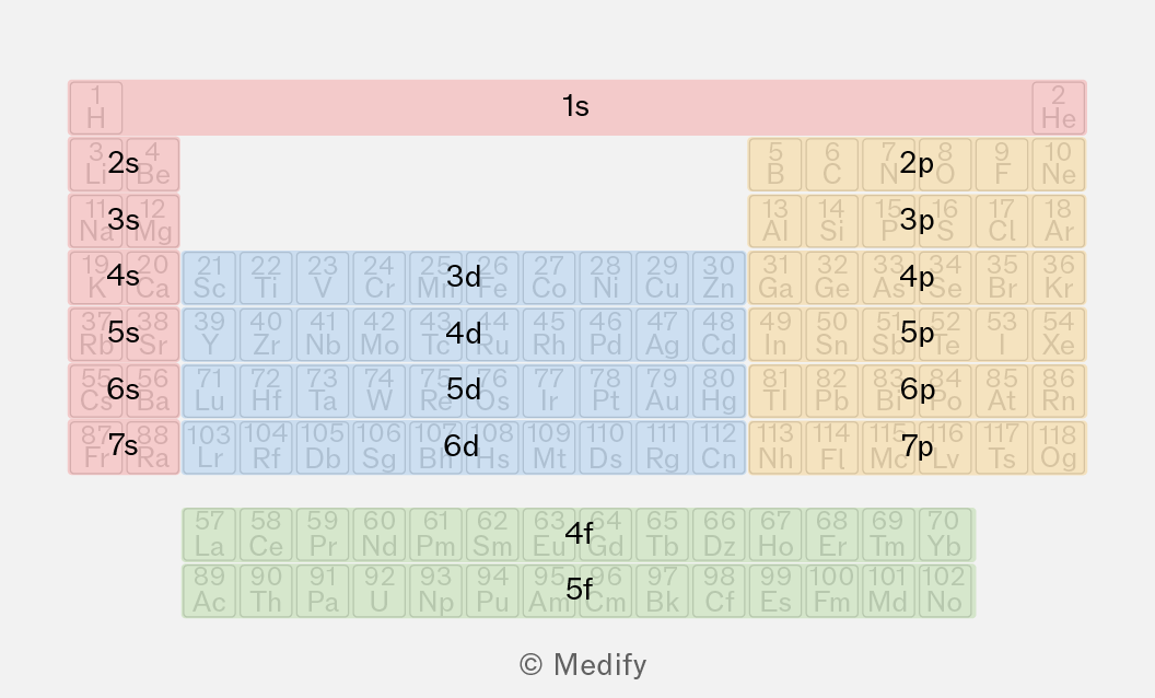
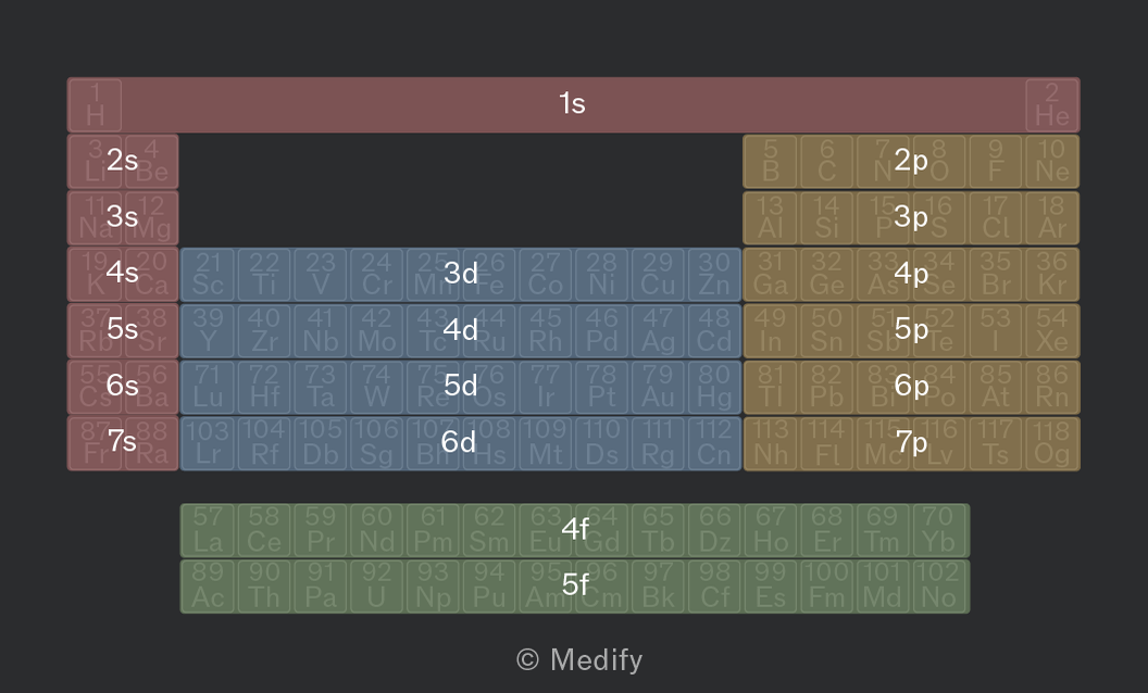
The energy levels of 3d and 4s subshells overlap.
The energy of 4s lies slightly below that of 3d therefore 4s is filled before 3d.
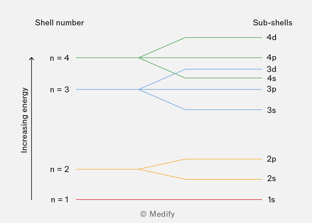
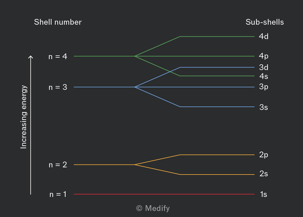
However, once filled, the energy of 3d falls below the 4s energy level.
When a transition metal is ionised, the 4s electrons are easier to remove than the 3d electrons.
Each period of the periodic table starts with one electron in a new electron shell.
The number before the orbital indicates the shell number, which also corresponds to the period number.
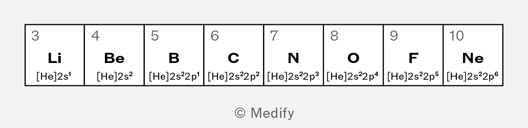
In period 2, the 2s subshell is filled first with two electrons.
After the 2s subshell is filled, electrons then occupy the 2p subshell, which can hold up to six electrons.
The trend in electron configuration across period 2 is shown below.
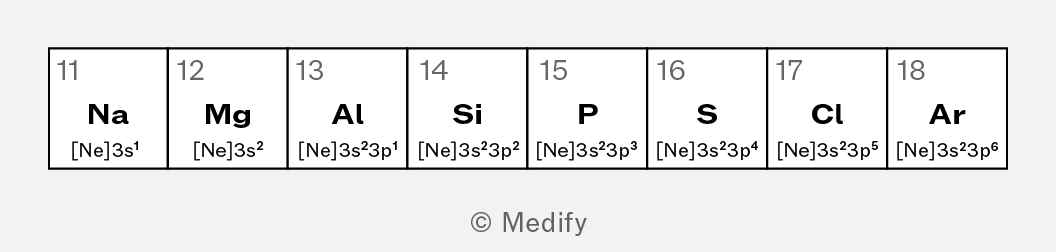
In the period 3, the 3s subshell is filled first with two electrons.
Once the 3s subshell is filled, electrons begin to occupy the 3p subshell, which can hold up to six electrons.
The trend in electron configuration across period 3 is shown below.
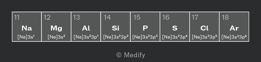
The block that an element is defined by is the orbital where the outermost electrons reside.
In d block elements, the outer electrons reside in the d orbital.
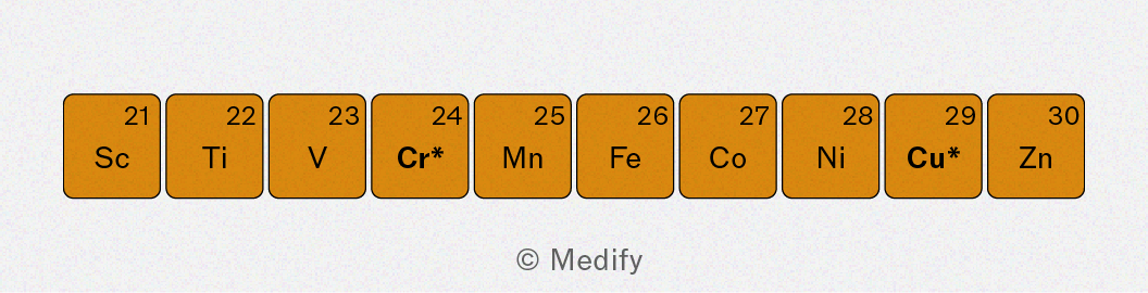
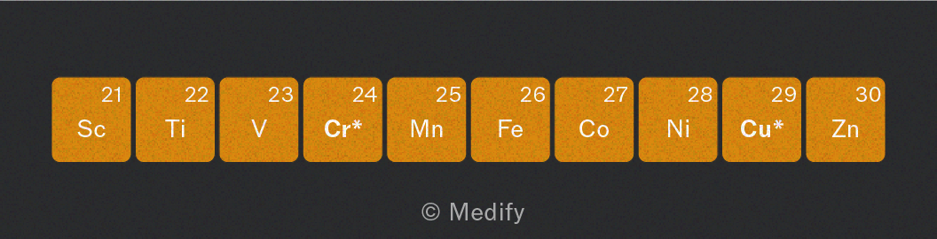
Following the rules of the Aufbau principle, the electrons in d-block elements fill the 4s orbital before the 3d orbital. The electron configurations for the neutral atoms are shown below.
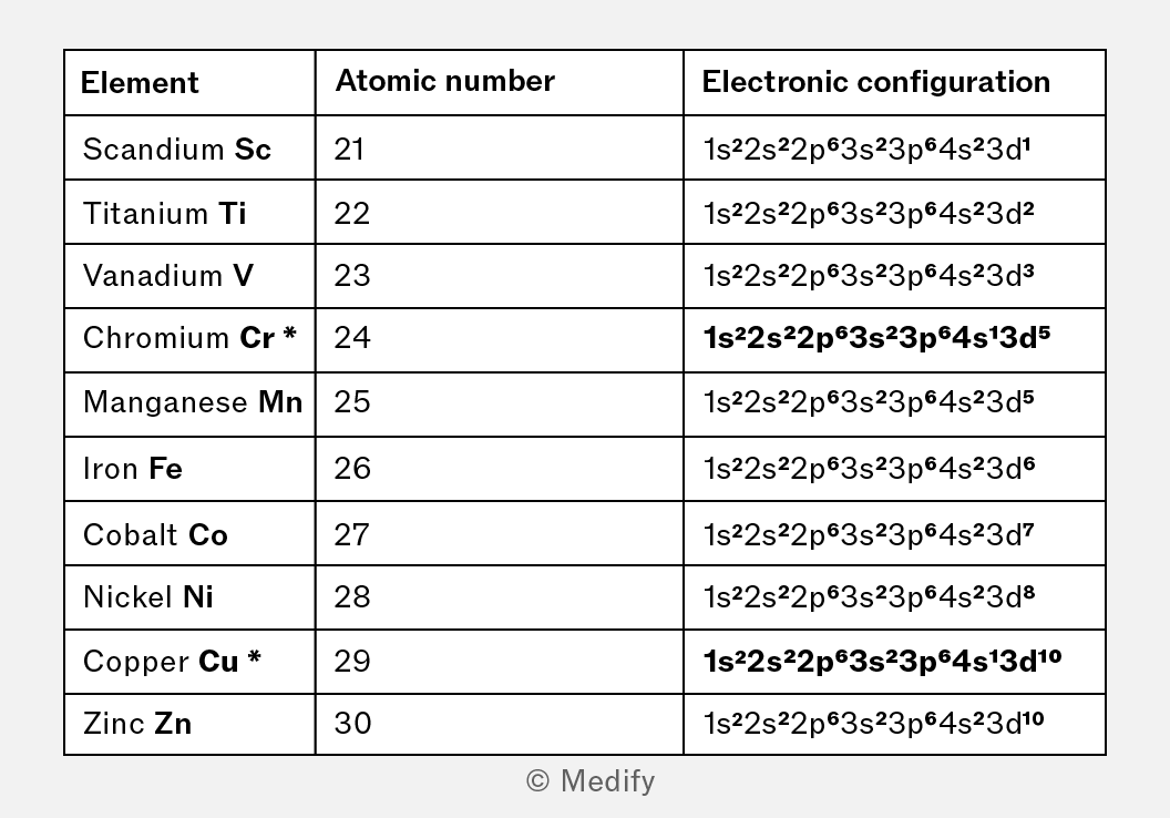
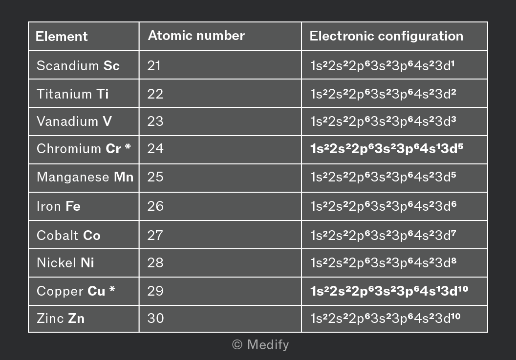
Remember the order by which orbitals are filled: 1s–2s–2p–3s–3p– 4s – 3d –4p–5s
You can shorten the electron configuration using the previous noble gas to represent the central shells up until 3p6. Scandium’s configuration thus becomes [] 4s23d1
Chromium and copper do not follow the general trend for electron configuration.
![A section of the periodic table displaying elements from scandium (Sc, atomic number 21) to zinc (Zn, atomic number 30). Highlighted elements include chromium (Cr, atomic number 24) and copper (Cu, atomic number 29), with their electron configurations shown: Cr as [Ar] 4s1 3d5 and Cu as [Ar] 4s1 3d10.](https://d3nzlfkeryzt6j.cloudfront.net/grfe3j4xa74828zlnb50oyy04mis)
![A section of the periodic table displaying elements from scandium (Sc, atomic number 21) to zinc (Zn, atomic number 30). Highlighted elements include chromium (Cr, atomic number 24) and copper (Cu, atomic number 29), with their electron configurations shown: Cr as [Ar] 4s1 3d5 and Cu as [Ar] 4s1 3d10.](https://d3nzlfkeryzt6j.cloudfront.net/vqwmdgq9xou94l2z8tiud4wruoc4)
In chromium, an electron from the 4s orbital is promoted to the 3d orbital, this is due to the half filled 3d orbitals in chromium being more stable as electron pair repulsion is decreased.
Similarly, in copper, an electron from the 4s orbital is promoted to the 3d orbital. This results in a complete 3d orbital. This is more energetically stable therefore more favourable.
Electron configuration of d block element ions
When forming positive ions, electrons are removed from the 4s orbitals first and then the 3d orbitals.
Scandium usually forms a 3+ ion.
: 1s22s22p63s23p64s23d1
is: 1s22s22p63s23p6
The three electrons have been lost, from the 4s orbitals first and then the 3d orbitals.
Iron can form and ions.
1s22s22p63s23p64s23d6
1s22s22p63s23p63d6 The two electrons have been lost from the 4s orbital.
1s22s22p63s23p63d5 The three electrons have been lost, from the 4s first, then the 3d orbitals.
Ionisation energy is the amount of energy required to remove one mole of electrons from one mole of gaseous atoms of an element to form one mole of gaseous 1+ ions:
Successive ionisation energies are associated with the repeated removal of electrons from increasingly positive ions.
First ionisation energy:
Second ionisation energy:
Third ionisation energy:
Each successive ionisation energy is higher than the previous one but the size of the increase is related to the position of the outermost electron within the subshells.
Successive ionisation energies can be used to make predictions about:
- the number of electrons in the outer shell
- the group number
- the identity of an element.
Successive ionisation energies for an unknown period 3 element are shown below:
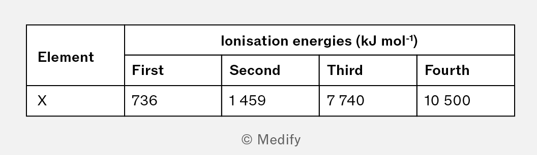
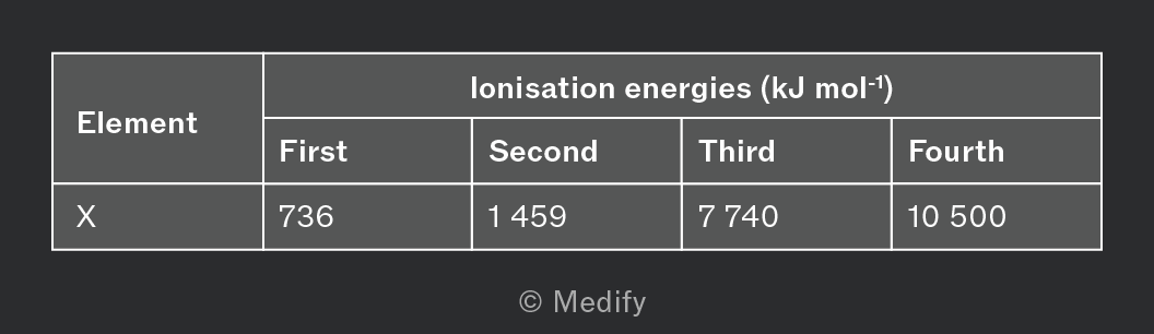
There is a steady rise between the first ionisation energy and second ionisation energy for the unknown element.
Between the second ionisation energy and third ionisation energy, there is comparatively a sharp rise indicating that the third electron is being removed from a new energy level.
From this information, we can therefore deduce that element X is in Group 2.
As it is in period 3, element X must be magnesium.
Question walkthrough
First ionisation energies
Using successive ionisation energies to deduce the identity of a compound