Carbohydrates (3.1.2)
On this page
Carbohydrates are biological molecules made from sugars.
Monosaccharides are the simplest form of sugar, consisting of a single monomer. Examples include glucose, fructose, and galactose.
Two monosaccharides join via a condensation reaction to form a disaccharide. Many monosaccharides join via condensation reactions to form a polysaccharide.
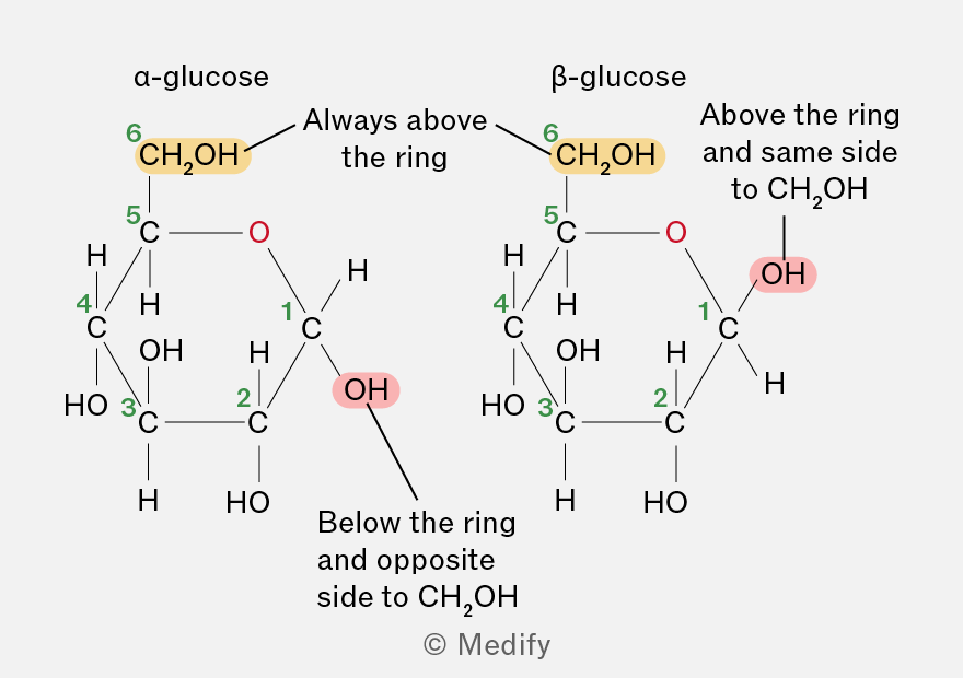
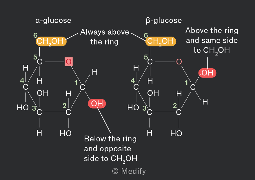
Glucose exists in two isomeric forms: -glucose and -glucose. They have the same chemical formula but different structures and properties.
In -glucose, the (hydroxyl) group on carbon 1 is on the opposite side of the ring from the group, whereas in -glucose it is on the same side.
Monosaccharides are linked by glycosidic bonds, which form in a condensation reaction between hydroxyl () groups on adjacent monosaccharides, releasing a molecule of water.
The name of the glycosidic bond indicates the carbon atoms involved in the linkage. For example, an 1,4 glycosidic bond forms between carbon 1 of one -glucose molecule and carbon 4 of another.
Two -glucose molecules combine to form the disaccharide maltose, joined by an 1,4 glycosidic bond.
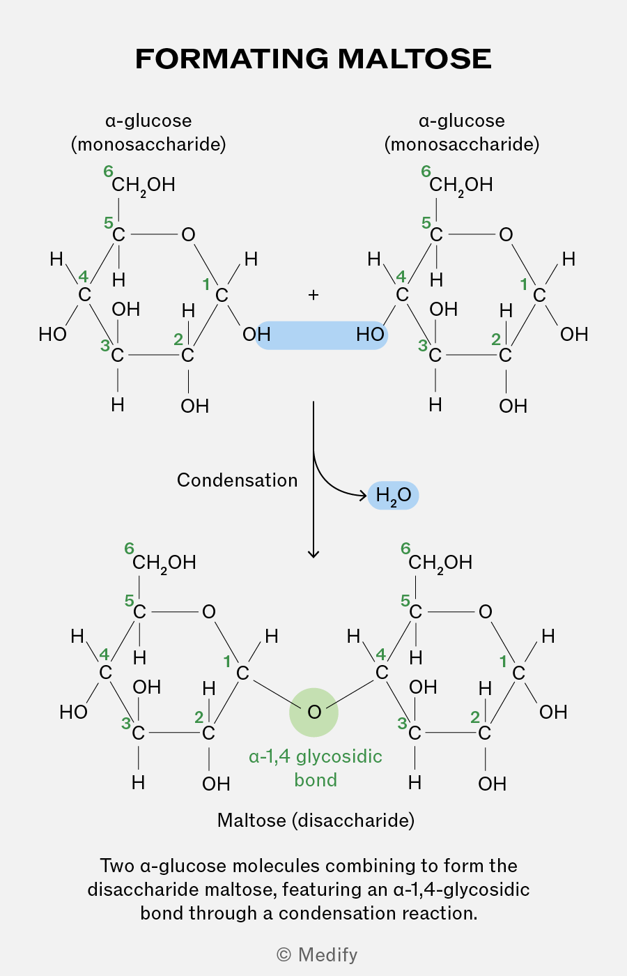
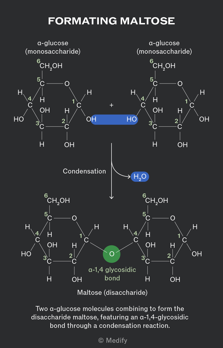
Two monosaccharides bonded together via a glycosidic bond form disaccharides:
- Maltose = -glucose + -glucose
- Sucrose = -glucose + -fructose
- Lactose = -galactose + glucose.
Starch is the insoluble, primary storage polysaccharide in plants and is formed from a few hundred to a few thousand -glucose monosaccharides bonded together. It is made from a mixture of two polysaccharides.
The two polysaccharides which form starch are:
- Amylose constitutes 10–30 of starch and has an unbranched, helix-shaped chain of -glucose molecules linked by 1,4 glycosidic bonds.
- Amylopectin constitutes 70–90 of starch and has both 1,4 glycosidic bonds and 1,6 glycosidic bonds, creating a branched molecule. Enzymes involved in breaking down these molecules can simultaneously work at multiple branch points, leading to rapid release of glucose for energy. More accessible hydroxyl groups make amylopectin more soluble in water.
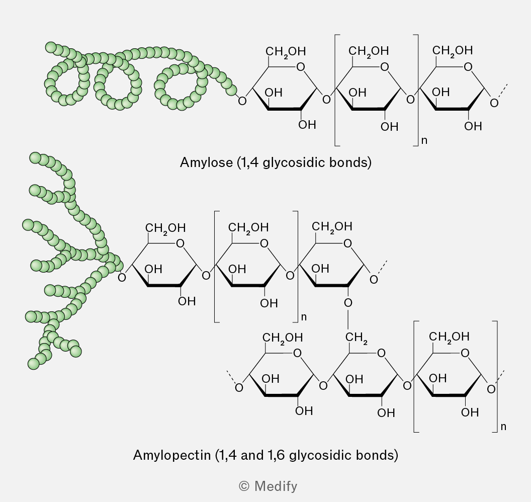
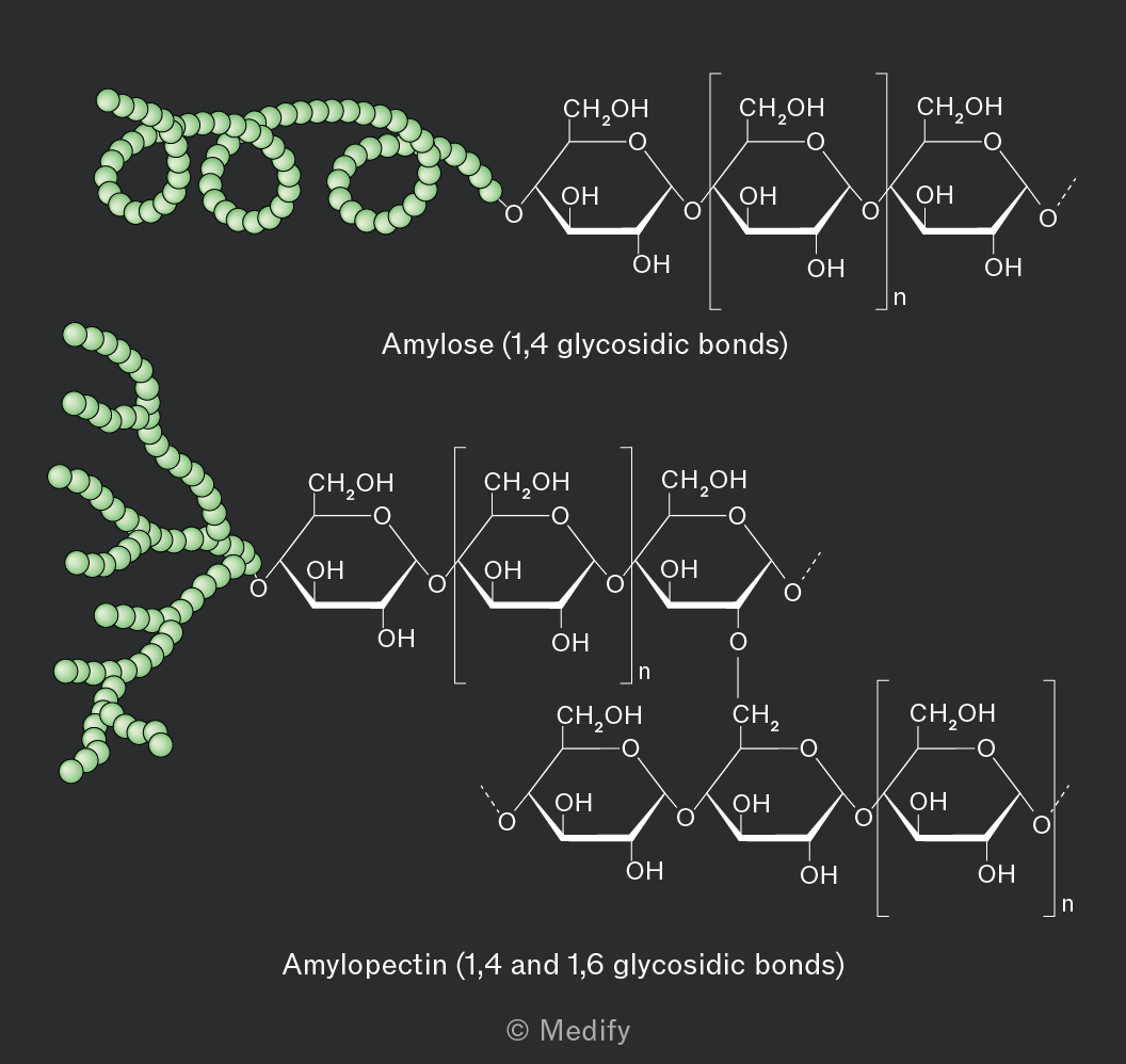
Glycogen is the primary storage polysaccharide in animals and fungi, characterised by a highly branched and uncoiled structure.
The highly branched structure of glycogen means there are numerous terminal glucose units leading to rapid release of glucose for energy. Branched polysaccharides with more accessible hydroxyl groups are also more soluble in water.
Glycogen’s structure is similar to amylopectin, but its branching is more extensive. This makes it more water-soluble and its terminal glucose molecules more accessible to enzymes.
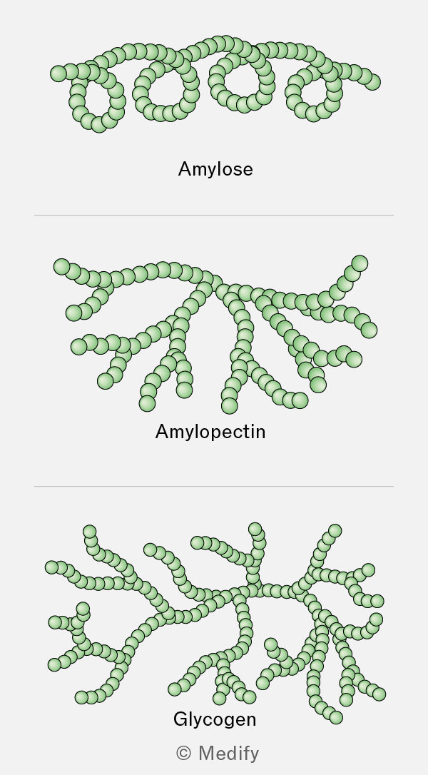
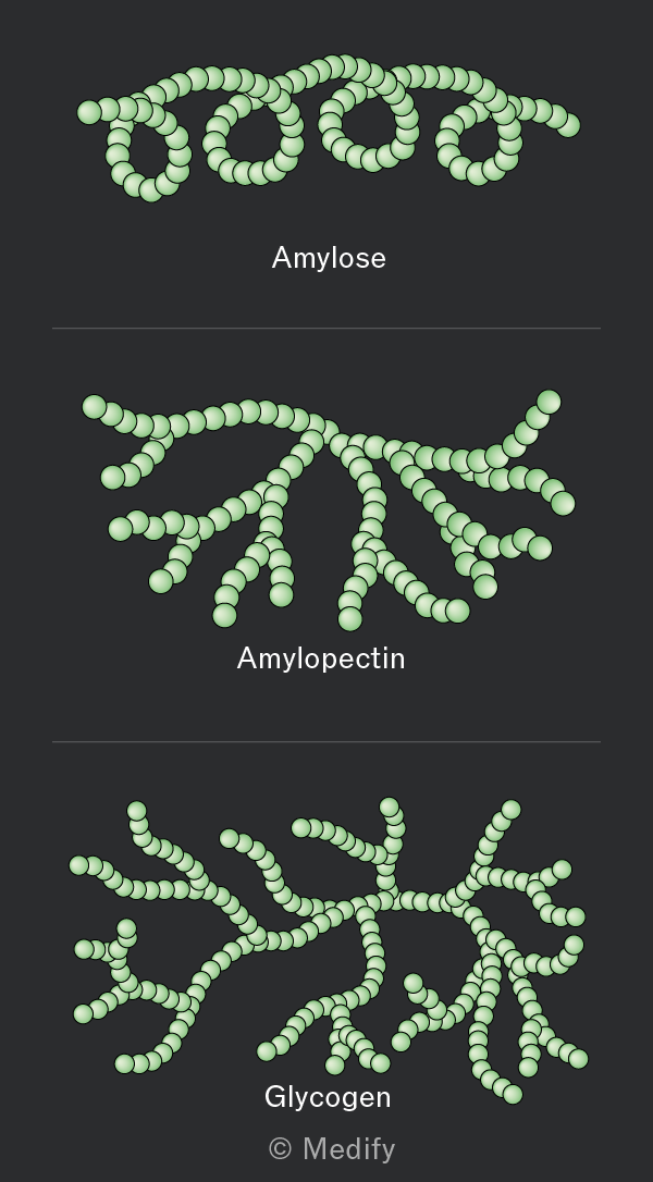
Amylose is coiled, making it insoluble and compact which is ideal for storage.
Amylopectin and glycogen are both highly branched molecules, making them compact and allowing more energy to be stored. Their branches provide multiple free ends where glucose molecules can be rapidly added in condensation reactions or removed in hydrolysis reactions, enabling quick adaptation to cellular energy demands.
Cellulose is a structural polysaccharide in plants. It is composed of long, linear chains of -glucose molecules joined by 1,4 glycosidic bonds.
Every other -glucose molecule in cellulose is inverted, producing a linear structure with extensive hydrogen bonding within and between polysaccharide chains.
Cellulose’s high tensile strength is a combined result of several key structural features:
- The strong 1,4 glycosidic bonds within a polysaccharide chain.
- There hydrogen bonding between polysaccharide chains within a microfibril.
- The alignment of microfibrils into macrofibrils.
- The alignment of macrofibrils into fibres.
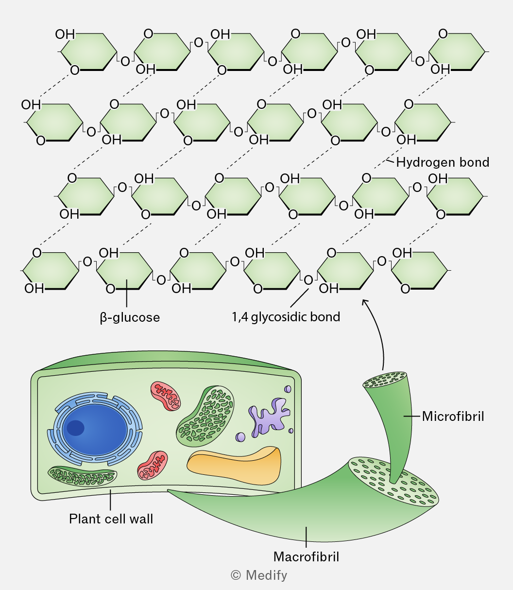
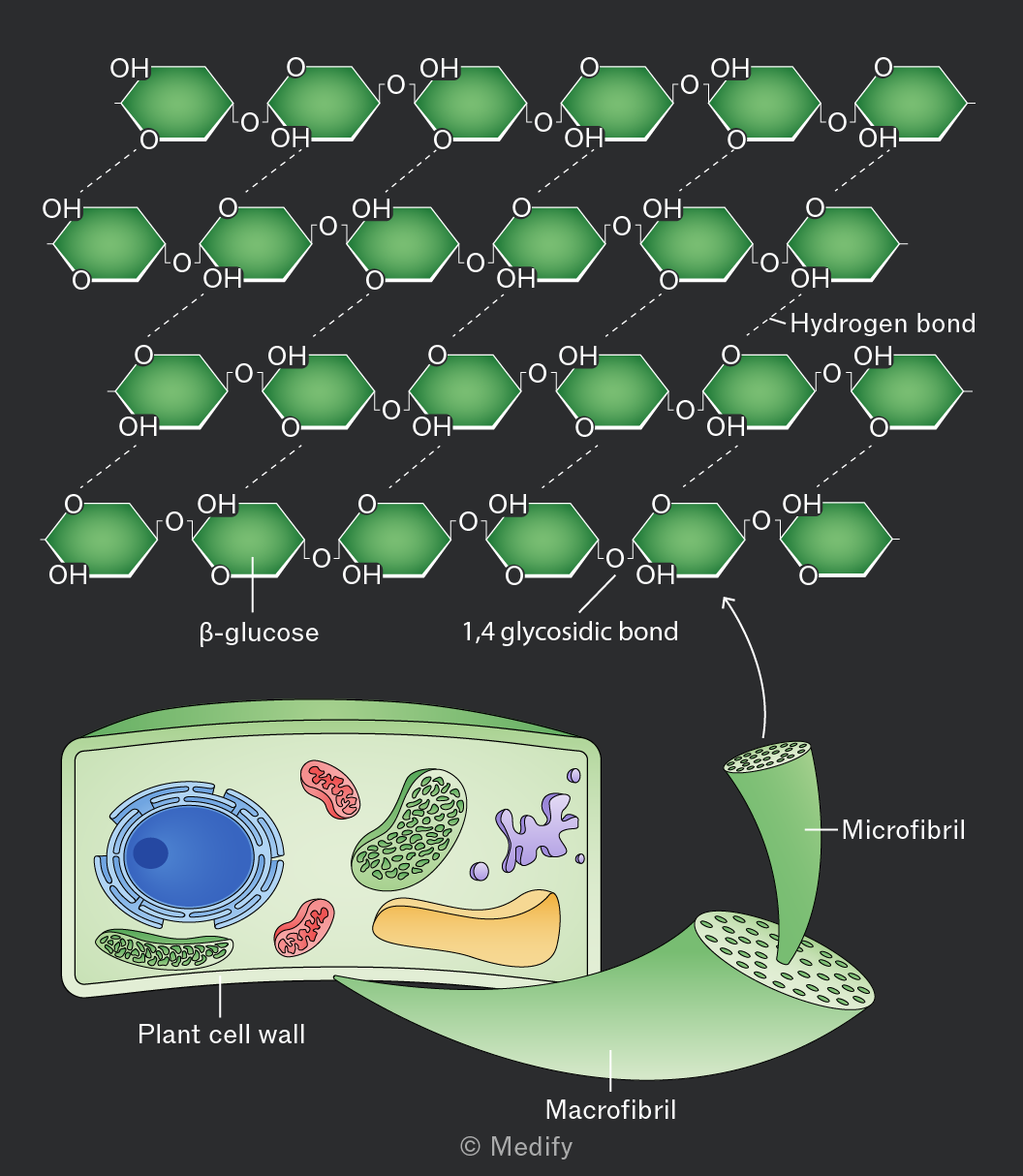
Individual cellulose chains are held together by hydrogen bonds to form microfibrils.
Microfibrils are organised into larger macrofibrils, further stabilised by additional hydrogen bonds.
The alignment of macrofibrils can vary depending on their role within a plant.
Cellulose is the main structural component of plant cell walls. The cell wall matrix includes other molecules such as lignin, to further increase cell wall strength.
Its structure gives it the following properties:
- Its high tensile strength enables cell walls to withstand turgor pressure and maximise leaf surface area for light absorption.
- Spaces between macrofibrils make cellulose fibres permeable to water and solutes, facilitating diffusion into and out of plant cells.
- Cellulose is a source of dietary fibre (roughage). Few organisms have the enzyme cellulase, which hydrolyses cellulose, meaning that it is not absorbed during digestion.
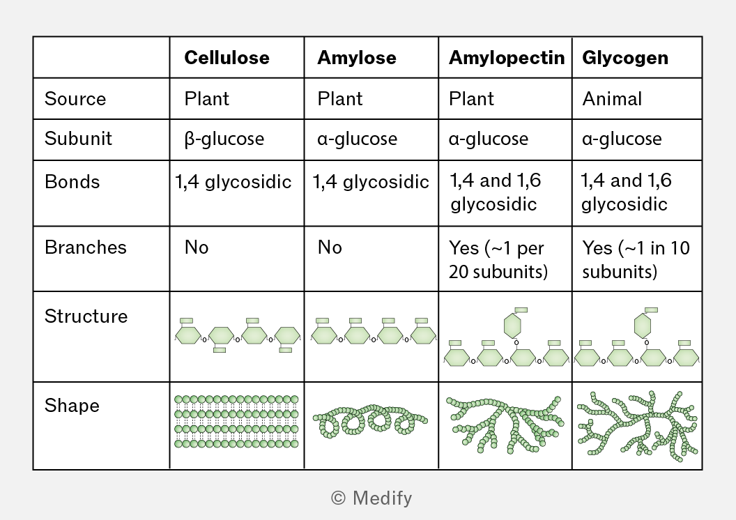
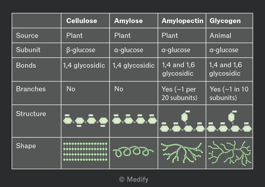
The bonding and structure of the key polysaccharides in plants and animals are summarised below.
Sugars can be classified as reducing or non-reducing depending on their structure and their functional groups. Reducing sugars can act as reducing agents by donating electrons.
Glucose, fructose, lactose, and maltose are reducing sugars, while sucrose is non-reducing.
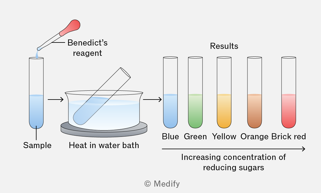
Benedict’s test detects reducing sugars in a sample.
Benedict’s reagent is a blue solution of () that contains copper(II) sulfate ions. When this reacts with reducing sugars, copper(II) is reduced to copper(I). The copper(I) oxide formed is insoluble in water and is detected as a brick-red precipitate.
Method:
- Add Benedict’s reagent (blue) to the sample. It is important that an excess of Benedict’s solution is used so that there is more than enough copper(II) sulfate present to fully react with the reducing sugars in the sample.
- Heat the test tube in boiling water or in a water bath for a few minutes.
Results:
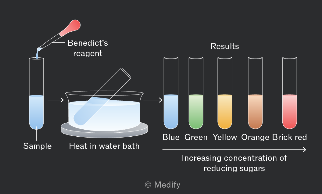
A colour change from blue to green, then yellow, orange, and finally brick-red, indicates the presence of reducing sugars in increasing concentrations.
Hydrolysing paired with Benedict’s test can be used to test for non-reducing sugars.
Method:
- Follow the protocol for detecting reducing sugars, and if the results are negative, proceed.
- Boil the sample with dilute hydrochloric acid. This breaks the glycosidic bonds in the sample, breaking the non-reducing sugars down into reducing sugars.
- Neutralise the sample with sodium hydrogencarbonate.
- Add an excess of Benedict’s solution.
- Heat the test tube in boiling water or in a water bath for a few minutes.
Results:
A colour change from blue to green, then yellow, orange, and finally brick-red indicates the presence of reducing sugars in increasing concentrations — i.e. the non-reducing sugars that have been broken down by hydrolysis into monosaccharides and therefore turned into reducing sugars.
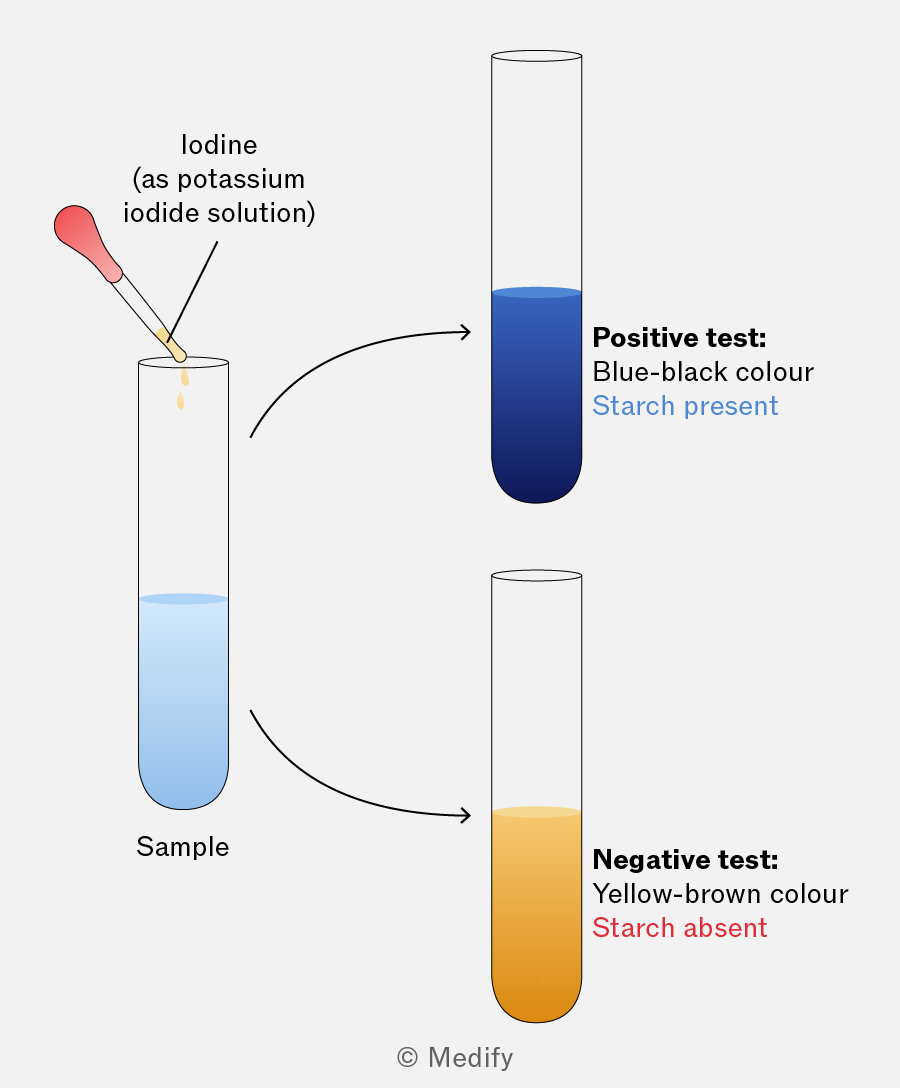
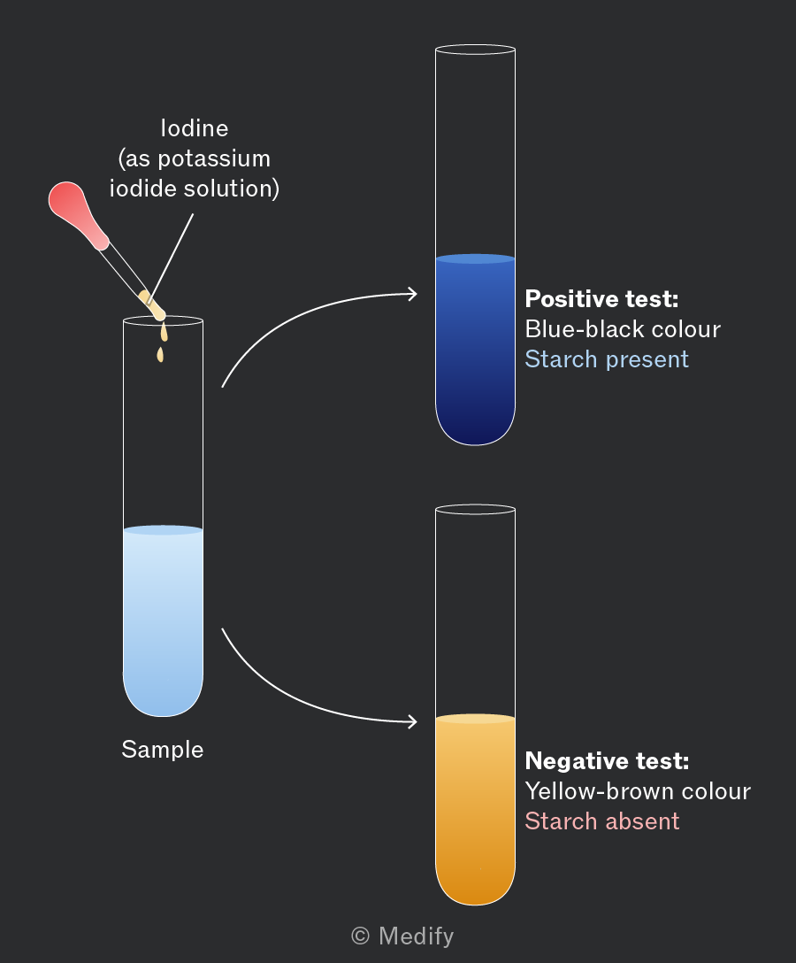
The iodine test is a qualitative test that detects starch in a sample.
Method:
- Add a few drops of iodine (in potassium iodide solution, which is insoluble in water) to the sample.
Results:
A blue-black colour indicates the presence of starch.