Biological molecules (3.1)›DNA replication (3.1.5.2)

DNA replication (3.1.5.2)

An overview of DNA replication (3.1.5.2) from AQA A level Biology including: semi-conservative replication and the process of replicating DNA
2 min

DNA replication is the process by which DNA is copied.

It follows a semi–conservative mechanism; each new DNA molecule consists of one original (parental) strand and one newly synthesised strand.

This ensures that each daughter cell receives an exact copy of the DNA, maintaining genetic continuity between generations of cells.
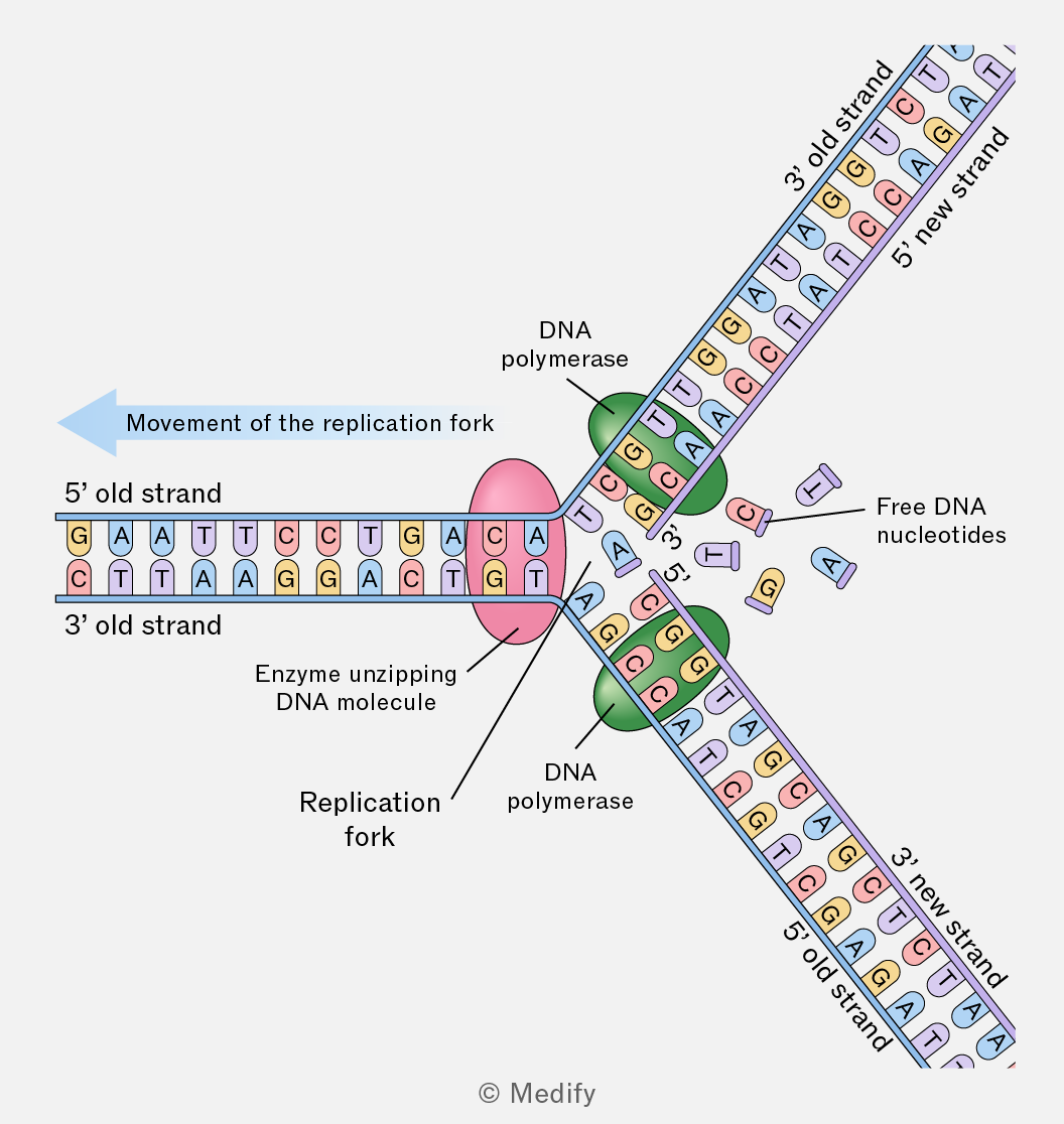
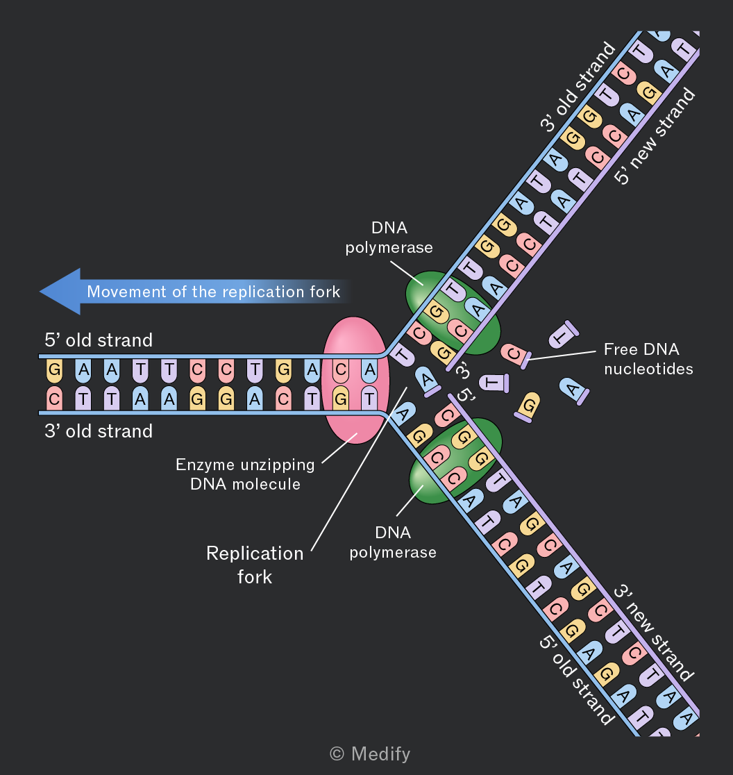
Diagram illustrating DNA replication. The image shows a replication fork with two strands: the 5' old strand and the 3' old strand. The enzyme unzipping the DNA molecule is labeled, and the DNA polymerase is shown on both the leading and lagging strands. Free DNA nucleotides A, T, C, and G are depicted near the replication fork. The 3' new strand and 5' new strand are being synthesized, with DNA polymerase facilitating the addition of nucleotides. An arrow labeled 'Movement of the replication fork' indicates the direction of replication. The DNA sequence includes bases such as G, A, T, C on the old strands and complementary bases on the new strands.
Add to favourites

Watson and Crick proposed the semi–conservative model of DNA replication. This replication mechanism was validated by the Meselson–Stahl experiment (1958) using two different isotopes of nitrogen: and
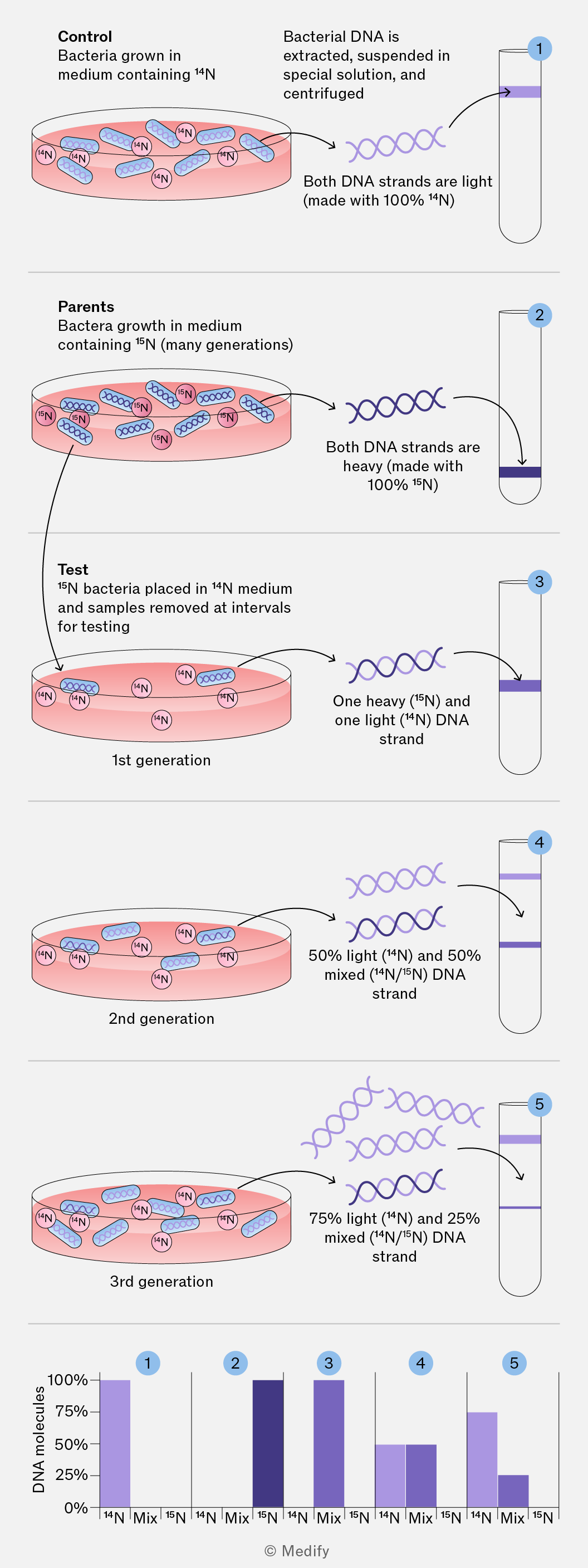
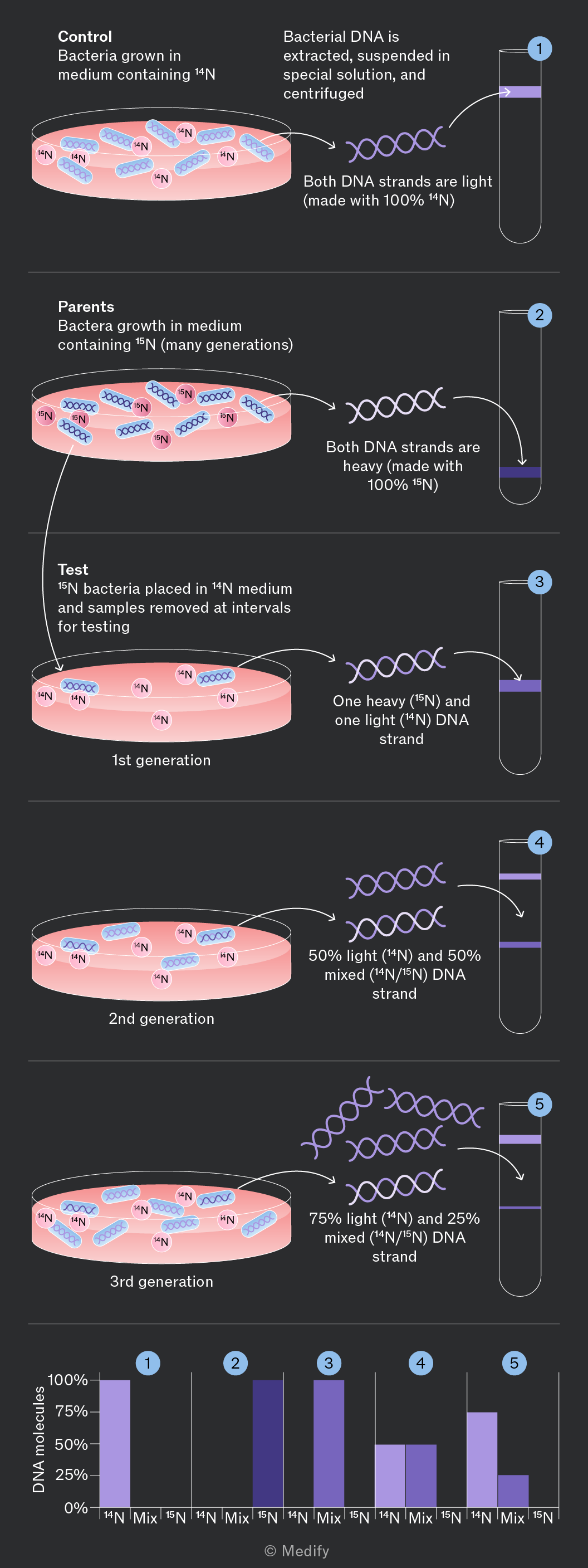
The image is a multi-panel diagram illustrating an experiment with bacteria grown in different nitrogen isotopes and the resulting DNA analysis. Panel 1 titled 'Control' shows bacteria grown in medium containing 14N, with an arrow pointing to a centrifuge tube containing DNA labeled 'Both DNA strands are light (made with 100% 14N)'. Panel 2 titled 'Parents' shows bacteria grown in medium containing 15N, with an arrow pointing to a centrifuge tube labeled 'Both DNA strands are heavy (made with 100% 15N)'. Panel 3 titled 'Test' shows 15N bacteria placed in 14N medium for '1st generation', with an arrow pointing to a centrifuge tube labeled 'One heavy (15N) and one light (14N) DNA strand'. Panel 4 shows '2nd generation' bacteria with an arrow to a centrifuge tube labeled '50% light (14N) and 50% mixed (14N/15N) DNA strand'. Panel 5 shows '3rd generation' bacteria with an arrow to a centrifuge tube labeled '75% light (14N) and 25% mixed (14N/15N) DNA strand'. Below is a bar graph showing 'DNA molecules' on the y-axis from 0% to 100%, with five numbered sections corresponding to each panel. Each section has three bars labeled '14N', 'Mix', and '15N', showing the distribution of DNA types. The bars indicate: 1 - 100% 14N, 2 - 100% 15N, 3 - 50% Mix, 50% 15N, 4 - 50% 14N, 50% Mix, 5 - 75% 14N, 25% Mix.
Add to favourites

DNA replication begins with DNA helicase unwinding the DNA double helix, separating the two strands by breaking the hydrogen bonds between complementary bases.

This separation creates two single–stranded templates that can both be used for synthesising new DNA strands.

Free DNA nucleotides bond to their complementary base pairs on the parent strands of DNA. DNA polymerase joins these nucleotides via condensation reactions, which form phosphodiester bonds.

Two identical daughter DNA molecules are created, each containing one parent strand and one newly synthesised strand.

Add to favourites