
Atoms and reactions
On this page
The structure of the atom:
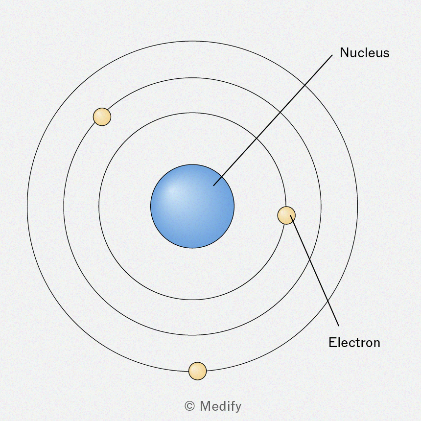
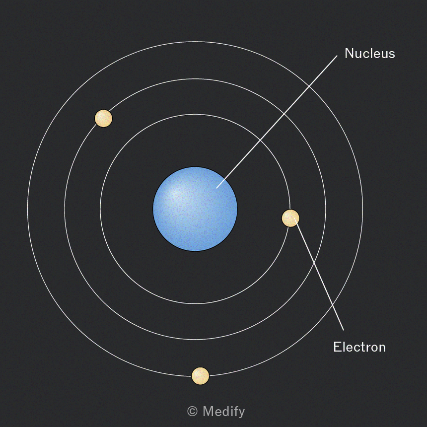
The nucleus contains the protons and neutrons.
The nucleus is surrounded by shells (orbitals) which contain the electrons.
Subatomic particles can be described by their relative charge and relative mass.
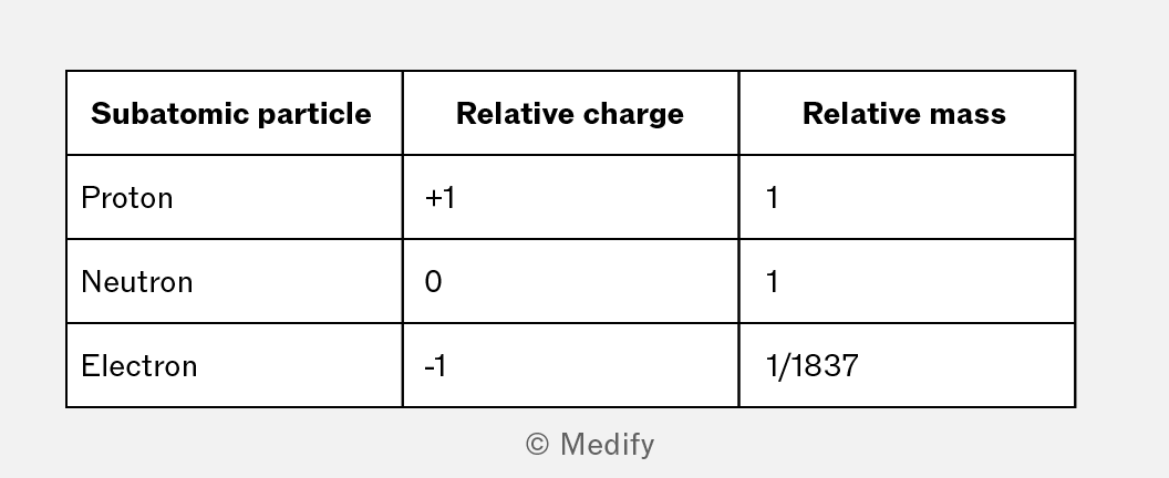
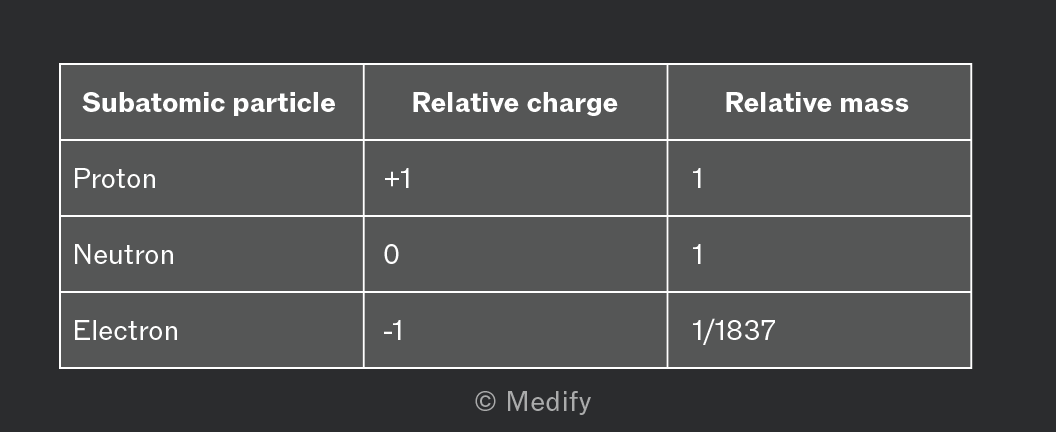
In a neutral atom, the number of protons is the same as the number of electrons.
Atomic number () = number of protons
Mass number () = number of neutrons + the atomic number
Ions are particles with a charge.
In an ion, the number of electrons = number of protons − the charge.
- The number of electrons in a
- The number of electrons in a
The number of neutrons = mass number − the atomic number.
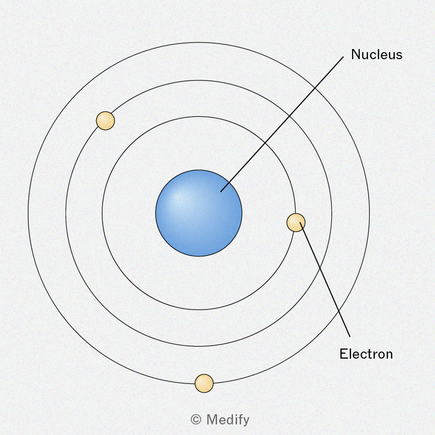
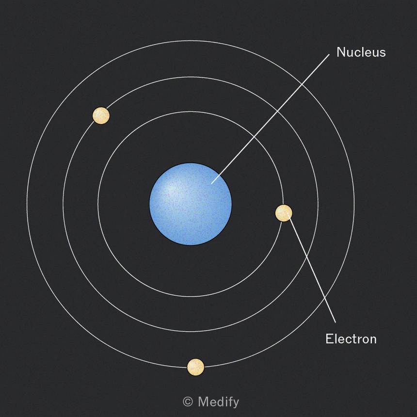
Bohr (1913):
- discovered energy levels
- related atomic spectra to electronic energy levels.
Dalton (1800s):
- discovered that atoms can be combined to make compounds
- believed atoms cannot be broken into smaller parts
- disproved by J.J. Thomson’s cathode ray tube experiments.

Thomson (1897):
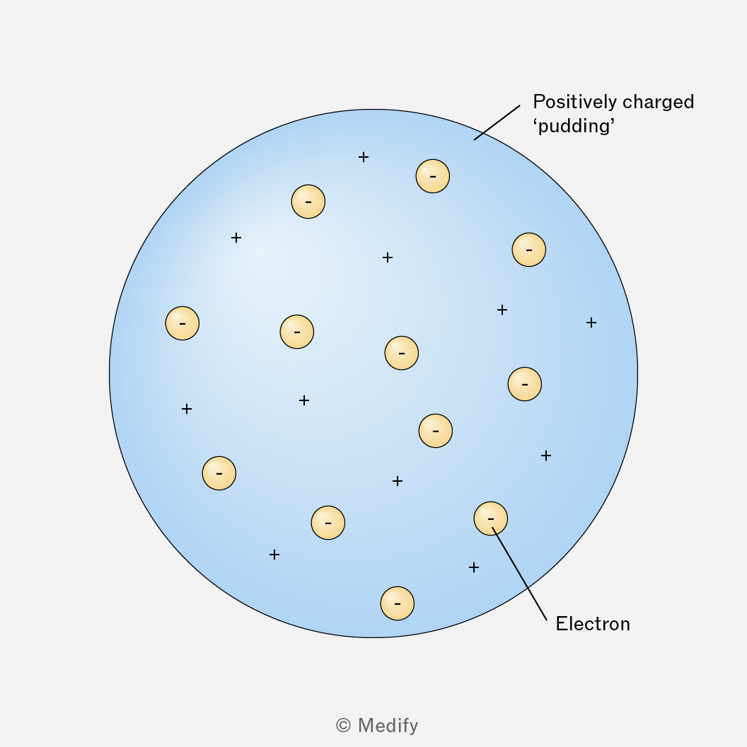
- discovered the electron
- believed that atoms were a sphere of positive mass embedded with electrons: the plum pudding model
- was disproved by Rutherford’s gold foil experiment.
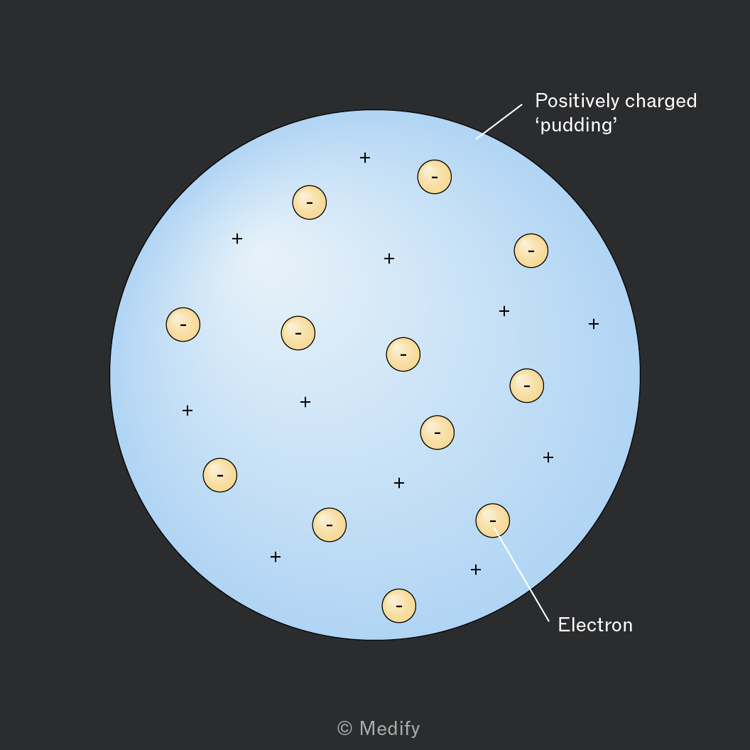
Rutherford (1905):
- discovered the nucleus with his gold foil experiment.
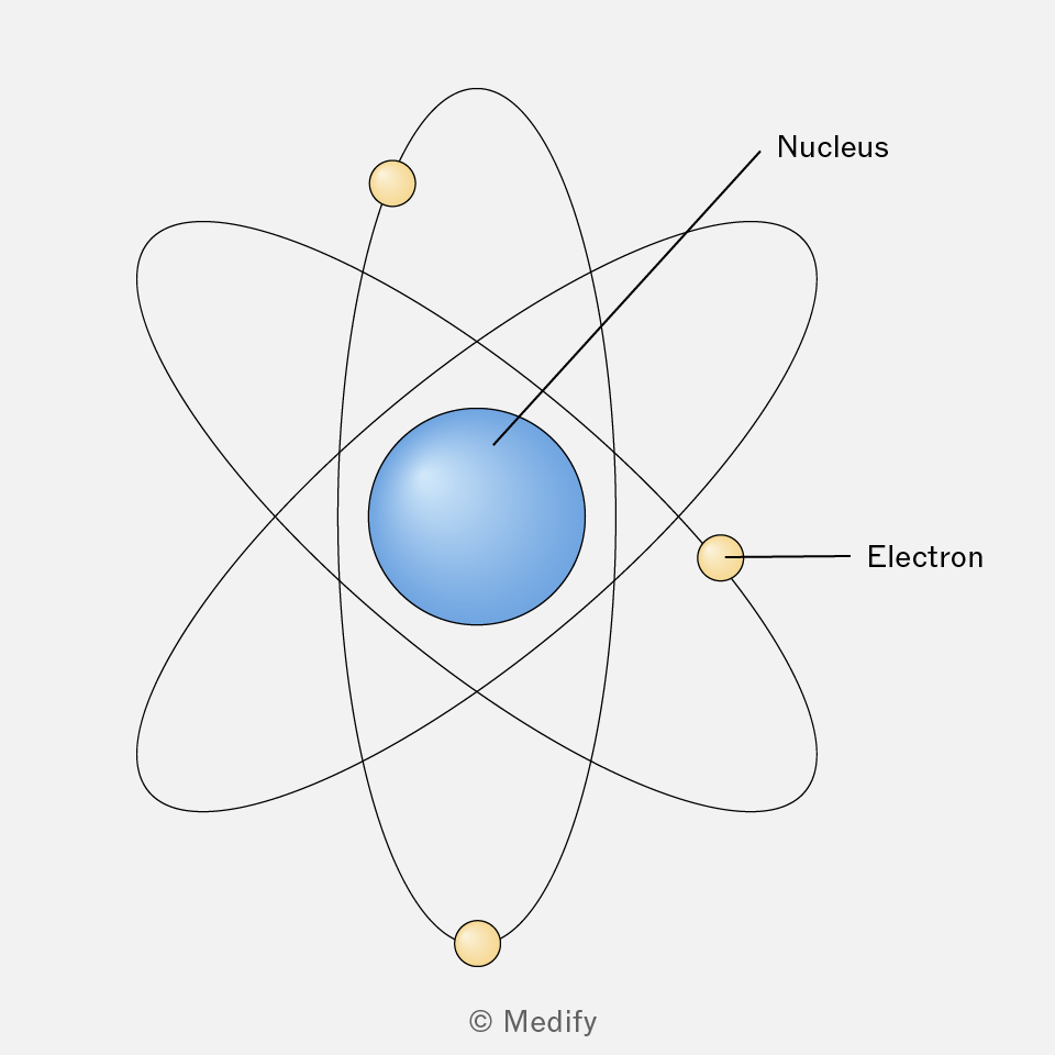
Rutherford’s gold foil experiment:
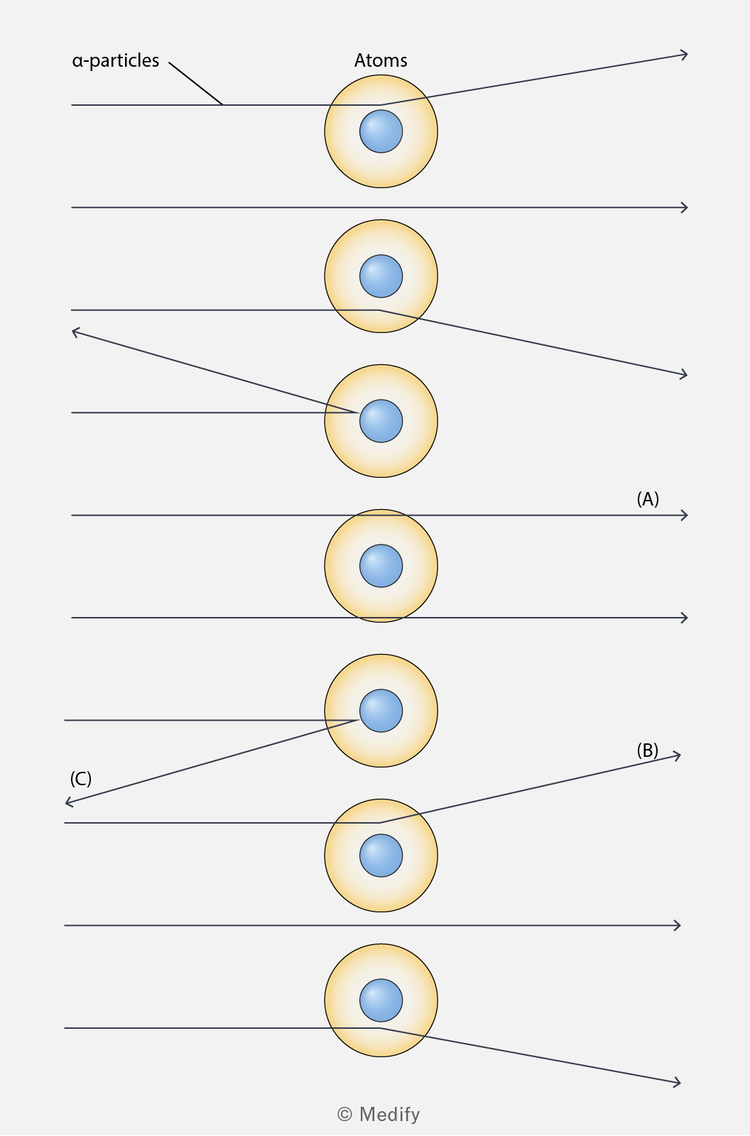
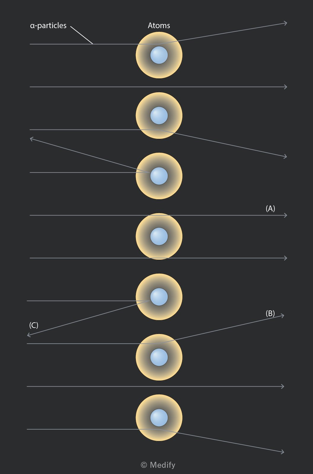
- A very thin piece of gold foil was bombarded with alpha particles.
- Most particles went straight through (A).
- Some were deflected (B) and a few were reflected (C).
- This proved that the vast majority of the mass of an atom is concentrated in a small central volume and that the central mass was positively charged.
Isotopes are atoms of the same element with different numbers of neutrons and different masses.
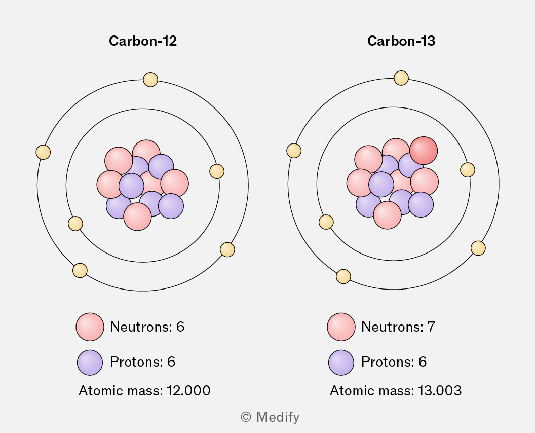
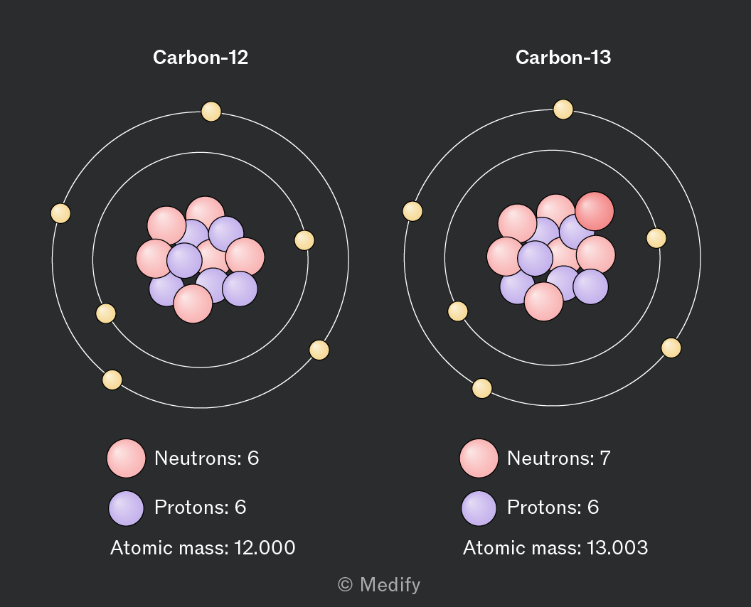
Carbon-12 and carbon-13 are isotopes of carbon.
- Both identify as carbon, with an atomic number of six.
- Carbon-12 has six neutrons and a relative atomic mass of 12 whereas carbon-13 has seven neutrons and a relative atomic mass of 13.
Electrons occupy specific energy levels outside the nucleus of an atom.
The arrangement of electrons in these quantised energy levels in an atom is referred to as its electron structure or electronic configuration.
Principal energy levels or principal quantum shells (n) are numbered based on their distance from the nucleus.
The first principle quantum shell is closest to the nucleus and described as n = 1.
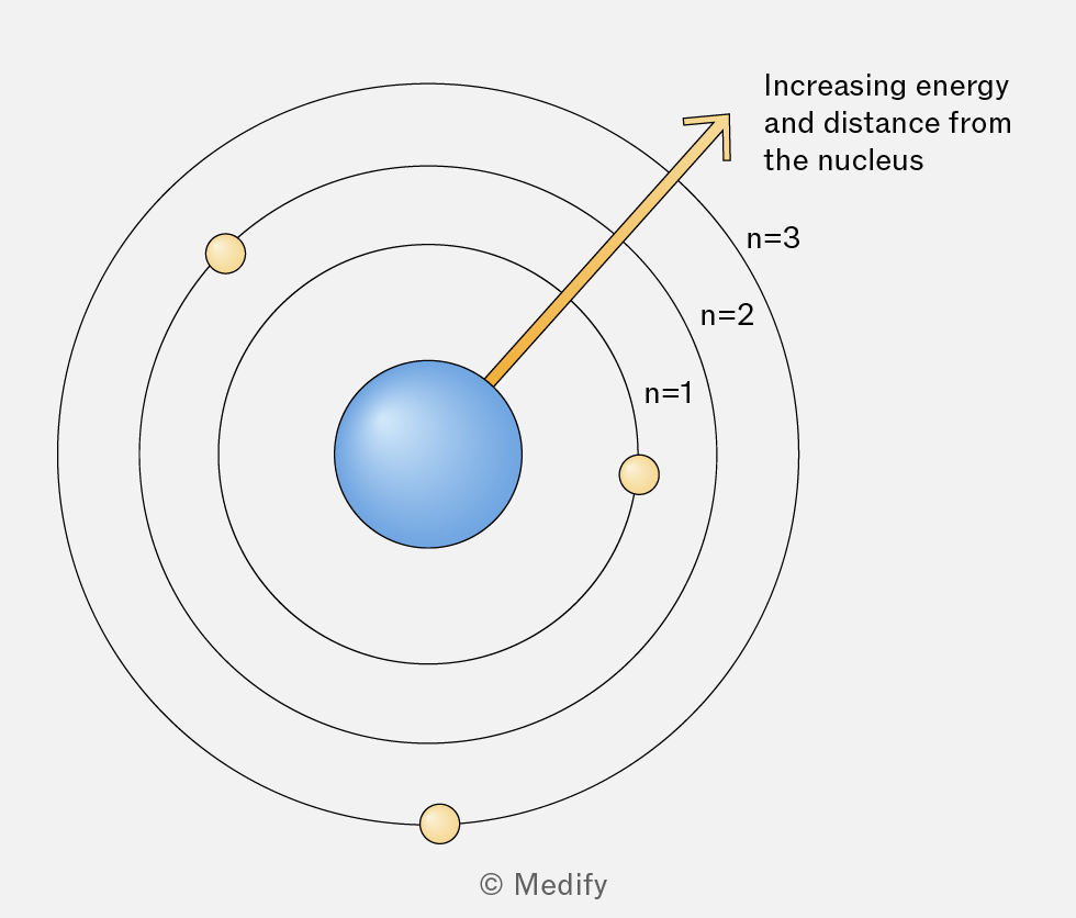
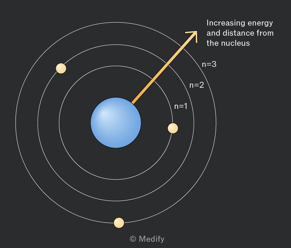
n is an integer value which increases as the shells move further from the nucleus.
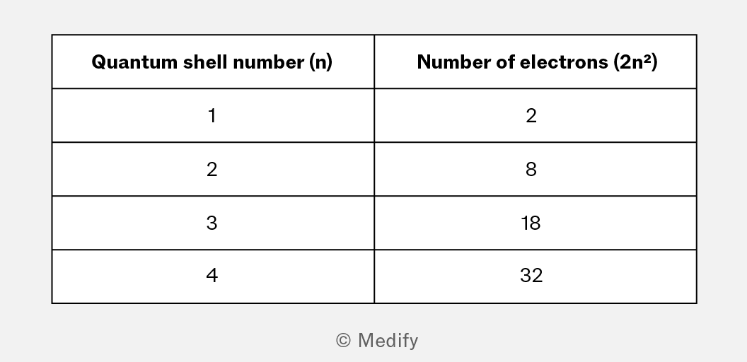
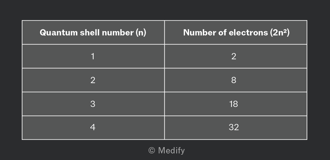
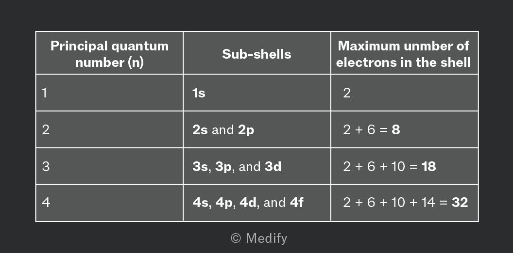
The maximum number of electrons that can fill a quantum shell is given by the formula e = 2n2
The number of electrons needed to fully occupy the first four quantum shells is shown below.
An atomic orbital has a three-dimensional shape indicating the area in which an electron is likely to be found.
The s orbital has a symmetrical, spherical shape, with the nucleus located at the centre.
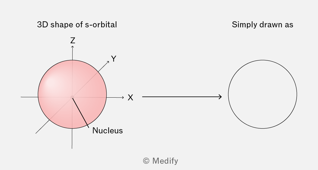
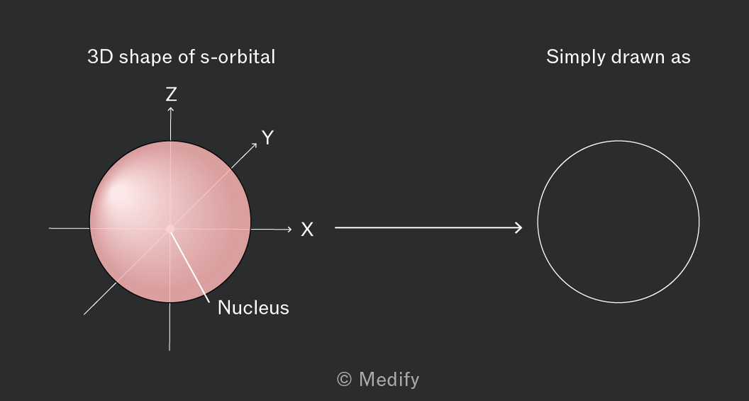
The s orbitals of all shells have the same spherical shape but differ in the size of their radius.
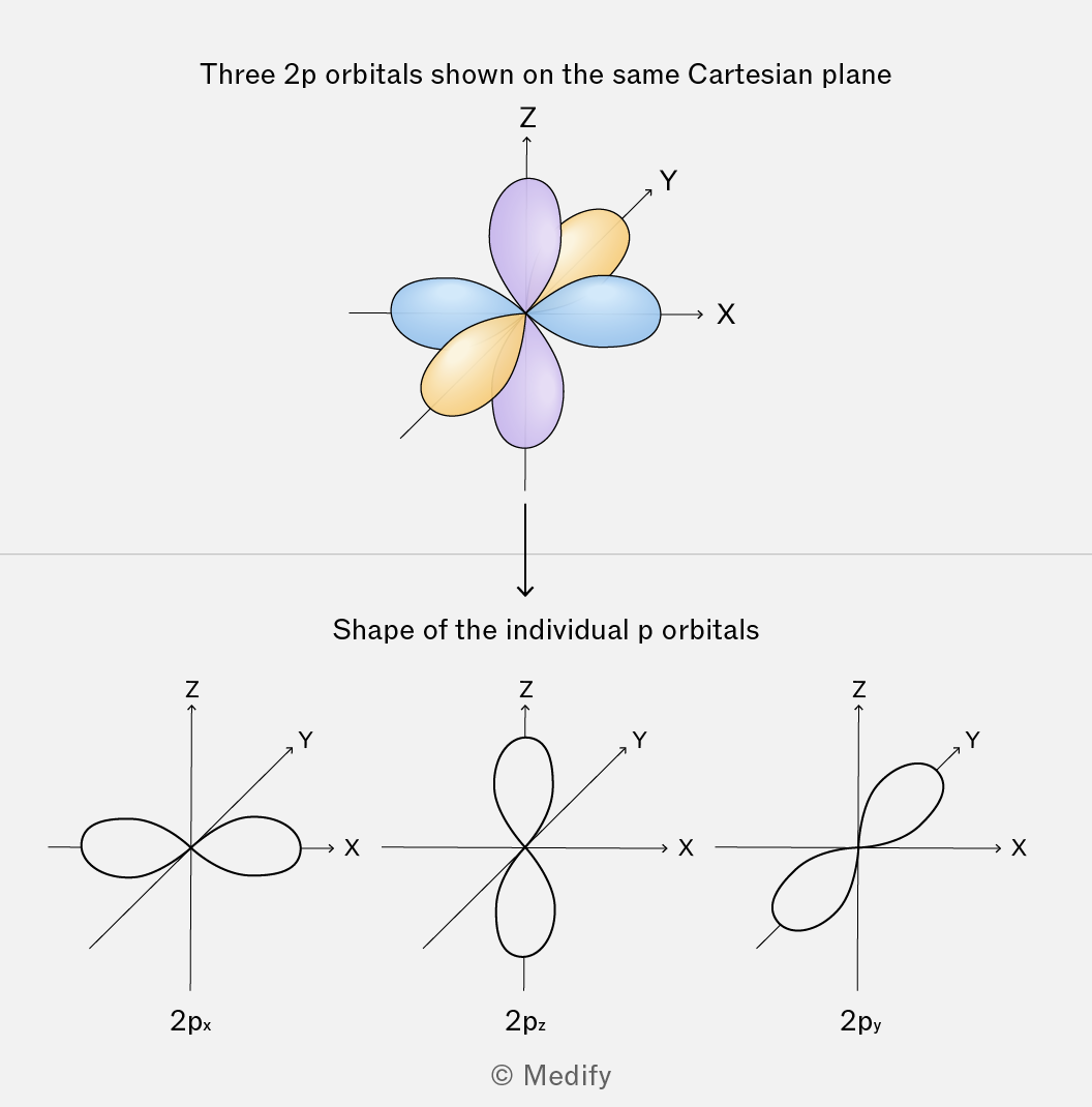
The p orbitals are described as dumb-bell shaped, with the nucleus located between the two halves.
There are three degenerate p orbitals in each p subshell. These orbitals have an identical shape and energy, however, their orientation in space differs. They lie at right angles to one another along the X, Y, and Z axes of a Cartesian plane.
The three p orbitals are thus distinguished as px, py, and pz.
Shells are divided into subshells which can be further divided into atomic orbitals.
The atomic orbital is a three-dimensional region around the nucleus of an atom, where there is a maximum probability of finding an electron.
Each orbital can have a maximum of 2 electrons, with opposite spins.
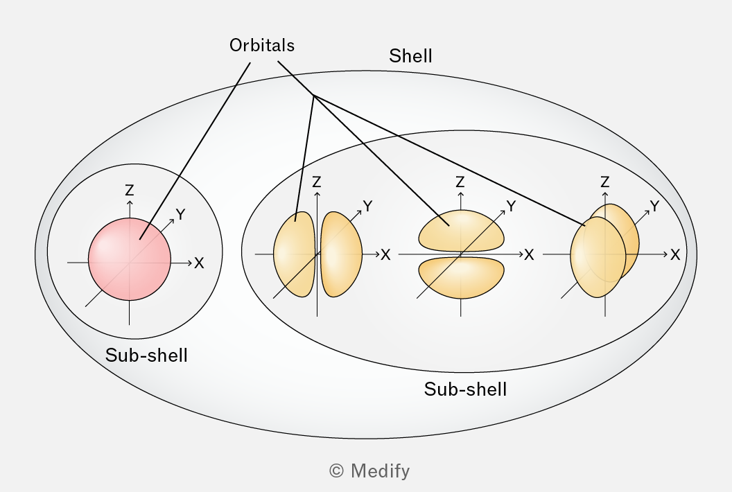
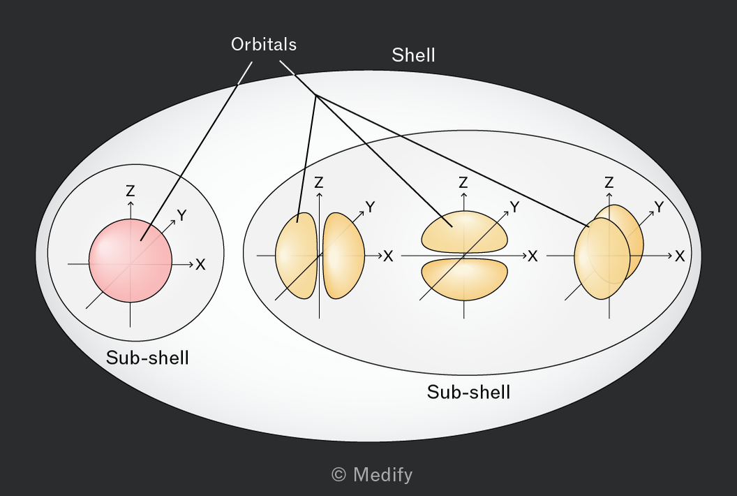
Within a shell, the atomic orbitals are grouped as subshells, distinguished by the letters s,p,d and f.
The s subshell has one atomic orbital, the p subshell has three orbitals, the d subshell has five orbitals while the f subshell has a total of seven atomic orbitals.
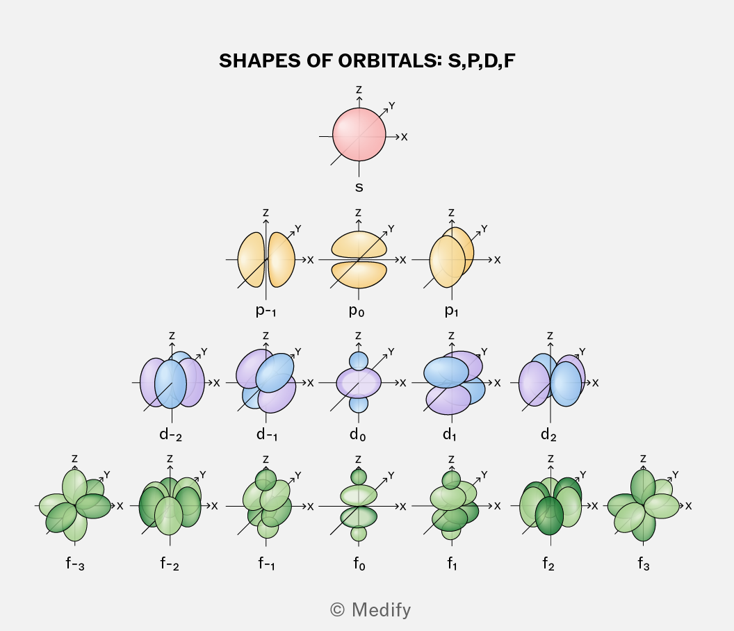
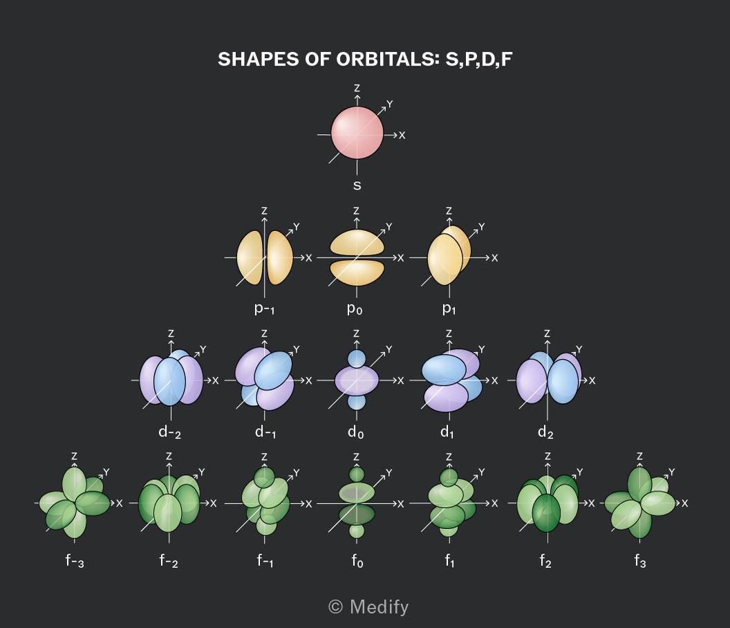
As the energy level of the shell increases, the size of the atomic orbitals within the subshells increases but their shapes remain similar.
The number of subshells per shell in an atom increases with the increase in principal quantum number.
The number of electrons per shell can be calculated by considering full occupancy of all the atomic orbitals present.
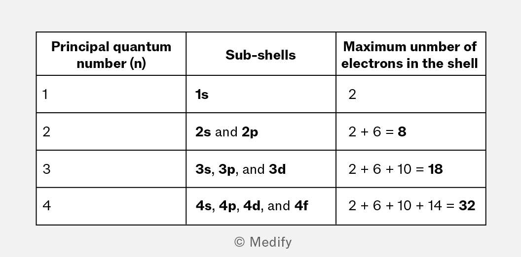
The existence of quantum shells was supported by experimental evidence involving characteristic atomic emission spectra of individual elements.
When electrons are excited, they move up an energy level. When they fall back to their original energy level they emit radiation at specific wavelengths, producing a line spectrum unique to each element.
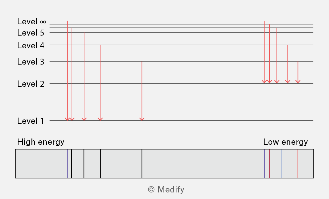
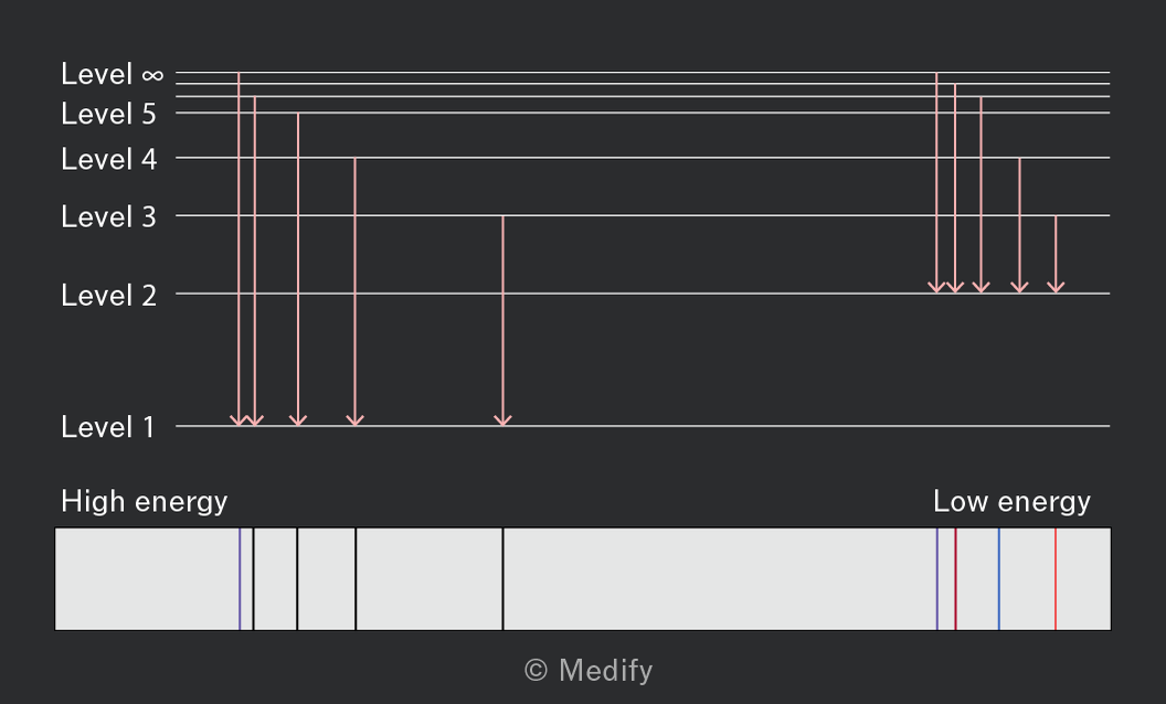
These discrete spectral lines reveal that only certain energy transitions are possible; electrons in an atom must occupy quantized energy levels. These are the ‘electron shells’.
Ionisation energy relates to the energy required to remove an electron from an atom or ion. The analysis of successive ionisation energies of an element provides evidence for the existence of quantum shells.
Ionisation energy increases with successive ionisations as the attraction of the outermost electron to the nucleus increases.
The increase in ionisation energy with successive ionisation is not steady.
This indicates that the reduced ionic radius and reduced electron shielding is more significant for some successions.
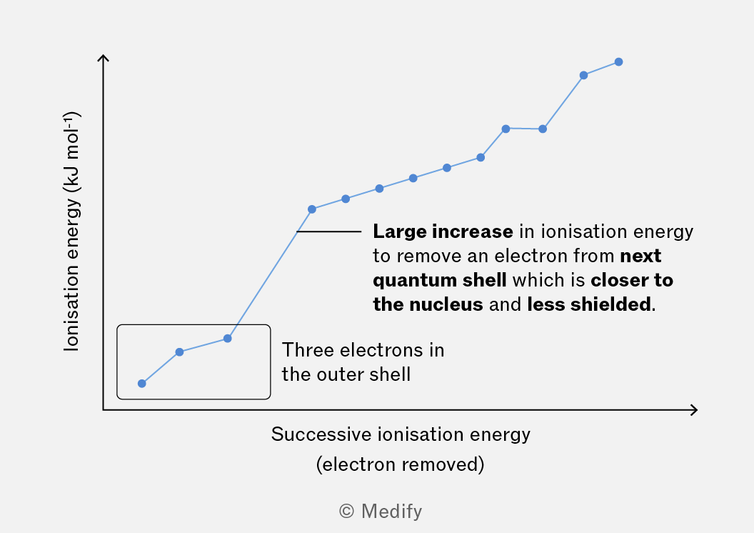
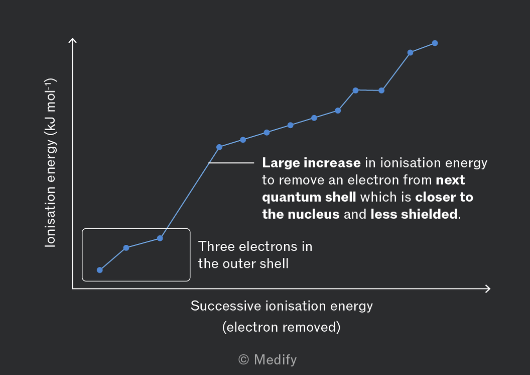
Elements in the same group show the largest jumps between the same successions; the elements have the same number of electrons in the highest energy level.
The change in the quantum shell the electron is being removed from accounts for this significant increase in ionisation energy.
The existence of quantum shells is supported by data from the first ionisation energies of elements of increasing atomic number.
Large drops in first ionisation energy are seen as you move between periods of the periodic table.
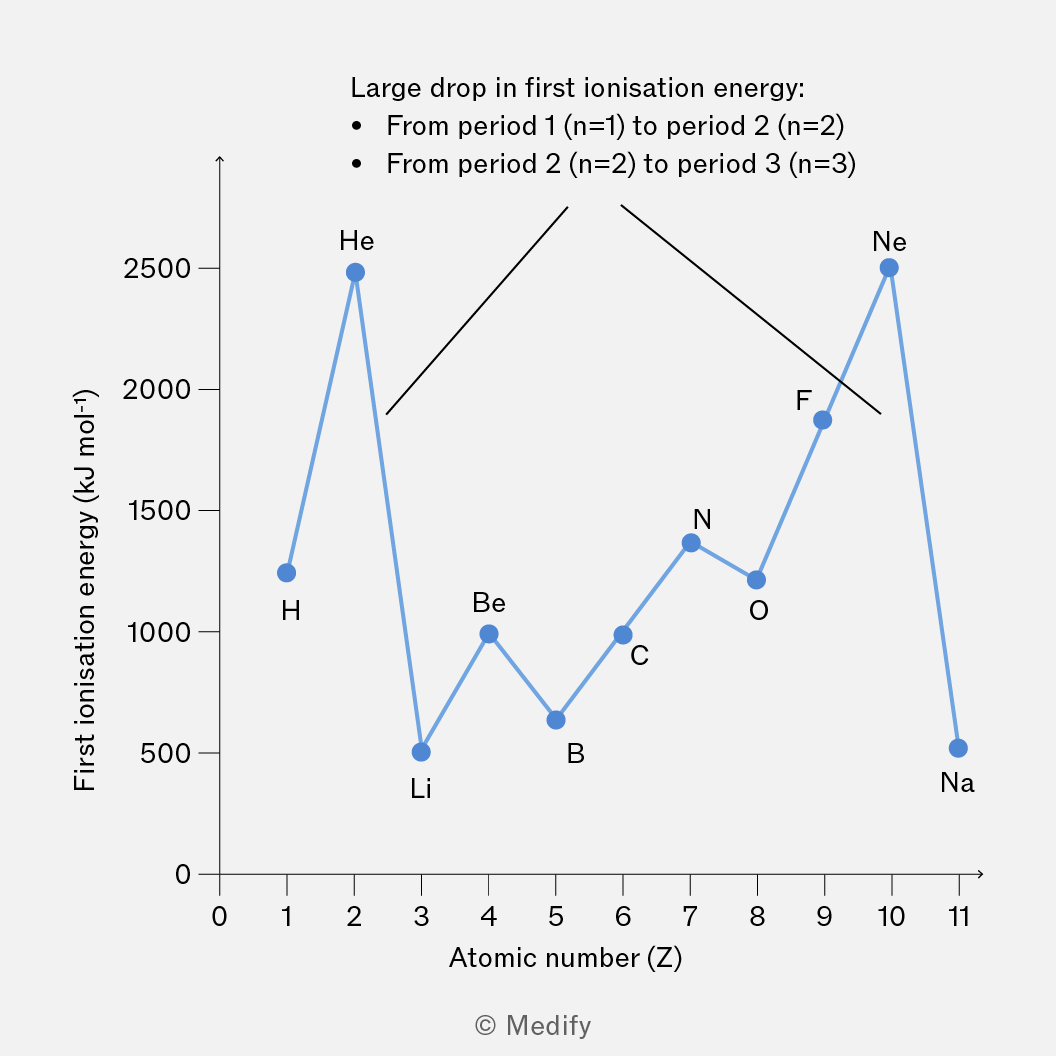
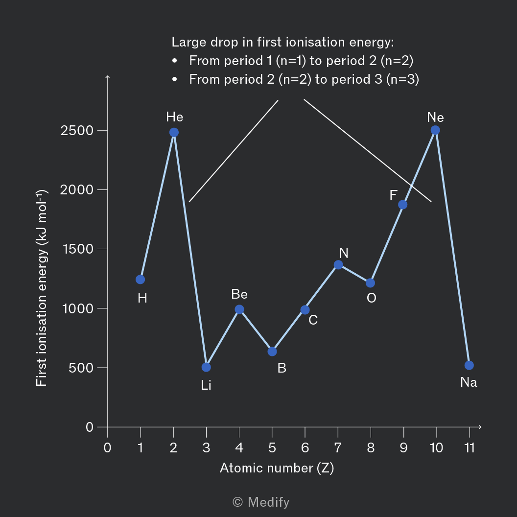
Electrons in higher energy quantum shells are further from the nucleus and experience more nuclear shielding, reducing the effective nuclear attraction. This reduces the energy required to remove an electron.
There is a general trend of increasing first ionisation energy across a period.
Ignoring subshells, as you move across the period you expect the nuclear charge to increase, causing the atomic radius to decrease, and the attraction to the outermost electron to increase; the first ionisation energy should increase.
Exceptions in the general trend can be explained by the existence of subshells.
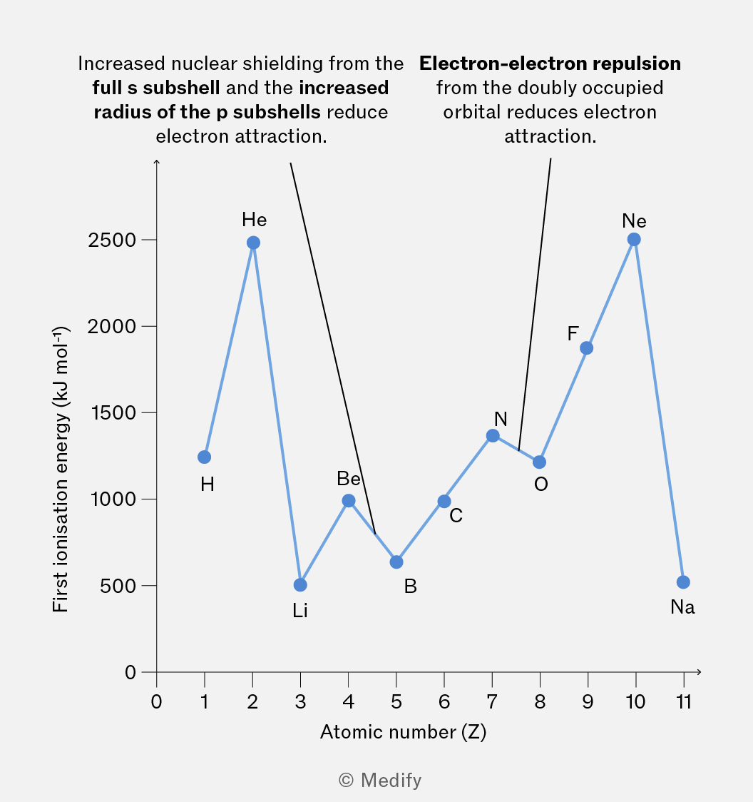
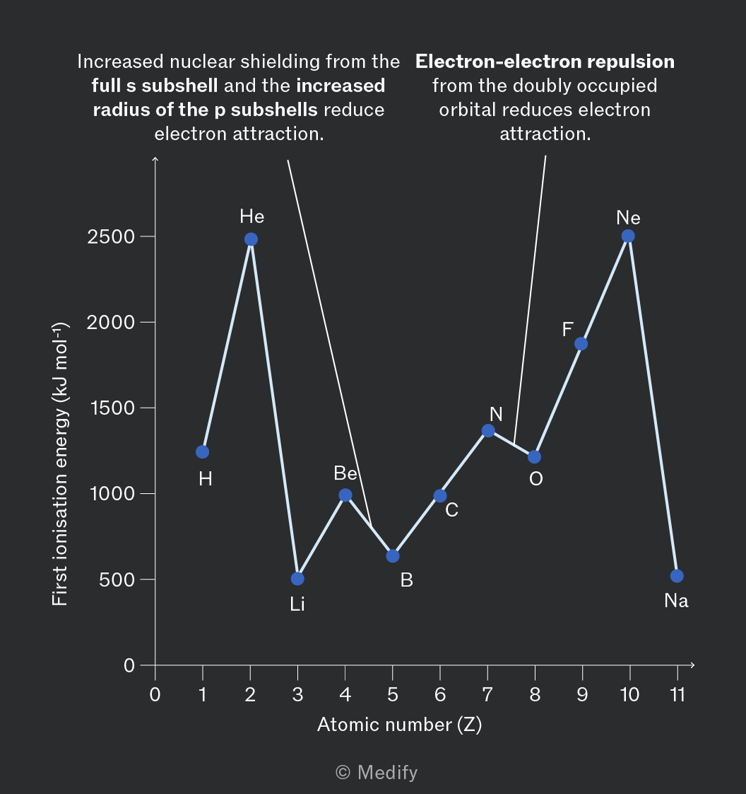
The drop in first ionisation energy from group 2 to group 3 (13) is due to the p subshell sitting at a higher energy level than the full s subshell. It is easier to remove an electron from the p subshell.
When subshells are half-filled each atomic orbital within the subshell is singly occupied. Moving along the period, following a half-filled subshell, a doubly occupied orbital is introduced. The impact of the increased electron–electron repulsion is greater than that of the increase in nuclear charge resulting in a weaker attraction and a drop in first ionisation energy from group 5 (15) to group 6 (16).
Balancing equations is a process to make sure there are the same number of atoms on each side of the symbol equation therefore obeying the law of conservation of mass.
When balancing equations ensure:
- the only numbers changes are the coefficients. These are placed in front of the formulae
- the formulae are not altered in any way, such as changing to to increase the number of oxygen atoms.
When balancing equations, we must:
- identify the number of atoms of each element in the reactants and products
- add the appropriate coefficient in front of the formulae to balance the number of atoms on each side
- make sure that the final balanced equation has the same number of each type of atom on both sides.
To convert a balanced (stoichiometric) symbol equation into an ionic equation we must:
- split compounds composed of aqueous ions into the individual ions, including any multiples
- leave intact in the equation any substance that does not contain aqueous ions or is covalently bonded
- cancel out spectator ions which appear on both sides of the equation and do not undergo any change.
Addition reactions are a type of chemical reaction where two or more molecules combine to form a single product.
A key example of an addition reaction is electrophilic addition to an alkene.
Elimination reactions remove atoms or small groups from a molecule, resulting in the formation of a double bond.
A key example of an elimination reaction is the elimination of water from an alcohol to form an alkene.
Substitution reactions involve replacing one atom or group with another.
Key examples of substitution reactions are electrophilic substitution on an aromatic ring or nucleophilic substitution on a haloalkane or alcohol.
Oxidation reactions involve:
- The loss of electrons
- Increase in oxidation state
- Addition of oxygen (sometimes)
- Removal of hydrogen (sometimes).
where:
- = oxidising agent
Formulae for oxidation reactions may include the symbol for an oxidising agent, .
Reduction reactions involve:
- The gain of electrons
- Decrease in oxidation state
- Removal of oxygen (sometimes)
- Addition of hydrogen (sometimes).
where:
- = reducing agent
Formulas for reduction reaction may include the symbol for a reducing agent, .
Hydrolysis reactions involve the breakdown of a compound by the addition of water, resulting in two products.
A key example of hydrolysis is the breakdown of an ester into a carboxylic acid and alcohol by water.
The term hydrolysis is also applied to other reactions where water is responsible for the breaking of a bond. In the hydrolysis of halogenoalkanes the water is a nucleophile breaking the bond.
Polymerisation reactions are reactions in which monomers are chemically bonded to form polymers.
where:
- represents the monomer
- is the number of monomers that combine to form the polymer
There are two types of polymerisation:
Addition polymerisation:
- Involves unsaturated monomers that contain double bonds. The double bonds open up, allowing the monomers to join together to form a long chain polymer.
Condensation polymerisation:
- Involves monomers that contain more than two functional groups. When these monomers react, a small molecule is eliminated as a by-product.
Free radical substitution of alkanes to produce haloalkanes.
There are three steps: initiation, propagation, and termination.
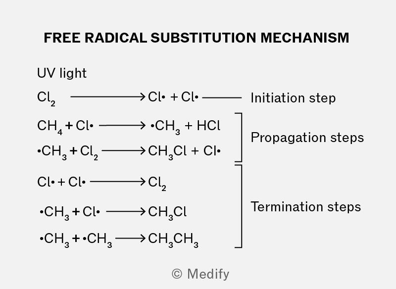
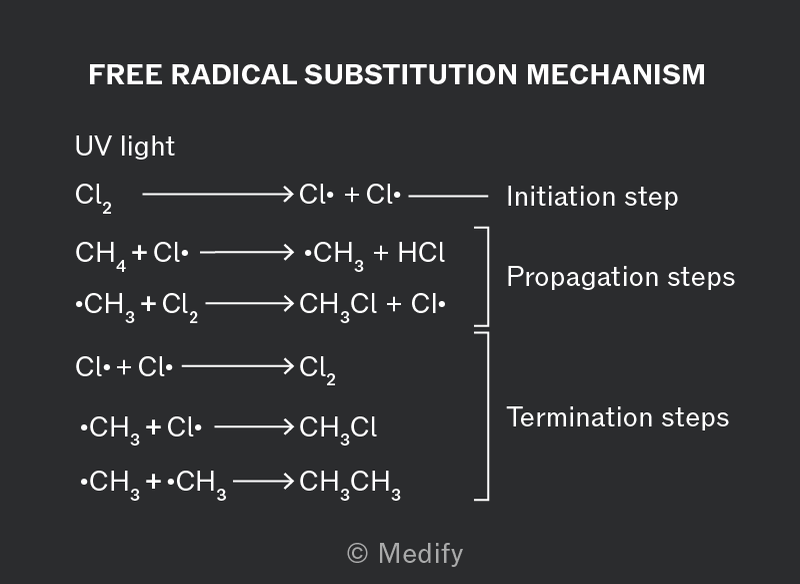
Initiation
Formation of halogen free radicals from a non radical molecule.
Conditions required: UV light.
Propagation
Step 1: The halogen radical reacts with the alkane, to produce an alkyl radical and .
Step 2: The alkyl radical then reacts with the halogen to produce a haloalkane and regenerates another halogen radical.
Termination
Two radicals combine, resulting in a non radical molecule being formed and no further reaction.